1.1.1 - Periodontium Components And Function
Why it matters
- Board stems often hide a basic anatomy question inside a diagnosis or treatment-planning scenario; if you do not know what tissue is being lost, you cannot choose the correct therapy.
- Many distractors exploit confusion between gingival findings and true periodontal attachment apparatus loss.
- Regeneration, resection, maintenance, implant planning, and restorative margin placement all depend on precise tissue relationships.
- Understanding normal function makes pathology interpretation faster: inflammation affects gingiva first, attachment loss involves PDL/cementum, and radiographic support changes reflect alveolar bone involvement.
Key Terms & Must-Know Facts
1.1.1.1 Key terms
- Periodontium: the tissues that surround and support the teeth.
- Gingiva: soft tissue covering the alveolar processes and surrounding cervical tooth portions.
- Free gingiva: unattached collar around the tooth forming the soft tissue wall of the sulcus.
- Attached gingiva: firm keratinized tissue bound to underlying periosteum/alveolar bone.
- Interdental gingiva: gingiva occupying the interproximal space.
- Periodontal ligament (PDL): specialized connective tissue between cementum and alveolar bone.
- Cementum: mineralized avascular tissue covering the root surface.
- Alveolar bone proper: bone lining the tooth socket where Sharpey fibers insert.
- Bundle bone: alveolar bone proper containing embedded PDL fiber bundles.
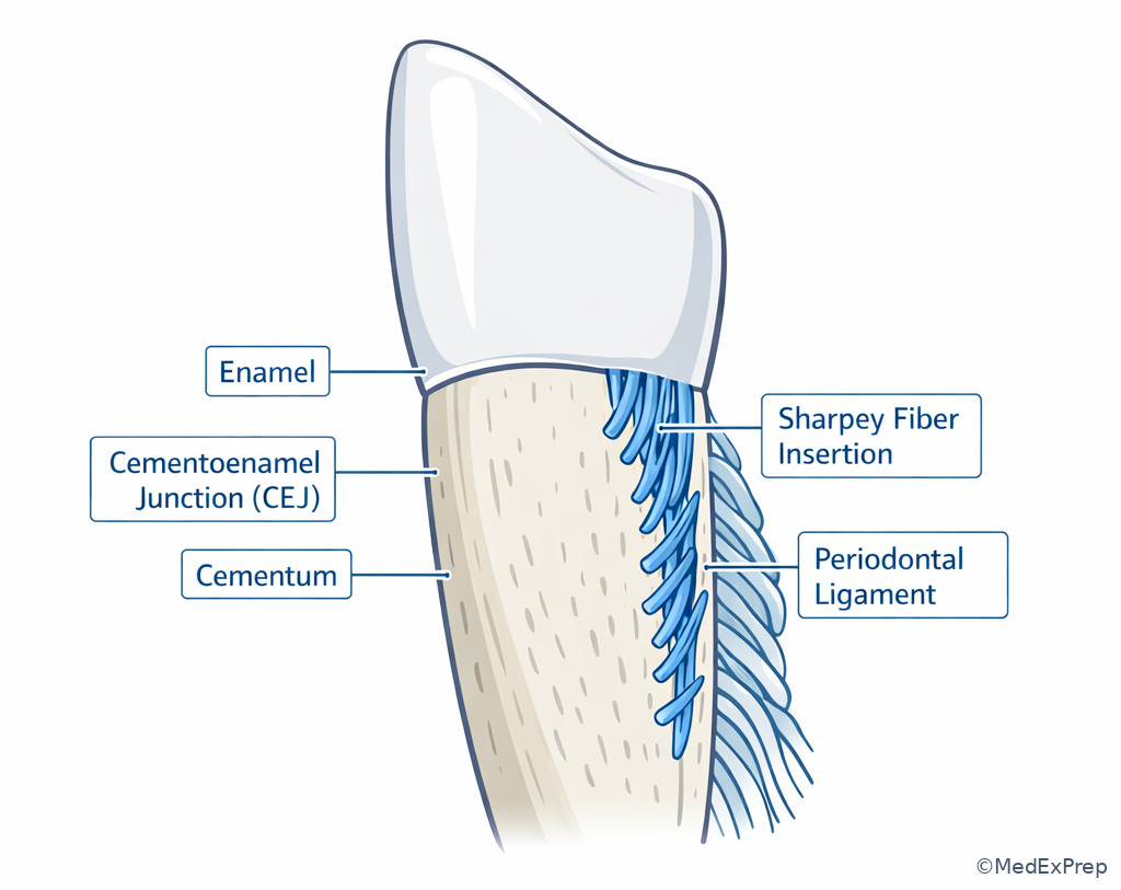
- Sharpey fibers: terminal portions of principal PDL fibers inserted into cementum and bone.
- Supracrestal tissue attachment: tissue coronal to the alveolar crest contributing to attachment around the tooth.
- Biotype/phenotype: tissue thickness and architecture influencing risk and treatment response.
- Alveolar process: tooth-dependent portion of the jaws that forms and persists with teeth.
1.1.1.2 Must-know facts
- The four principal components are gingiva, periodontal ligament, cementum, and alveolar bone.
- Only gingiva is directly visible clinically in routine examination; the other supporting tissues are inferred from probing, radiographs, and surgical exposure.
- The PDL is the key shock absorber and force-transmitting tissue.
- Cementum does not remodel like bone and serves as the root-side attachment surface for PDL fibers.
- Alveolar bone exists because teeth exist; tooth loss leads to alveolar process resorption.
- Inflammation limited to gingiva does not equal periodontitis; periodontitis requires loss of periodontal attachment apparatus.
- Radiographs show mineralized tissues, not the gingiva or direct attachment level.
- Clinical attachment concepts require understanding the cementoenamel junction as the reference point.
- Many surgical goals differ by tissue target: soft tissue positioning, root preparation, PDL preservation, or bone recontouring/regeneration.
Exam takeaway: Identify which tissue is involved before deciding whether a finding represents gingivitis, attachment loss, or loss of support.
graph TD
A[Periodontium] --> B[Gingiva]
A --> C[Periodontal ligament]
A --> D[Cementum]
A --> E[Alveolar bone]
B --> B1[Seal and protect]
C --> C1[Attach and absorb force]
D --> D1[Root attachment surface]
E --> E1[Socket support and remodeling]
C --> C2[Links root to bone]
D --> C
E --> C
See Fig 1 for the core structure-function map you should hold in mind during every periodontal diagnosis stem.
Core content
1.1.1.3 Foundational map: what counts as the periodontium?
A Explanation
The first board-style decision conflict is simple but constantly tested: Is the stem describing a surface soft-tissue problem, or is it describing loss of the tooth-supporting apparatus? The rule that resolves this is whether the finding involves only gingiva or extends to the attachment system formed by cementum, PDL, and alveolar bone. The periodontium includes four components: gingiva, periodontal ligament, cementum, and alveolar bone. Gingiva protects and seals. The other three create attachment and support. If a patient has red swollen marginal tissue with no attachment loss, do not overcall periodontitis. If a stem reports CAL, radiographic bone loss, furcation involvement, or mobility from reduced support, then the supporting periodontium is involved.
WHY THIS IS TESTED: candidates often choose therapy based on visible inflammation alone and ignore the deeper apparatus actually being assessed in periodontal diagnosis.
Another useful choose-versus-reject rule: choose “supporting tissues” when the question asks about tooth retention, mobility, force distribution, regenerative goals, or osseous change; reject “gingiva alone” because gingiva does not by itself anchor the tooth in bone. Gingiva is part of the periodontium, but not the whole attachment system. This distinction becomes critical later when you study supracrestal tissue attachment and phenotype, because the visible margin may appear stable while deeper support is compromised.
Exam takeaway: On exam stems, visible inflammation alone does not prove loss of attachment; support depends on deeper tissues.
B Worked example
A 38-year-old woman is referred before restorative treatment of a fractured maxillary premolar. She has controlled hypothyroidism, a history of orthodontic treatment, and occasional vaping. The general dentist notes “periodontal disease” because of generalized marginal erythema and 4 mm probing depths on several posterior teeth. Radiographs show intact crestal bone levels. There is mild bleeding on probing, but no recession, no mobility, and no CAL from the CEJ. A tempting clue is the posterior 4 mm sulci, which may look like early periodontitis.
Reasoning chain: Identify the task: distinguish gingival inflammation from attachment loss. Extract key facts: erythema, bleeding, no radiographic bone loss, no CAL, no mobility. Apply rule: visible inflammation without attachment loss indicates gingival disease rather than periodontitis. Eliminate distractors: periodontitis would require support loss; trauma from occlusion would not explain generalized BOP and erythema; endo-perio is unsupported. Verify: the affected tissue is primarily gingiva, not the deeper attachment apparatus.
C Exam trap
D Checkpoint
Question: A 29-year-old man with plaque accumulation, intermittent bleeding during brushing, and generalized erythematous gingival margins has probing depths of 3–4 mm but no radiographic bone loss, no recession, and no CAL. Which periodontal component is primarily diseased in this scenario?
- Gingiva - Tempting because the signs are superficial inflammatory findings; correct because the stem lacks evidence of attachment apparatus loss.
- Periodontal ligament - Tempting because probing relates to the sulcus, but wrong because no attachment loss or mobility suggests PDL destruction.
- Cementum - Tempting because root surface attachment matters in periodontal disease, but wrong because the stem gives no evidence of root attachment loss.
- Alveolar bone - Tempting because periodontal disease often implies bone involvement, but wrong because radiographs show intact crestal support.
Answer: A. Gingiva.
1.1.1.4 Gingiva: barrier, seal, and clinical landmark
A Explanation
The next decision conflict is when should you think of gingiva as a protective tissue versus a diagnostic landmark that can mislead you? The single resolving rule is this: gingiva is the only component directly visible in routine examination, but it does not by itself tell you the full status of attachment. Gingiva covers the alveolar process and surrounds the cervical tooth region. Its major functional roles are barrier protection, resistance to mechanical insult, and formation of the soft tissue wall of the sulcus. Clinically you must separate free gingiva, attached gingiva, and interdental gingiva because contour, color, stippling, consistency, and bleeding are interpreted at this level.
Choose gingival interpretation when the stem asks about margin position, recession, inflammatory enlargement, plaque-related redness, or restorative margin tolerance. Reject “deeper support loss” if the only data are color and contour changes. The gingiva also matters because the location of the gingival margin can produce pseudo-pocketing or mask recession. This is why probing depth alone is never enough; you must integrate the margin position and CEJ reference point. If the margin is coronal from enlargement, probing may be deeper without attachment loss. If the margin is apical from recession, probing depth may be modest even though attachment loss is significant.
WHY THIS IS TESTED: exam questions frequently pair a visible soft tissue abnormality with one deeper finding, and the correct answer depends on not equating sulcus depth with attachment loss.
Exam takeaway: Gingival contour and margin position affect probing interpretation, so use the CEJ and support findings before labeling periodontitis.
B Worked example
A 52-year-old man preparing for a crown on a mandibular molar presents with hypertension, xerostomia from medication use, and inconsistent flossing. The tissue around the molar is enlarged and bleeds on probing. Probing depths are 5 mm circumferentially, but the CEJ is not apical to the margin, there is no recession, and radiographs show normal interproximal crest levels. The tempting-but-wrong clue is the uniform 5 mm probing depth, which invites a diagnosis of established periodontitis.
Reasoning chain: Identify task: determine whether deep probing reflects gingival enlargement or attachment loss. Extract key facts: enlarged tissue, no recession, normal bone, CEJ relationship not showing apical attachment loss. Apply rule: coronal displacement of the gingival margin can create pseudo-pocketing. Eliminate distractors: PDL and bone loss are unsupported; cementum destruction is inferred only when attachment is lost. Verify: primary tissue process is gingival enlargement/inflammation.
C Exam trap
D Checkpoint
Question: A 46-year-old woman with plaque accumulation and generalized gingival enlargement has multiple 5 mm probing depths. Which additional finding most strongly supports pseudo-pocketing rather than true periodontal attachment loss?
- Bleeding on probing - Tempting because inflamed tissues often bleed, but wrong because BOP can occur in both gingivitis and periodontitis.
- Normal crestal bone levels on radiographs - Tempting because radiographs are imperfect, but correct because preserved support strongly argues against established attachment loss in this stem.
- Plaque accumulation at the margin - Tempting because plaque drives enlargement, but wrong because plaque can also coexist with periodontitis.
- Posterior molar involvement - Tempting because molars are often periodontal problem sites, but wrong because site location alone does not distinguish pseudo-pocketing.
Answer: B. Normal crestal bone levels on radiographs.
- The only periodontal component directly visible on routine exam is gingiva.
- True or False: probing depth alone proves attachment loss. False.
- Name the three common clinical gingival subdivisions: free, attached, interdental.
Exam takeaway: Recognize free gingiva, attached gingiva, mucogingival junction, sulcus, and interdental papilla to interpret probing and recession correctly.
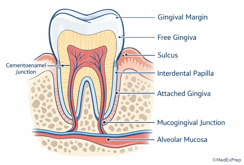
See Fig 3 when reviewing how soft tissue landmarks can change the meaning of a probing measurement.
1.1.1.5 Periodontal ligament and cementum: the living attachment unit
A Explanation
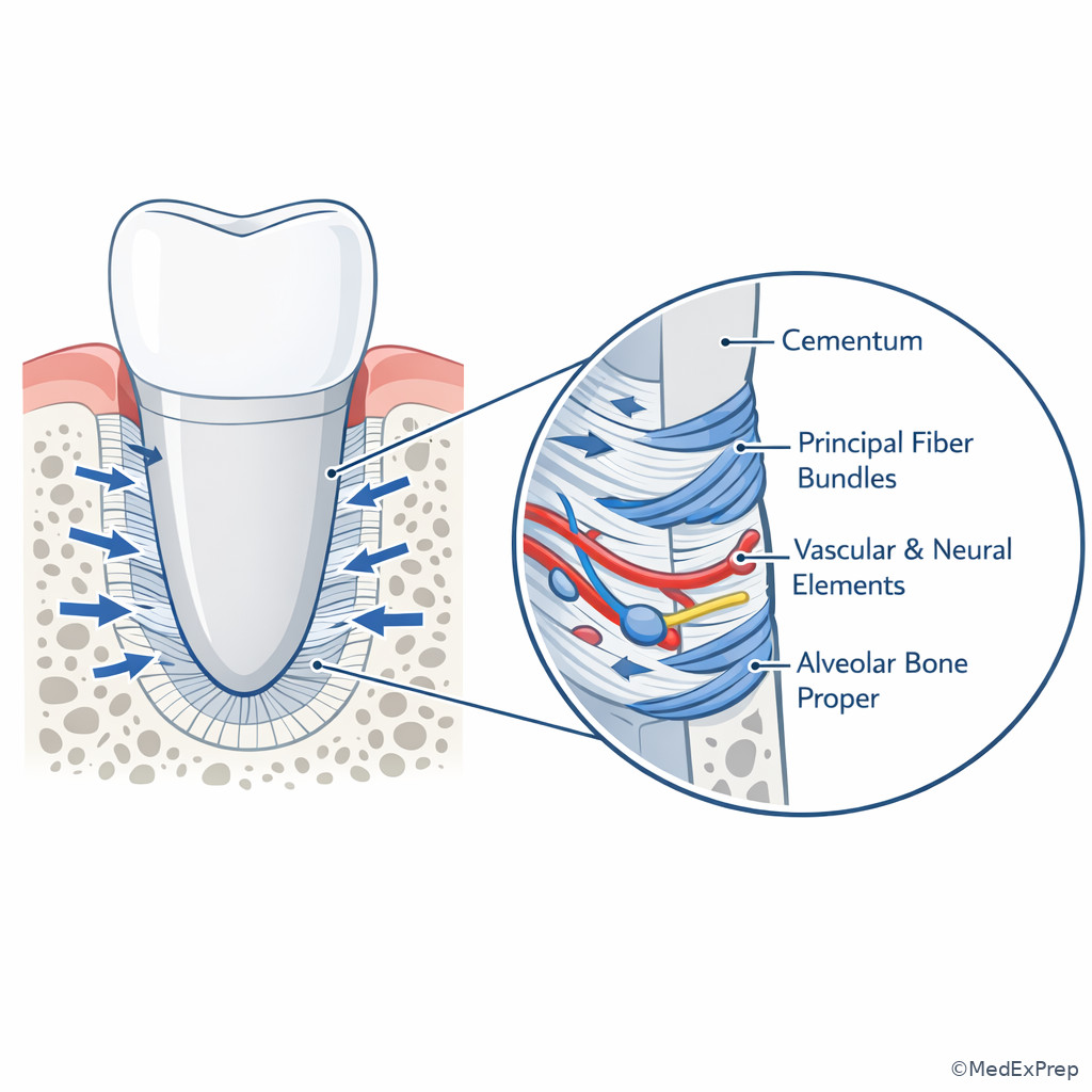
Now move deeper. The decision conflict becomes what actually anchors the tooth, and which tissue should you think about when the stem mentions force distribution, mobility, regeneration, or root instrumentation? The single resolving rule is that the PDL-cementum complex is the root-side attachment unit. The periodontal ligament is a specialized vascular connective tissue occupying the space between root cementum and alveolar bone. Its functions include tooth attachment, shock absorption, sensory input, nutritive support, and cellular participation in remodeling and healing. Cementum is the mineralized root covering that receives insertion of Sharpey fibers. It is avascular, does not have the same remodeling behavior as bone, and is indispensable as the root attachment surface.
Choose the PDL when the exam asks about mobility, occlusal force dissipation, proprioception, orthodontic tooth movement biology, or regenerative tissue goals. Choose cementum when the stem asks what covers the root, where PDL fibers insert on the tooth side, or why root planing decisions matter biologically. Reject alveolar bone alone as the sole attachment tissue because the tooth is not fused directly to bone in health; the PDL intervenes. Reject gingiva when the issue is support rather than soft tissue seal.
WHY THIS IS TESTED: board items often compress multiple tissues into one phrase like “attachment apparatus,” and candidates must know the root-side versus bone-side roles to answer regeneration or prognosis questions correctly.
Exam takeaway: When the stem asks what links tooth to bone, think cementum plus PDL rather than gingiva or bone alone.
B Worked example
A 61-year-old man with a history of bruxism, controlled type 2 diabetes, and a planned fixed prosthesis presents with localized grade I mobility of a mandibular canine. Probing depths are 3 mm, gingival tissues are minimally inflamed, and radiographs show mild horizontal bone loss but intact root structure. The referring dentist asks which tissue primarily allows physiologic tooth movement and force absorption. A tempting wrong clue is the radiographic bone loss, which may push the candidate to answer alveolar bone.
Reasoning chain: Identify task: determine the functional tissue responsible for suspension and force distribution. Extract key facts: mobility question, force absorption, living attachment system. Apply rule: the PDL is the shock absorber and suspensory ligament. Eliminate distractors: cementum is needed for fiber insertion but does not itself absorb force; alveolar bone supports the socket but health requires the intervening ligament; gingiva does not suspend teeth. Verify: answer centers on PDL function within the PDL-cementum-bone unit.
C Exam trap
D Checkpoint
Question: Which periodontal component directly covers the root and serves as the tooth-side insertion surface for principal PDL fibers?
- Alveolar bone proper - Tempting because fibers insert there too, but wrong because it is the socket-side insertion, not the root covering.
- Cementum - Tempting because it is less emphasized than bone, and correct because it is the root surface attachment substrate.
- Attached gingiva - Tempting because it is firm and close to the tooth, but wrong because it is a soft tissue covering, not the root covering.
- Periosteum - Tempting because it lines bone surfaces, but wrong because it is not the tooth-side insertion tissue.
Answer: B. Cementum.
Exam takeaway: Visualize the ligament space, fiber orientation, and cementum-to-bone connection to answer mobility and regeneration questions.
1.1.1.6 Alveolar bone: tooth-dependent support and radiographic context
A Explanation
The decision conflict here is when should you rely on alveolar bone findings, and when should you avoid over-interpreting radiographs? The single rule is that alveolar bone is the mineralized socket support of the tooth, but radiographs show only one part of the story. The alveolar process forms with tooth eruption and is maintained by the presence of teeth. The alveolar bone proper lines the socket and receives PDL fiber insertion; surrounding cortical and cancellous bone provide additional architecture. Bone supports the tooth indirectly through the PDL. This is why radiographic crestal level is a support indicator, not a direct picture of soft tissue attachment.
Choose alveolar bone when the question concerns radiographic bone loss patterns, socket anatomy, osseous support, extraction consequences, or regenerative/resective osseous planning. Reject using bone alone to diagnose attachment level because radiographs do not reveal the gingival margin, sulcus, or exact histologic attachment. A patient may have clinical inflammation with normal radiographs, or early attachment changes not fully visible on imaging.
WHY THIS IS TESTED: many stems include a radiograph because it tempts the candidate to anchor on bone and ignore clinical attachment data. The best answer usually integrates both.
Exam takeaway: Radiographic bone supports diagnosis of periodontal breakdown only when interpreted alongside clinical findings.
B Worked example
A 67-year-old woman referred for implant planning lost tooth no. 19 five years ago. She has well-controlled osteoporosis, a history of smoking cessation 10 years earlier, and a fixed bridge on the contralateral side. The ridge is reduced in height and width. The question asks why the alveolar process has diminished after tooth loss. A tempting wrong clue is her osteoporosis, which may encourage a systemic explanation as the primary answer.
Reasoning chain: Identify task: explain local anatomic change after extraction. Extract key facts: site is edentulous for years, ridge reduction localized to missing-tooth area. Apply rule: alveolar process is tooth-dependent and resorbs after tooth loss. Eliminate distractors: systemic factors may modify remodeling but do not replace the fundamental principle that alveolar bone exists to support teeth. Verify: this is a function/anatomy question about tooth dependence of the alveolar process.
C Exam trap
D Checkpoint
Question: Which statement best explains the alveolar process in board-style periodontal reasoning?
- It is a permanent jaw structure independent of tooth presence - Tempting because jaw bone seems static, but wrong because alveolar process is tooth-dependent.
- It directly fuses to the root in health - Tempting because bone supports the tooth, but wrong because the PDL separates root and bone.
- It forms to support teeth and tends to resorb after tooth loss - Tempting because this is a classic principle, and correct because it explains socket support and post-extraction ridge change.
- It is clinically visible without surgery or radiography - Tempting because crestal changes may influence gingival form, but wrong because bone itself is not directly visible in routine non-surgical exam.
Answer: C. It forms to support teeth and tends to resorb after tooth loss.
- The alveolar process is tooth-dependent.
- True or False: the tooth is fused directly to socket bone in health. False.
- Name the key mineralized support tissue visible on routine radiographs: alveolar bone.
Exam takeaway: Use the layered relationship of gingiva, cementum, PDL, and alveolar bone to localize where disease or therapy is acting.
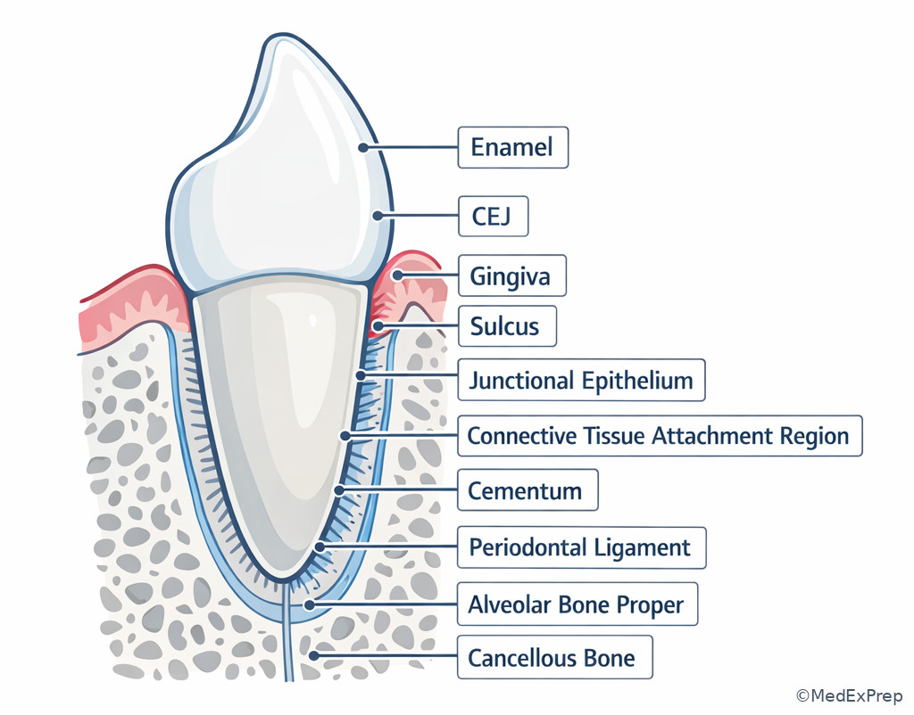
See Fig 4 for the spatial model that underlies most probing, bone-loss, and regenerative reasoning questions.
1.1.1.7 Structure-function integration: how anatomy drives diagnosis, prognosis, and therapy selection
A Explanation
The final foundational-to-integration conflict is how do you use anatomy to choose the right periodontal reasoning pathway? The single resolving rule is: first localize the tissue involved, then ask what function has been lost, then choose the most biologically appropriate next step. If gingiva is inflamed, prioritize etiologic control and margin interpretation. If PDL-cementum attachment is compromised, think attachment loss and regenerative implications. If alveolar support is reduced, think prognosis, defect morphology, and long-term maintenance. The periodontium functions as a coordinated unit, so destruction rarely remains academically isolated for long, but the earliest and most testable exam question is always “which tissue matters most here?”
This is also where anatomy connects to specialty-level care. Flap surgery accesses root and bone. Regeneration seeks new attachment apparatus, not merely a long junctional epithelial seal. Mucogingival procedures may change gingival position or phenotype without directly rebuilding lost bone support. Supportive periodontal therapy monitors recurrence at the tissue level: inflammation in gingiva, attachment change in PDL/cementum relationship, and mineralized support change in bone.
WHY THIS IS TESTED: board cases reward candidates who use anatomy as a framework for treatment sequencing rather than jumping from a single finding to a procedure name.
Exam takeaway: The best next step follows the tissue involved—soft tissue control for gingival problems, attachment-focused thinking for deeper support loss.
B Worked example
A 55-year-old woman seeks periodontal clearance before replacing a missing maxillary lateral incisor. She has controlled rheumatoid arthritis, a history of smoking 15 cigarettes per day, and a thin scalloped soft tissue appearance. On adjacent teeth there is 2 mm recession, minimal inflammation, noncarious cervical wear, 3 mm probing depths, no radiographic bone loss, and no mobility. The distracting clue is smoking history, which might tempt a candidate to assume deeper periodontal support loss.
Reasoning chain: Identify task: determine which periodontal component/function should guide next thinking. Extract facts: recession and thin soft tissue appearance, no bone loss, no deep pockets, no mobility. Apply rule: this is primarily a gingival/phenotype and margin-position issue, not a current deep support-loss issue. Eliminate distractors: alveolar bone loss is not demonstrated; PDL compromise is not suggested by mobility or CAL progression pattern; cementum matters secondarily because root surface is exposed, but the dominant decision lane is soft tissue/phenotype planning. Verify: anatomy points toward gingival phenotype and mucogingival considerations, which will be expanded in the next lessons.
C Exam trap
D Checkpoint
Question: A stem asks which tissue must be re-established for true periodontal regeneration rather than soft tissue repair alone. Which answer best fits that concept?
- New junctional epithelium only - Tempting because epithelial healing is common, but wrong because regeneration requires more than epithelial closure.
- New attachment apparatus including PDL insertion on new cementum with supporting bone - Tempting because it sounds comprehensive, and correct because regeneration targets the lost attachment unit.
- Thicker attached gingiva only - Tempting because soft tissue augmentation can improve stability, but wrong because it does not alone recreate the attachment apparatus.
- Reduced gingival inflammation only - Tempting because inflammation control is essential, but wrong because regeneration is a structural, not merely inflammatory, endpoint.
Answer: B. New attachment apparatus including PDL insertion on new cementum with supporting bone.
Exam takeaway: Match the dominant tissue problem to the correct periodontal decision pathway before choosing therapy.
graph TD
A[Clinical finding] --> B{Primary tissue involved?}
B -->|Gingiva| C[Inflammation or margin issue]
B -->|PDL/Cementum| D[Attachment apparatus issue]
B -->|Bone| E[Support/defect issue]
C --> F[Etiology control or soft tissue planning]
D --> G[Assess CAL, regeneration potential, prognosis]
E --> H[Assess pattern, defect morphology, support level]
G --> I[Integrated periodontal treatment plan]
H --> I
F --> I
See Fig 5 and Fig 6 for the core “localize first, then decide” workflow that should frame future treatment-planning questions.
Tables
| Component | Primary composition | Main function | Best exam clue |
|---|---|---|---|
| Gingiva | Keratinized/nonkeratinized soft tissue with epithelial and connective tissue components | Barrier, seal, sulcus wall, protection | Visible margin changes, BOP, enlargement, recession |
| Periodontal ligament | Vascular cellular connective tissue with principal fibers | Tooth suspension, force absorption, proprioception, remodeling | Mobility, force questions, regeneration logic |
| Cementum | Mineralized avascular root covering | Root-side attachment surface for PDL fibers | What covers root? where fibers insert on tooth? |
| Alveolar bone | Mineralized socket-supporting bone | Osseous support and socket architecture | Radiographic crestal support, edentulous ridge change |
| Comparison point | Gingival finding only | Attachment apparatus involvement | Supportive bone involvement |
|---|---|---|---|
| Typical data source | Visual exam, BOP, tissue contour | CEJ-based CAL, probing interpretation, mobility context | Radiographs, defect architecture, support level |
| What it means | Inflammation or margin-position change may be present | Root attachment relationship has changed | Mineralized support has been reduced or reshaped |
| Common board trap | Calling gingivitis periodontitis | Ignoring root-side biology in regeneration questions | Using radiograph alone as a diagnosis |
| Best next reasoning step | Ask if there is true CAL | Ask what tissue was lost and can be restored | Ask whether bone pattern changes prognosis or therapy |
| Related upcoming topic | Keratinized Tissue and Phenotype | Supracrestal Tissue Attachment | Bone Loss Mechanisms |
| Stem wording | Most likely tissue focus | Why | Reject this tempting distractor |
|---|---|---|---|
| Red, edematous, bleeding margin | Gingiva | Surface inflammatory tissue signs | Periodontitis if no CAL/bone loss is shown |
| Physiologic mobility, force absorption, proprioception | PDL | Core ligament functions | Alveolar bone as sole answer |
| Root surface attachment substrate | Cementum | Fiber insertion on tooth side | Attached gingiva |
| Radiographic crestal support or defect form | Alveolar bone | Mineralized socket support | Direct measurement of soft tissue attachment |
| Post-extraction ridge shrinkage | Alveolar process | Tooth-dependent bone resorption | Generalized systemic disease as first explanation |
| True regeneration endpoint | New attachment apparatus | Requires PDL, cementum, and supporting bone | Long junctional epithelium alone |
| Clinical clue | Looks similar | But actually suggests | Discriminating clue |
|---|---|---|---|
| 5 mm probing with enlarged tissue | Periodontal pocket | Pseudo-pocketing | Coronal margin position with no support loss |
| Minimal probing depth with recession | Healthy attachment | Possible attachment loss | Apical margin relative to CEJ |
| Radiographic bone intact despite BOP | No disease | Likely gingival inflammation | Inflammation can exist without osseous loss |
| Localized ridge resorption after extraction | Systemic skeletal disorder only | Loss of tooth-dependent alveolar process | Change confined to edentulous site |
| Mobility question | Bone-only support issue | PDL-centered function question | Suspension and shock absorption language |
Exam takeaway: Use clinical examination for gingiva and integrated clinical-radiographic reasoning for deeper tissues.
graph LR
A[Visible directly] --> B[Gingiva]
C[Inferred clinically] --> D[PDL status]
C --> E[Cementum attachment]
F[Seen mainly on imaging] --> G[Alveolar bone]
B --> H[Color contour bleeding recession]
D --> I[Mobility force response probing context]
E --> J[Root-side attachment logic]
G --> K[Crest level defect pattern ridge form]
Exam takeaway: Understand alveolar bone proper, crest, cancellous bone, and tooth dependence for ridge-loss and support questions.
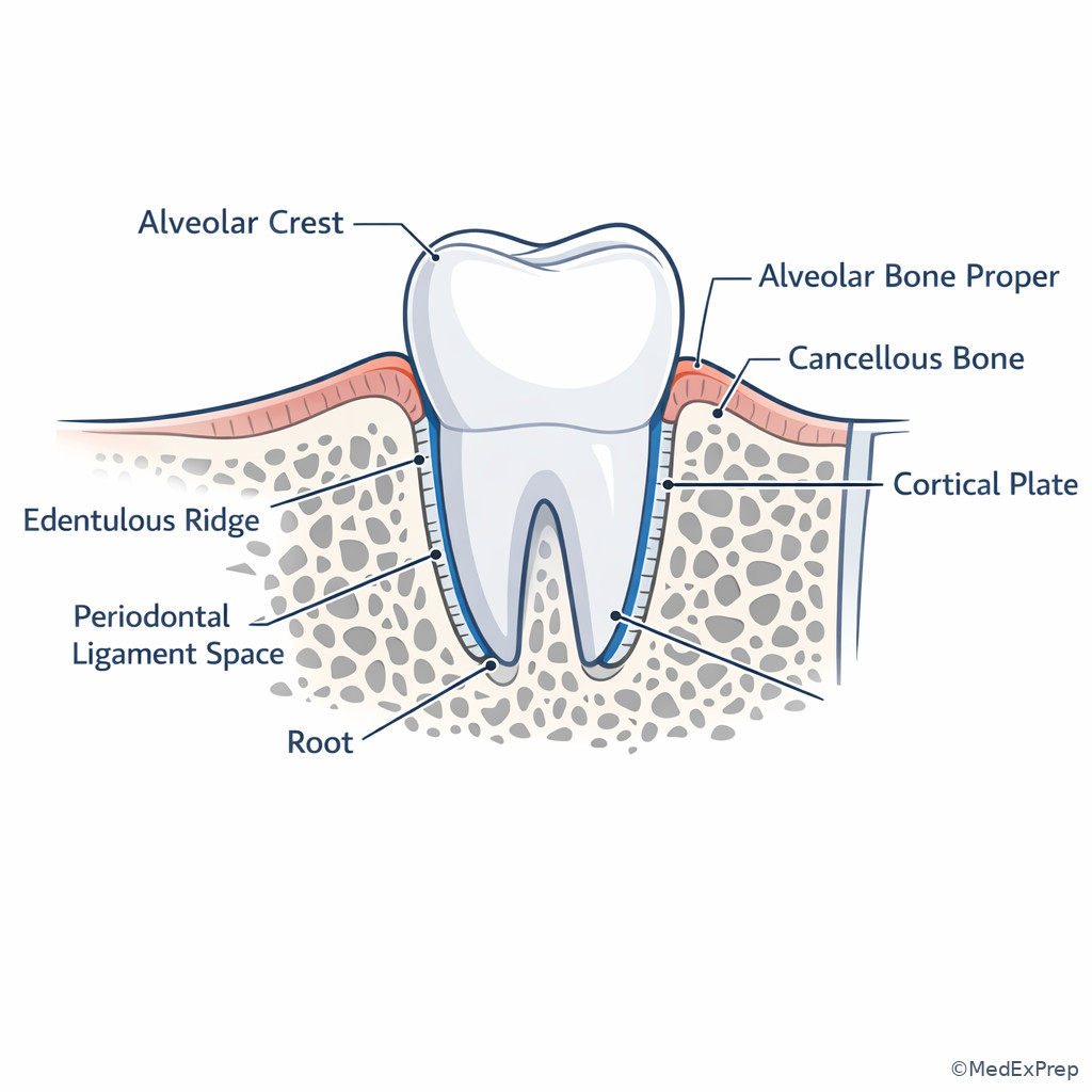
Exam takeaway: Cementum is the root covering that enables fiber insertion and should not be confused with enamel or bone.
Exam Traps & Differentiators
- Most common wrong answer: equating deep probing with periodontitis. Why: candidates anchor on the number instead of interpreting margin position, CEJ, and bone support.
- Looks similar but isn’t: gingival enlargement with pseudo-pocketing versus true periodontal pocketing. One requires support-loss evidence; the other may be coronal tissue enlargement only.
- Looks similar but isn’t: recession with shallow probing versus health. The shallow number is misleading when the margin has migrated apically.
- Looks similar but isn’t: radiographic crest change versus definitive attachment diagnosis. Bone image findings inform support, but clinical attachment interpretation still matters.
- Looks similar but isn’t: root coverage or soft tissue augmentation versus regeneration. Soft tissue improvement is not the same as re-creating the full attachment apparatus.
| If the stem says... | Think... | Not automatically... |
|---|---|---|
| Red, enlarged, bleeding tissue | Gingival inflammation or enlargement | Periodontitis |
| Mobility, force dissipation, proprioception | PDL function | Bone alone |
| Root covering or fiber insertion on tooth side | Cementum | Enamel or gingiva |
| Edentulous ridge resorption | Loss of tooth-dependent alveolar process | Only generalized skeletal disease |
| Need for new attachment apparatus | Regeneration target includes PDL/cementum/bone | Soft tissue healing alone |
If the stem says generalized inflamed margins with intact bone, think gingival disease rather than support loss.
If the stem says “what tissue suspends the tooth,” think periodontal ligament.
If the stem says “what covers the root and accepts fiber insertion,” think cementum.
If the stem says “why did the ridge resorb after extraction,” think tooth-dependent alveolar process loss.
If the stem says “true regeneration,” think new attachment apparatus, not epithelial repair alone.
Algorithm / Approach
Exam takeaway: Start with the clue type, then verify with the right confirming data rather than guessing from one finding.
flowchart TD
A[Board stem] --> B{Main clue type}
B -->|Color contour bleeding| C[Gingiva]
B -->|Mobility force proprioception| D[PDL]
B -->|Root covering attachment surface| E[Cementum]
B -->|Radiographic crest ridge defect| F[Alveolar bone]
C --> G[Check CEJ and CAL]
D --> H[Check mobility and support context]
E --> I[Check regeneration/root questions]
F --> J[Check clinical correlation]
G --> K[Choose best answer]
H --> K
I --> K
J --> K Rapid Review
- Gingiva → visible protective tissue; inflammation here alone does not prove periodontitis.
- Free gingiva → forms the soft tissue wall of the sulcus.
- Attached gingiva → firm bound tissue; think stability and resistance, not root attachment.
- PDL → shock absorber and proprioceptive tissue; key answer for mobility/function stems.
- Cementum → root-side fiber insertion surface; not enamel, not bone.
- Alveolar bone proper → socket lining receiving Sharpey fibers on the bone side.
- Alveolar process → tooth-dependent; resorbs after tooth loss.
- Radiographs → show mineralized support, not direct gingival attachment.
- Pseudo-pocket → deep probing from coronal gingival enlargement without true attachment loss.
- Recession → shallow probing can still coexist with attachment loss if the margin is apical.
- Regeneration → requires new attachment apparatus, not epithelial coverage alone.
- Periodontium question strategy → localize tissue first, then decide function lost.
Self-check quiz
1. A 31-year-old woman presents for specialty evaluation before orthodontic retreatment. She has generalized plaque accumulation, mild marginal erythema, and bleeding during brushing. Probing depths are 3–4 mm, there is no recession, and bitewings show intact crestal bone. Which periodontal component is most directly represented by the visible inflamed tissue?
- Gingiva
- Periodontal ligament
- Cementum
- Alveolar bone
2. A 44-year-old man with bruxism, controlled hypertension, and mild fremitus asks which tissue allows normal tooth suspension and force absorption. Clinical examination shows no acute inflammation and only slight mobility of a mandibular incisor. Which periodontal component is the best answer?
- Attached gingiva
- Periodontal ligament
- Cementum
- Alveolar crest
3. A 53-year-old woman taking a calcium channel blocker presents with generalized gingival enlargement, bleeding on probing, and several 5 mm probing depths. She has no recession, no mobility, and radiographs show preserved crestal support. What is the most appropriate interpretation of the probing depths?
- They confirm periodontitis because depths of 5 mm represent true pockets
- They likely reflect pseudo-pocketing due to coronal gingival enlargement
- They indicate primary cementum loss without gingival involvement
- They prove traumatic occlusion as the main disease process
4. A 62-year-old man with a missing mandibular first molar for 8 years is being evaluated for implant site development. He has controlled diabetes, a remote smoking history, and localized ridge narrowing at the edentulous site. Which explanation best accounts for the ridge change?
- Alveolar process resorption after tooth loss
- PDL contraction around the absent root
- Cementum overgrowth on the socket wall
- Interdental gingival migration into cancellous bone
5. A 48-year-old woman is referred for evaluation of a mandibular premolar before crown lengthening. She has mild plaque accumulation, 2 mm facial recession, probing depths of 3 mm, and no radiographic bone loss. Which finding most strongly prevents you from calling this “periodontal health with no attachment loss”?
- Bleeding on probing at one site
- Apical displacement of the gingival margin relative to the CEJ
- Posterior tooth location
- Absence of mobility
6. A 57-year-old man with controlled type 2 diabetes, 20-pack-year smoking history, and a planned fixed prosthesis has a mandibular canine with mild mobility, 3 mm probing depths, minimal inflammation, and slight horizontal bone loss radiographically. The stem asks which tissue most directly explains physiologic tooth movement and force dissipation. What is the best answer?
- Alveolar bone proper
- Periodontal ligament
- Cementum
- Attached gingiva
7. A 50-year-old woman with controlled rheumatoid arthritis, thin scalloped gingiva, and a restorative plan for maxillary anterior veneers has 2 mm facial recession on a canine, 2–3 mm probing depths, no radiographic bone loss, and no mobility. Which tissue/problem lane should guide the most appropriate next periodontal reasoning?
- Primary osseous defect management
- PDL failure with advanced support loss
- Soft tissue margin/phenotype evaluation
- Immediate regenerative therapy for vertical bone loss
8. A 60-year-old man presents after initial periodontal therapy with reduced inflammation in a site previously affected by intrabony loss. A board-style question asks which outcome would represent true periodontal regeneration rather than repair alone. Which is the most appropriate answer?
- Long junctional epithelium with reduced bleeding
- Coronal soft tissue coverage of the root
- New attachment apparatus with new cementum, functionally oriented PDL, and supporting bone
- Thicker attached gingiva with shallower probing
Answer key
1. Correct answer: A. Gingiva.
- A. Gingiva: Tempting because the stem centers on visible inflamed tissue; correct because erythema and BOP without support loss localize primarily to gingiva. Single clue: visible marginal inflammation with intact bone. Source: Newman and Carranza, Clinical Periodontology.
- B. Periodontal ligament: Tempting because probing makes candidates think “attachment,” but incorrect because no mobility, CAL, or support loss is described. Single clue: only superficial inflammatory findings are given. Source: Newman and Carranza.
- C. Cementum: Tempting because it is part of the attachment apparatus, but incorrect because the stem does not ask about root covering or fiber insertion. Single clue: no root-side attachment issue is presented. Source: Lindhe, Clinical Periodontology and Implant Dentistry.
- D. Alveolar bone: Tempting because periodontal disease often implies bone, but incorrect because radiographs show intact crestal support. Single clue: preserved bone levels. Source: Newman and Carranza.
2. Correct answer: B. Periodontal ligament.
- A. Attached gingiva: Tempting because it provides firmness around the tooth, but incorrect because it does not suspend the tooth or absorb occlusal forces. Single clue: the question asks about force absorption. Source: Newman and Carranza.
- B. Periodontal ligament: Tempting and correct because the PDL is the suspensory apparatus responsible for physiologic mobility, proprioception, and force distribution. Single clue: “tooth suspension and force absorption.” Source: Lindhe.
- C. Cementum: Tempting because fibers insert into it, but incorrect because it is the root-side substrate rather than the dynamic shock absorber. Single clue: function asked is ligamentous, not simply attachment surface. Source: Lindhe.
- D. Alveolar crest: Tempting because it is supportive bone near the tooth, but incorrect because bone does not provide physiologic suspension by itself. Single clue: tooth is not ankylosed in health. Source: Newman and Carranza.
3. Correct answer: B. They likely reflect pseudo-pocketing due to coronal gingival enlargement.
- A. They confirm periodontitis because depths of 5 mm represent true pockets: Tempting because 5 mm sounds pathologic, but incorrect here because coronal tissue enlargement can deepen probing without support loss. Single clue: preserved crestal support and no recession. Source: Newman and Carranza.
- B. They likely reflect pseudo-pocketing due to coronal gingival enlargement: Correct because drug-associated enlargement and normal bone support explain deeper probing without attachment loss. Single clue: enlargement plus preserved support. Source: Lindhe.
- C. They indicate primary cementum loss without gingival involvement: Tempting because a root-related structure is part of attachment, but incorrect because the stem is dominated by enlarged inflamed gingiva. Single clue: visible gingival overgrowth is the main abnormality. Source: Newman and Carranza.
- D. They prove traumatic occlusion as the main disease process: Tempting because deeper pockets can coexist with occlusal issues, but incorrect because the stem lacks mobility/fremitus features and provides a stronger soft tissue explanation. Single clue: generalized enlargement on medication. Source: Lindhe.
4. Correct answer: A. Alveolar process resorption after tooth loss.
- A. Alveolar process resorption after tooth loss: Correct because the alveolar process is tooth-dependent and diminishes when the tooth is lost. Single clue: localized long-standing edentulous ridge change. Source: Newman and Carranza.
- B. PDL contraction around the absent root: Tempting because the PDL influences surrounding structures, but incorrect because the ligament is lost with the extracted tooth. Single clue: there is no root or PDL remaining at the site. Source: Lindhe.
- C. Cementum overgrowth on the socket wall: Tempting because cementum is near the root, but incorrect because cementum covers roots, not socket walls after extraction. Single clue: edentulous ridge anatomy is osseous, not cemental. Source: Lindhe.
- D. Interdental gingival migration into cancellous bone: Tempting because soft tissue form changes after extraction, but incorrect because the primary mechanism is osseous remodeling of the tooth-dependent alveolar process. Single clue: ridge narrowing is a bone support issue. Source: Newman and Carranza.
5. Correct answer: B. Apical displacement of the gingival margin relative to the CEJ.
- A. Bleeding on probing at one site: Tempting because it signals inflammation, but incorrect because BOP alone does not establish attachment loss. Single clue: the question asks what prevents calling the site free of attachment loss. Source: Newman and Carranza.
- B. Apical displacement of the gingival margin relative to the CEJ: Correct because recession means the margin has moved apically, which may reflect attachment loss even with modest probing depths. Single clue: 2 mm facial recession. Source: Lindhe.
- C. Posterior tooth location: Tempting because posterior sites can have more periodontal issues, but incorrect because location is not the key discriminator here. Single clue: the diagnosis turns on margin position, not tooth type. Source: Newman and Carranza.
- D. Absence of mobility: Tempting because mobility suggests advanced support loss, but incorrect because lack of mobility does not rule out attachment loss. Single clue: recession already signals apical margin change. Source: Lindhe.
6. Correct answer: B. Periodontal ligament.
- A. Alveolar bone proper: Tempting because it receives Sharpey fibers and support is reduced radiographically, but incorrect because the specific function tested is force dissipation and physiologic movement. Single clue: “physiologic tooth movement and force dissipation.” Source: Newman and Carranza.
- B. Periodontal ligament: Correct because the PDL is the suspensory, sensory, and shock-absorbing tissue between root and bone. Single clue: mobility/function wording. Source: Lindhe.
- C. Cementum: Tempting because it anchors fibers on the root side, but incorrect because it is not the primary dynamic absorber of occlusal load. Single clue: the question emphasizes function, not root covering. Source: Lindhe.
- D. Attached gingiva: Tempting because it provides firmness around the tooth, but incorrect because it does not explain physiologic movement. Single clue: gingiva is not the suspensory tissue. Source: Newman and Carranza.
7. Correct answer: C. Soft tissue margin/phenotype evaluation.
- A. Primary osseous defect management: Tempting because restorative planning can involve bone concerns, but incorrect because there is no radiographic bone loss or defect evidence. Single clue: intact osseous support. Source: Newman and Carranza.
- B. PDL failure with advanced support loss: Tempting because recession can be mistaken for severe periodontal breakdown, but incorrect because probing and mobility findings do not support advanced PDL failure. Single clue: no mobility and shallow probing. Source: Lindhe.
- C. Soft tissue margin/phenotype evaluation: Correct because recession and thin scalloped tissue identify a soft tissue/phenotype reasoning lane rather than active deep support-loss management. Single clue: recession with no bone loss. Source: Zucchelli and Mounssif; Newman and Carranza.
- D. Immediate regenerative therapy for vertical bone loss: Tempting because anterior esthetic cases can provoke aggressive treatment thinking, but incorrect because the stem provides no vertical defect. Single clue: absent radiographic bone loss. Source: Cortellini and Tonetti; Lindhe.
8. Correct answer: C. New attachment apparatus with new cementum, functionally oriented PDL, and supporting bone.
- A. Long junctional epithelium with reduced bleeding: Tempting because clinical improvement is real and common after therapy, but incorrect because this is repair, not true regeneration. Single clue: epithelial adaptation alone is insufficient. Source: Lindhe; Cortellini and Tonetti.
- B. Coronal soft tissue coverage of the root: Tempting because coverage may look clinically successful, but incorrect because soft tissue position does not prove re-creation of the attachment apparatus. Single clue: the question asks specifically about regeneration. Source: Newman and Carranza.
- C. New attachment apparatus with new cementum, functionally oriented PDL, and supporting bone: Correct because regeneration requires reconstruction of the lost supporting tissues. Single clue: inclusion of cementum, PDL, and bone together. Source: Lindhe; Cortellini and Tonetti.
- D. Thicker attached gingiva with shallower probing: Tempting because this may improve long-term soft tissue stability, but incorrect because it does not alone prove regenerated attachment apparatus. Single clue: soft tissue improvement is not enough. Source: Newman and Carranza.
Keep going with the full Periodontics Board Prep Course (Perio)
You just studied the way members do, in the same lesson interface. Full access unlocks every lesson, the question bank and flashcards for a full year.
One payment · 1 year of access · No auto-renewal