1.1.1 - External And Middle Ear Anatomy
Why it matters
- Many ENT stems are anatomy questions disguised as infection, hearing loss, trauma, or postoperative complication questions.
- The external-versus-middle ear distinction is the first branch point in conductive hearing loss reasoning.
- Safe procedures and correct urgency decisions depend on knowing what lies behind each tympanic membrane quadrant and each middle ear wall.
- Cholesteatoma, facial nerve injury, CSF leak risk, and vascular anomalies are all tested through anatomic localization rather than memorized lists.
Key Terms & Must-Know Facts
1.1.1.1 Key terms
- Auricle/pinna — cartilaginous external ear funnel except the lobule.
- Concha — bowl leading into the external auditory canal.
- External auditory canal (EAC) — lateral cartilaginous and medial bony canal ending at the tympanic membrane.
- Ceruminous glands — modified apocrine glands in the cartilaginous canal only.
- Tympanic membrane (TM) — pars tensa and pars flaccida with a cone of light and malleus landmarks.
- Umbo — central point where malleus handle attaches to the TM.
- Pars flaccida — superior, relatively lax TM segment; retraction here raises cholesteatoma concern.
- Pars tensa — larger, taut portion of the TM.
- Mesotympanum — main middle ear cavity opposite the TM.
- Epitympanum — attic above the level of the TM; common cholesteatoma origin site.
- Hypotympanum — inferior middle ear space near the jugular bulb.
- Ossicular chain — malleus, incus, stapes transmitting sound to the oval window.
- Eustachian tube — ventilates and drains middle ear into nasopharynx.
- Round window — pressure-relief membrane inferior/posterior to oval window.
- Middle ear cleft — eustachian tube, middle ear, aditus, antrum, and mastoid air cells as one continuous mucosal system.
1.1.1.2 Must-know facts
- The lateral EAC is cartilaginous; the medial EAC is bony and more pain-sensitive when inflamed.
- Furuncles arise in the cartilaginous canal because hair follicles and ceruminous glands are located there.
- The middle ear is an air-filled space lined by respiratory-type mucosa and connected to the mastoid and nasopharynx.
- The facial nerve traverses the temporal bone in close relation to the middle ear; dehiscence increases procedural risk.
- Pars flaccida retraction is more concerning for acquired cholesteatoma than a simple central pars tensa perforation.
- The posterior superior quadrant is where ossicles and attic disease matter most on exam stems.
- The anteroinferior TM is the usual safe target for myringotomy/tympanostomy because it avoids ossicles and most critical posterior structures.
- Middle ear pathology causes conductive hearing loss; inner ear pathology causes sensorineural hearing loss, covered in the next topic.
- A red mass behind the inferior TM raises concern for vascular anomaly such as high riding jugular bulb or aberrant carotid, not just otitis media.
| Structure | High-yield location | Why boards test it |
|---|---|---|
| Auricle | Lateral external ear | Trauma, hematoma, skin cancer, perichondritis localization |
| Cartilaginous EAC | Lateral one-third | Furuncle, cerumen, otitis externa pain source |
| Bony EAC | Medial two-thirds | Exostoses, osteoma, instrumentation pain |
| Pars flaccida | Superior TM | Retraction pocket and cholesteatoma risk |
| Pars tensa | Main TM portion | Perforation pattern and myringotomy orientation |
| Epitympanum | Attic above TM | Cholesteatoma origin and ossicular erosion |
| Hypotympanum | Inferior middle ear | Jugular bulb relationship |
| Eustachian tube | Anterior middle ear to nasopharynx | Ventilation failure causing effusion/retraction |
Core content
Exam takeaway: Start by deciding whether the stem localizes to canal/TM surface or to the air-filled middle ear cleft.
flowchart LR
A[Chief clue] --> B{Pain with auricle/tragus motion?}
B -->|Yes| C[External canal process likely]
B -->|No| D{Bulging or effusion behind TM?}
D -->|Yes| E[Middle ear process likely]
D -->|No| F{Retraction pocket or attic debris?}
F -->|Yes| G[Epitympanic disease / cholesteatoma risk]
F -->|No| H[Consider other otologic localization] Exam takeaway: Know the canal segments, tympanic membrane landmarks, ossicles, eustachian tube, and mastoid continuity to localize pathology fast.
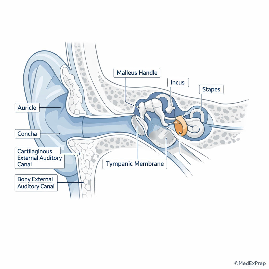
1.1.1.3 Foundational map: external ear, canal, and tympanic membrane landmarks
A Explanation
The first decision conflict in otology is whether the abnormality is on the outside of the tympanic membrane or behind it. Choose external ear/canal pathology when the stem emphasizes auricular tenderness, pain with tragal or pinna movement, canal edema, debris, cerumen, or a focal furuncle. Reject that choice when the exam instead describes a neutral external ear with a bulging, immobile, retracted, perforated, or opacified tympanic membrane. The single resolving rule is simple: manipulation pain and canal findings localize laterally; TM contour and middle ear air-space findings localize medially. See Fig 1 and Fig 2.
Board stems also expect you to know the basic external anatomy. The auricle is mostly elastic cartilage except the lobule. The lateral canal is cartilaginous and contains hair follicles and ceruminous glands, explaining why furunculosis occurs there. The medial canal is bony, skin is thin, and instrumentation is particularly painful. The tympanic membrane has a superior pars flaccida and larger pars tensa; the malleus handle and umbo orient the otoscopic exam. The cone of light is less important diagnostically than the actual TM position and landmarks. WHY THIS IS TESTED: many stems hide an anatomic localization question inside “otalgia,” “hearing loss,” or “ear fullness.”
Exam takeaway: Use the symptom-exam pairing rather than the disease label to choose the correct compartment.
B Worked example
A 42-year-old man with diabetes and psoriasis presents with 4 days of severe left ear pain after swimming. He reports muffled hearing and says his child recently had an upper respiratory infection, which makes acute otitis media sound tempting. Temperature is 37.4°C. Pulling on the auricle reproduces marked pain. Otoscopy shows edematous canal skin with debris; the tympanic membrane is only partly seen and is not bulging. The question asks for the most likely anatomic site of disease.
Reasoning chain: Identify the task: localize anatomy, not name every possible diagnosis. Extract key facts: diabetes is noise that raises concern for severity, but the discriminating facts are pain with auricle movement and canal edema. Apply rule: those findings point to the external canal. Eliminate distractors: recent family URI is a tempting but wrong clue for middle ear infection; lack of bulging TM argues against middle ear as the primary compartment. Verify: muffled hearing can still occur from canal swelling causing conductive symptoms.
C Exam trap
D Checkpoint
Question: A 27-year-old woman has right-sided otalgia after frequent earbud use and Q-tip cleaning. The canal is tender and mildly swollen; the tympanic membrane is intact without bulging or retraction. Which structure is most likely the primary site of disease?
- Cartilaginous external auditory canal
- Epitympanum
- Eustachian tube orifice
- Round window niche
Answer: A
- A. Tempting because the stem emphasizes instrumentation, tenderness, and superficial canal disease; correct because hair-bearing, gland-containing lateral canal is the usual site of traumatic otitis externa/furunculosis.
- B. Tempting because attic disease is high yield; wrong because epitympanic problems present with retraction pockets, debris behind the superior TM, or chronic middle ear findings rather than canal tenderness.
- C. Tempting because eustachian dysfunction causes fullness; wrong because it produces middle ear pressure problems, not localized canal tenderness on exam.
- D. Tempting because it is a true ear structure; wrong because round window pathology is inner/middle ear adjacent and not suggested by an external canal examination.
1.1.1.4 Foundational map: middle ear compartments, walls, and ossicular chain
A Explanation
The second foundational conflict is whether a middle ear question is really about space or about chain. Choose a space problem when the stem describes effusion, ventilation failure, retraction, attic disease, mastoid continuity, or spread of infection. Choose an ossicular problem when the stem emphasizes conductive hearing loss with normal-appearing canal and relatively limited inflammatory findings, trauma, fixation, or erosion. The single resolving rule is: air-space disease distorts pressure and TM position; chain disease impairs sound transmission to the oval window.
The middle ear is divided conceptually into epitympanum, mesotympanum, and hypotympanum. The epitympanum houses the malleus head and incus body and is the key attic space in acquired cholesteatoma. The mesotympanum lies opposite the TM and contains much of the ossicular chain. The hypotympanum is important because inferior vascular variants may appear as retrotympanic masses. The ossicular chain transmits vibration from TM to malleus to incus to stapes footplate at the oval window; the round window moves reciprocally. The middle ear also communicates posteriorly with the mastoid via the aditus and antrum, and anteriorly with the eustachian tube. See Fig 2.
Exam takeaway: Ask whether the stem is testing ventilation of the cavity or transmission through the ossicles.
B Worked example
A 59-year-old woman with allergic rhinitis and chronic reflux reports months of left ear fullness and decreased hearing after repeated flights. She has no auricular tenderness. Otoscopy shows a retracted TM with visible malleus prominence and amber fluid behind the membrane. The stem asks which anatomic system most directly explains her symptoms. A tempting wrong clue is her prior minor temporal bone trauma years ago, which could suggest ossicular chain injury.
Reasoning chain: Identify task: choose the anatomic system responsible. Extract key facts: retracted TM, visible middle ear fluid, no external canal findings, pressure-related onset. Apply rule: this is a middle ear space/ventilation problem involving the eustachian tube and middle ear cleft, not a primary ossicular chain lesion. Eliminate distractors: old trauma would fit ossicular discontinuity only if the TM were relatively normal and effusion/retraction absent. Verify: chronic poor ventilation causes negative pressure and effusion, both classic cavity findings.
C Exam trap
D Checkpoint
Question: Which middle ear compartment is most classically associated with acquired attic cholesteatoma origin?
- Hypotympanum
- Epitympanum
- Eustachian tube lumen
- Round window recess only
Answer: B
- A. Tempting because inferior middle ear abnormalities can be visible on otoscopy; wrong because hypotympanum is more relevant to jugular bulb relationships than classic attic cholesteatoma origin.
- B. Tempting because “attic” is a known board buzzword; correct because the epitympanum is the attic and is closely tied to pars flaccida retraction pockets.
- C. Tempting because eustachian dysfunction contributes to disease; wrong because dysfunction promotes retraction but the typical origin site of attic cholesteatoma is epitympanic/pars flaccida region.
- D. Tempting because it sounds anatomically specific; wrong because round window recess is not the classic starting point tested in attic cholesteatoma stems.
- The most common site of a furuncle in the ear canal is the cartilaginous lateral canal.
- True or False: A retracted tympanic membrane suggests positive middle ear pressure. False.
- Name the 3 middle ear compartments: epitympanum, mesotympanum, hypotympanum.
Exam takeaway: Link each middle ear region to the structure or complication most likely to appear in board stems.
flowchart TD
A[Middle ear cleft] --> B[Epitympanum]
A --> C[Mesotympanum]
A --> D[Hypotympanum]
A --> E[Eustachian tube]
A --> F[Aditus / antrum / mastoid]
B --> G[Pars flaccida retraction and cholesteatoma]
C --> H[Effusion and ossicles behind TM]
D --> I[Jugular bulb relationship]
E --> J[Ventilation failure]
F --> K[Spread of chronic disease]
Exam takeaway: Distinguish attic, mesotympanic, and hypotympanic disease to predict the likely pathology and complication.
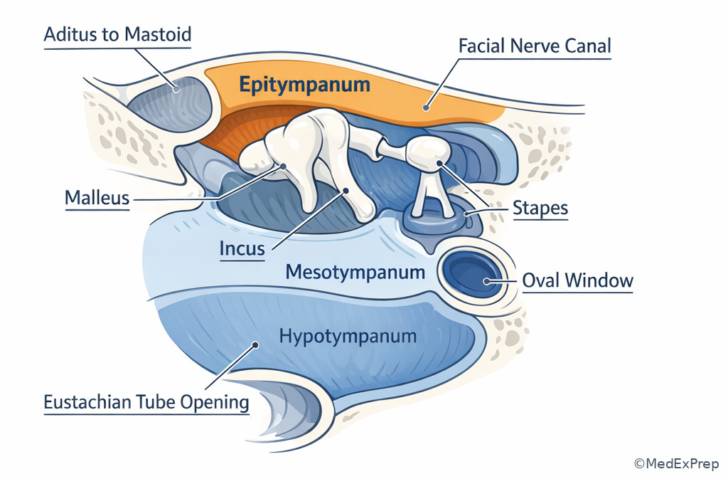
1.1.1.5 Applied anatomy: tympanic membrane quadrants and what lies at risk
A Explanation
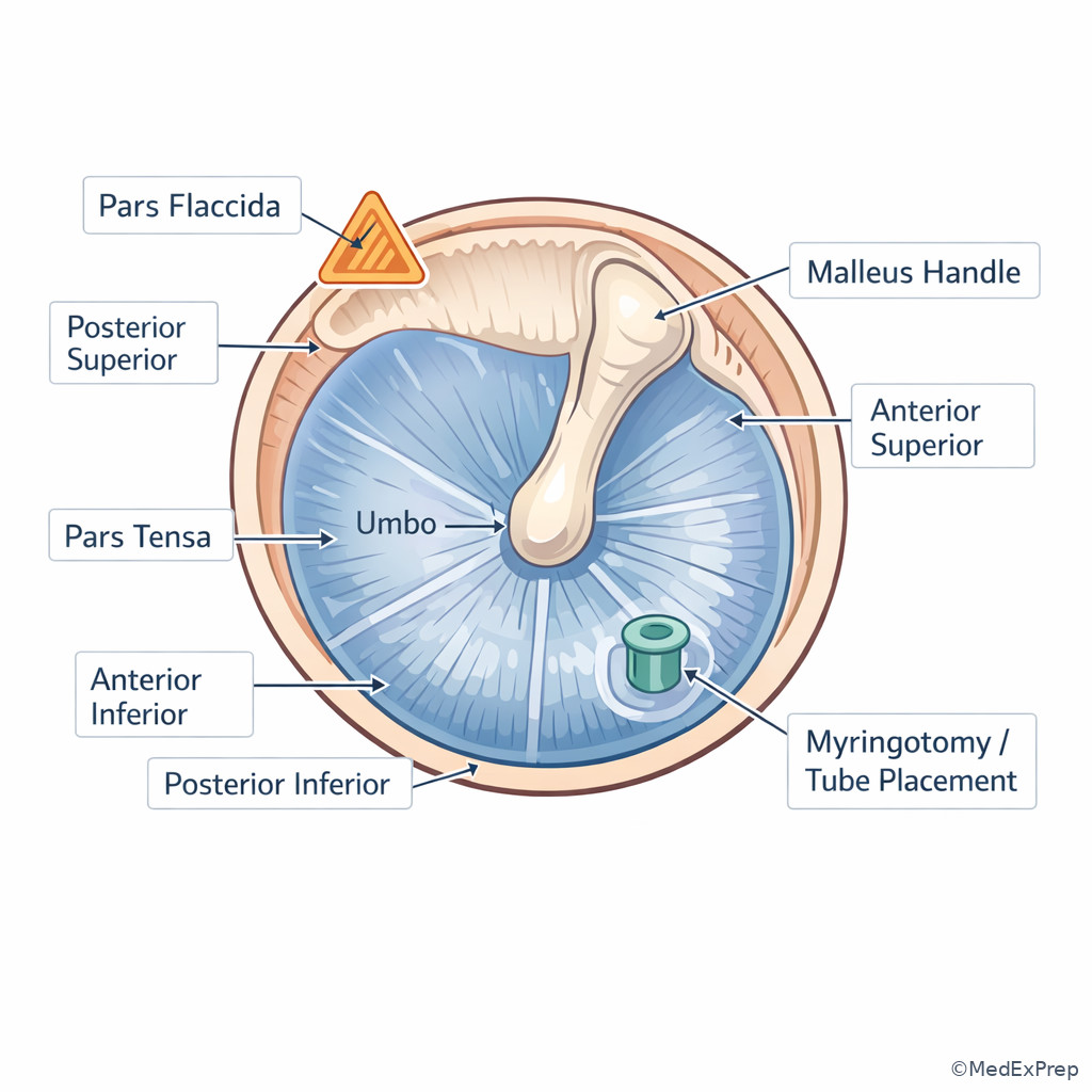
When a stem asks about a TM perforation, myringotomy site, or retraction pocket, the decision conflict is safe access versus dangerous neighborhood. Choose the anteroinferior quadrant for standard myringotomy/tube placement because it provides dependent drainage and avoids the ossicular chain and many posterior superior risks. Reject posterior superior manipulations when the stem implies routine tube placement; that quadrant is where attic disease, the incudostapedial region, and cholesteatoma-related erosion are more relevant. The single resolving rule is: posterior superior TM findings deserve structural caution; anteroinferior is the usual safe working zone.
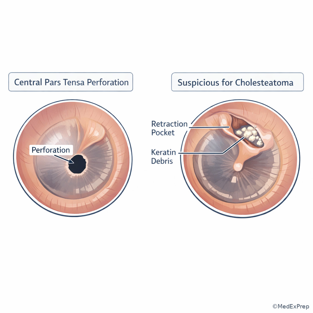
This topic is tested because candidates must translate a simple otoscopic image into procedural judgment. Superior retraction pockets, especially involving pars flaccida or posterior superior pars tensa, raise concern for retraction-pocket cholesteatoma. A small central pars tensa perforation is a different problem from a marginal or attic lesion. Similarly, a retrotympanic mass in the inferior quadrant changes the differential toward vascular anomalies. In procedure questions, the exam usually rewards broad safety principles over exact operative nuance. See Fig 4.
Exam takeaway: The quadrant tells you whether the question is routine access, ossicular risk, or cholesteatoma risk.
B Worked example
A 7-year-old boy with recurrent effusions, allergic rhinitis, and mild asthma is scheduled for tympanostomy tubes. His mother mentions a distant relative had hearing loss after ear surgery, a distracting but emotionally compelling clue. The question asks for the most appropriate tympanic membrane quadrant for uncomplicated tube placement. Otoscopy shows a dull intact TM with serous effusion but no attic retraction or superior debris.
Reasoning chain: Identify task: procedural landmark choice. Extract key facts: uncomplicated effusion, no suspicious superior pathology, routine tube placement. Apply rule: choose anteroinferior quadrant because it is safer relative to the ossicular chain and permits drainage. Eliminate distractors: posterior superior sounds precise and therefore tempting, but that is the danger area for ossicular and attic-related structures. Verify: this is a standard board-style anatomy-safety question, not a trick about family history.
C Exam trap
D Checkpoint
Question: A superior retraction pocket with keratin debris most strongly raises concern for which process?
- Simple otitis externa
- Acquired cholesteatoma
- High-riding jugular bulb
- Otosclerosis
Answer: B
- A. Tempting because debris can be present in the canal; wrong because the finding is a superior TM retraction pocket, not canal skin inflammation.
- B. Tempting because this is the classic attic association; correct because pars flaccida/superior retraction with keratin debris is a high-yield cholesteatoma clue.
- C. Tempting because vascular anomalies can be seen through the TM; wrong because those present as a retrotympanic mass, often inferiorly, not a keratin-filled retraction pocket.
- D. Tempting because it causes conductive loss; wrong because otosclerosis does not create visible superior retraction-pocket debris on otoscopy.
Exam takeaway: Posterior superior findings are structurally worrisome, while anteroinferior access is the routine safe zone.
Exam takeaway: Superior retraction with debris is not managed like a simple central pars tensa perforation.
1.1.1.6 Applied anatomy: eustachian tube, mastoid continuity, and middle ear ventilation
A Explanation
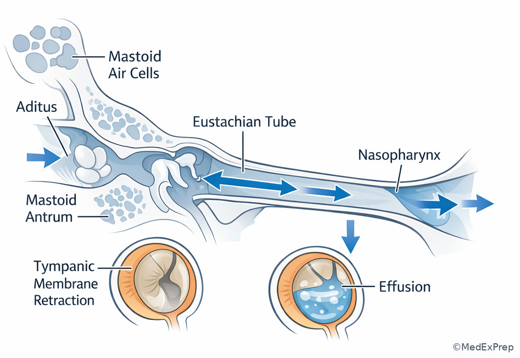
Many exam stems require choosing between infection as an isolated ear event versus disease of the entire middle ear cleft. Choose the latter when the stem describes recurrent effusions, retraction, mastoid extension, chronic inflammatory change, or symptoms linked to upper respiratory or nasopharyngeal dysfunction. Reject the idea that the middle ear is a sealed box. The single resolving rule is: the middle ear, aditus, mastoid, and eustachian tube behave as one connected mucosal system. See Fig 4.
The eustachian tube equalizes pressure and allows drainage from the middle ear to the nasopharynx. When it fails, negative pressure develops, the TM retracts, and effusion may accumulate. Posteriorly, the aditus opens into the mastoid antrum and mastoid air cell system; this continuity explains why chronic or acute disease can involve the mastoid. On boards, the point is not to memorize every cell group but to understand the pathway. A child with recurrent nasal inflammation and middle ear effusions, or an adult with unilateral persistent effusion needing nasopharyngeal evaluation, is being tested on this continuity. In upcoming topics, tympanograms will convert these anatomic ideas into data patterns.
Exam takeaway: Pressure failure starts at the eustachian tube but the visible consequences appear across the connected middle ear system.
B Worked example
A 51-year-old man with tobacco exposure, chronic allergic symptoms, and recent weight loss presents with unilateral right ear fullness for 8 weeks. He had one urgent care visit labeled “ear infection,” but pain is minimal. Otoscopy shows a dull retracted TM with effusion. The distracting clue is a recent swimming trip, which might suggest otitis externa. The stem asks for the most important anatomic pathway to consider in the evaluation.
Reasoning chain: Identify task: choose the anatomic link that explains persistent unilateral effusion. Extract key facts: unilateral adult effusion, chronicity, little pain, retracted TM, systemic red flags. Apply rule: the eustachian tube connects middle ear to nasopharynx, so obstructive nasopharyngeal pathology must be considered. Eliminate distractors: swimming explains canal problems, not isolated persistent middle ear effusion with retraction. Verify: this is a classic anatomy-driven oncologic safety stem.
C Exam trap
D Checkpoint
Question: Which structure directly connects the anterior middle ear to the nasopharynx and is central to pressure equalization?
- Aditus ad antrum
- Eustachian tube
- Facial recess
- Round window
Answer: B
- A. Tempting because it is a true middle ear connection; wrong because it connects the epitympanum to mastoid antrum, not the nasopharynx.
- B. Tempting because it fits the physiology described; correct because it is the drainage and pressure equalization pathway from middle ear to nasopharynx.
- C. Tempting because it is a real surgical space; wrong because it is not the pressure-equalizing conduit tested here.
- D. Tempting because it is involved in sound mechanics; wrong because it is a membranous window, not a drainage/ventilation pathway.
- The middle ear connects posteriorly to the mastoid through the aditus ad antrum.
- True or False: The middle ear is anatomically isolated from the mastoid. False.
- Persistent unilateral adult middle ear effusion should prompt consideration of nasopharyngeal obstruction.
Exam takeaway: Treat eustachian tube, middle ear, and mastoid findings as a connected system when localizing disease spread.
flowchart LR
A[Nasopharynx] --> B[Eustachian tube]
B --> C[Middle ear]
C --> D[Aditus]
D --> E[Mastoid antrum]
E --> F[Mastoid air cells]
C --> G[TM retraction or effusion]
E --> H[Mastoid extension if disease progresses]
Exam takeaway: Understanding this pathway explains effusion, retraction, mastoid extension, and unilateral adult red-flag presentations.
1.1.1.7 Integration: adjacent critical structures and high-stakes board localization
A Explanation
The final integration step is choosing whether a middle ear finding represents a routine otologic problem or a nearby-structure hazard. Choose hazard thinking when the stem mentions facial weakness, pulsatile retrotympanic mass, CSF leak concern, intracranial extension, or atypical severe disease. Reject a simple benign explanation if one adjacent structure changes the risk category. The single resolving rule is: the middle ear is bordered by neural, vascular, and intracranial structures, so “where” determines “how dangerous.”
High-yield neighbors include the facial nerve canal, tegmen superiorly, jugular bulb inferiorly, carotid canal anteriorly, and inner ear windows medially. You are not expected to reproduce microsurgical anatomy in detail for a foundational lesson, but you must recognize the implications. A bluish or red inferior retrotympanic mass suggests vascular anomaly rather than ordinary effusion. Facial weakness with chronic middle ear disease raises concern for aggressive infection or cholesteatoma-related erosion. Clear otorrhea after trauma can imply CSF leak through temporal bone injury involving adjacent skull base structures. See Fig 5 for quadrant-based risk framing, and note how board stems often present “hearing loss” only as a distractor while the real issue is danger recognition.
Exam takeaway: One red-flag neighbor such as facial nerve, vascular mass, or skull-base involvement should change the entire management path.
B Worked example
A 63-year-old woman with hypertension, prior neck radiation, and chronic conductive hearing symptoms presents with pulsatile tinnitus. Otoscopy reveals a reddish-blue mass visible behind the inferior TM. She has no fever and only mild aural fullness. A tempting clue is her history of recurrent childhood ear infections, which could push a candidate toward chronic effusion. The stem asks for the most appropriate next step.
Reasoning chain: Identify task: management based on anatomy. Extract key facts: pulsatile tinnitus, inferior retrotympanic mass, no infectious picture. Apply rule: think vascular neighbor such as high-riding jugular bulb or aberrant carotid; avoid myringotomy or blind aspiration. Eliminate distractors: a history of old infections does not explain a pulsatile retrotympanic mass. Verify: anatomy changes the safety profile more than the hearing symptom does.
C Exam trap
D Checkpoint
Question: A patient with chronic middle ear disease develops new ipsilateral facial weakness. Which adjacent structure most directly changes the urgency of the evaluation?
- Facial nerve canal
- Styloid process
- Temporomandibular joint disc
- Parotid tail
Answer: A
- A. Tempting because it is the classic nearby critical structure; correct because middle ear/temporal bone disease with facial weakness suggests involvement of the facial nerve pathway and raises urgency.
- B. Tempting because it is anatomically nearby in the region; wrong because it does not explain facial weakness emerging from middle ear disease in the tested way.
- C. Tempting because TMJ can refer otalgia; wrong because it does not convert chronic middle ear disease into a cranial neuropathy emergency.
- D. Tempting because facial nerve issues can involve the parotid more distally; wrong because the stem specifically frames middle ear disease, pointing to intratemporal facial nerve adjacency.
Exam Traps & Differentiators
| Looks similar | Tempting overlap | Single discriminator | Think instead |
|---|---|---|---|
| Otitis externa vs otitis media | Both cause ear pain and conductive symptoms | Pinna/tragus movement pain and canal edema | External canal disease |
| Effusion vs ossicular discontinuity | Both cause conductive hearing loss | Retracted or fluid-filled TM | Ventilation/cavity problem |
| Central perforation vs attic cholesteatoma | Both may show TM abnormality and hearing loss | Superior retraction pocket with keratin debris | Acquired cholesteatoma risk |
| Middle ear effusion vs vascular anomaly | Both may appear as retrotympanic opacity | Pulsatile red-blue inferior mass | High jugular bulb/aberrant carotid evaluation |
| Routine chronic ear disease vs urgent complication | Both may present with hearing loss and drainage | New facial weakness or neurologic sign | Adjacent structure threat |
Most common wrong answer and why: Candidates often select a disease label before localizing the compartment. On this topic, that reverses the tested reasoning order. The exam usually wants: Where is it? then What is it?
If the stem says pain with auricle movement, think external canal disease.
If the stem says retracted TM with amber fluid, think middle ear ventilation failure via eustachian tube dysfunction.
If the stem says superior retraction pocket with debris, think attic cholesteatoma until proven otherwise.
If the stem says pulsatile retrotympanic mass, think vascular anomaly and avoid blind incision.
If the stem says chronic ear disease plus facial weakness, think intratemporal facial nerve involvement and escalate.
If the stem says unilateral persistent adult effusion, think nasopharyngeal obstruction along the eustachian tube pathway.
| TM finding | Primary anatomic implication | Board consequence |
|---|---|---|
| Bulging TM | Positive pressure/inflammatory middle ear process | Middle ear, not canal, localization |
| Retracted TM | Negative middle ear pressure | Eustachian tube dysfunction/effusion pathway |
| Superior retraction pocket | Pars flaccida/attic involvement | Cholesteatoma concern |
| Central pars tensa perforation | Non-attic TM defect | Different risk profile than attic disease |
| Inferior pulsatile mass behind TM | Hypotympanic vascular relationship | Avoid blind myringotomy |
| Differential/classification target | Best clue | Reject because |
|---|---|---|
| External ear/canal disease | Tragus/pinna motion pain, canal edema, debris | Bulging or retracted TM shifts medially |
| Middle ear pressure disease | Retracted TM, effusion, fullness | Normal TM with trauma history may suggest chain problem instead |
| Attic disease/cholesteatoma | Pars flaccida or posterior superior retraction with debris | Simple canal debris lacks retraction pocket anatomy |
| Vascular retrotympanic anomaly | Pulsatile red-blue inferior mass | Routine effusion is not pulsatile |
| Adjacent facial nerve involvement | New facial weakness with middle ear disease | Uncomplicated effusion does not cause cranial neuropathy |
Algorithm / Approach
Rapid Review
- Cartilaginous EAC → hair follicles and ceruminous glands; furuncle localizes here.
- Bony EAC → thin adherent skin; instrumentation is disproportionately painful.
- Pinna/tragus motion pain → external canal process, not primary middle ear disease.
- Retracted TM → negative middle ear pressure from ventilation failure.
- Bulging TM → middle ear inflammatory pressure process.
- Pars flaccida → attic cholesteatoma risk zone.
- Posterior superior TM abnormality → think ossicular/attic proximity before routine intervention.
- Anteroinferior quadrant → routine myringotomy/tube safety zone.
- Epitympanum → “attic,” not generic middle ear space.
- Aditus ad antrum → posterior link between middle ear and mastoid.
- Persistent unilateral adult effusion → evaluate the eustachian tube/nasopharyngeal pathway.
- Inferior pulsatile retrotympanic mass → vascular anomaly until proven otherwise.
- Facial weakness with ear disease → adjacent intratemporal facial nerve concern.
- Conductive symptoms with obvious canal edema → do not overcall middle ear disease.
Self-check quiz
1. A 24-year-old man presents with ear pain after aggressive cotton-swab use. He has mild hearing reduction, no fever, and marked pain when the tragus is pressed. The tympanic membrane is intact without bulging. Which anatomic site is the most likely source of his symptoms?
- Cartilaginous external auditory canal
- Epitympanum
- Oval window
- Eustachian tube
2. A 6-year-old child with chronic serous effusions is scheduled for routine tympanostomy tube placement. Which tympanic membrane quadrant is most appropriate in an uncomplicated case?
- Posterior superior
- Anterior superior
- Anteroinferior
- Posterior central near the umbo
3. A 58-year-old woman with allergic rhinitis and frequent air travel presents with months of ear fullness and conductive hearing loss. Otoscopy shows a retracted tympanic membrane with amber fluid behind it. Which anatomic pathway best explains the process that requires management?
- Facial recess to round window pathway
- Eustachian tube to nasopharynx pathway
- Oval window to vestibule pathway
- Styloid process to parotid pathway
4. A 35-year-old man with chronic ear fullness and conductive hearing loss has a superior tympanic membrane retraction pocket containing keratin debris. He also has seasonal allergies and recently had swimmer's ear, a distracting detail. Which diagnosis is most likely?
- Otitis externa
- Acquired cholesteatoma
- Otosclerosis
- Patulous eustachian tube
5. A 49-year-old man with tobacco exposure, weight loss, and unilateral ear fullness has a persistent middle ear effusion on the right. He denies significant otalgia and recently returned from a beach vacation, which makes external ear disease tempting. What is the most appropriate next anatomic concern to clarify?
- External canal hair follicle infection
- Nasopharyngeal obstruction affecting the eustachian tube
- Round window rupture
- Temporomandibular joint inflammation
6. A 67-year-old woman with hypertension and chronic conductive symptoms presents with pulsatile tinnitus. Otoscopy reveals a reddish-blue mass behind the inferior tympanic membrane. She had recurrent otitis media as a child, which makes effusion seem plausible. What is the most appropriate next step?
- Perform office myringotomy for presumed effusion
- Start topical drops for otitis externa
- Recognize possible vascular retrotympanic anomaly and avoid blind incision
- Diagnose otosclerosis and reassure
7. A 44-year-old man with chronic otorrhea, poorly controlled diabetes, and longstanding conductive hearing loss develops new ipsilateral facial weakness. Otoscopy shows chronic middle ear disease. Which anatomic relationship most directly explains why this finding changes urgency?
- Middle ear proximity to the intratemporal facial nerve
- Middle ear proximity to the temporomandibular joint
- Middle ear proximity to the thyroid cartilage
- Middle ear proximity to the submandibular gland
8. A 32-year-old woman has conductive hearing loss after blunt temporal trauma 3 months ago. The external canal is normal and the tympanic membrane appears intact without retraction or effusion. She also reports nasal congestion from allergies, a tempting clue toward eustachian dysfunction. Which anatomic category is the best localization for the likely cause?
- Middle ear ventilation failure in the cavity
- Ossicular chain transmission abnormality
- External canal obstruction
- Nasopharyngeal mass causing eustachian dysfunction
Answer key
1. Correct answer: A
- A. Cartilaginous external auditory canal — Tempting because tragal tenderness and instrumentation history point laterally; correct because the cartilaginous canal contains hair follicles and glands and is the classic site of traumatic external canal inflammation. Single clue: pain with tragus pressure. Sources: Cummings Otolaryngology; Flint et al.
- B. Epitympanum — Tempting because middle ear disease can reduce hearing; incorrect because epitympanic disease would not be defined by external manipulation pain with a normal TM. Single clue: intact nonbulging TM. Sources: Cummings Otolaryngology; Lalwani.
- C. Oval window — Tempting because it is involved in hearing transmission; incorrect because inner/middle ear windows do not produce focal tragal tenderness from swab trauma. Single clue: provoked external tenderness. Sources: Glasscock-Shambaugh Surgery of the Ear.
- D. Eustachian tube — Tempting because it can cause fullness; incorrect because eustachian dysfunction produces pressure symptoms and TM changes rather than localized canal pain on manipulation. Single clue: normal TM contour. Sources: Cummings Otolaryngology.
2. Correct answer: C
- A. Posterior superior — Tempting because it sounds precise and close to the middle ear; incorrect because that region is structurally riskier due to attic/ossicular proximity. Single clue: uncomplicated routine tube placement. Sources: Cummings Otolaryngology; Pediatric Otolaryngology texts.
- B. Anterior superior — Tempting because it is anterior; incorrect because superior positions are less favored than inferior drainage/safety zones in routine placement. Single clue: standard myringotomy target. Sources: Flint et al.
- C. Anteroinferior — Tempting because it is the classic board answer; correct because it is the usual safer zone relative to posterior superior structural risks and facilitates drainage. Single clue: “routine uncomplicated case.” Sources: Cummings Otolaryngology.
- D. Posterior central near the umbo — Tempting because it sounds central and accessible; incorrect because proximity to the ossicular chain makes it less appropriate for routine placement. Single clue: need to avoid key posterior structures. Sources: Glasscock-Shambaugh.
3. Correct answer: B
- A. Facial recess to round window pathway — Tempting because both are real middle ear structures; incorrect because they do not explain chronic pressure equalization failure with effusion. Single clue: retracted TM plus amber fluid. Sources: Cummings Otolaryngology.
- B. Eustachian tube to nasopharynx pathway — Tempting because flights and rhinitis point to pressure dysfunction; correct because impaired eustachian tube function causes negative pressure, TM retraction, and effusion. Single clue: pressure-related chronic retraction. Sources: Bluestone and Klein, Otitis Media; Cummings.
- C. Oval window to vestibule pathway — Tempting because it transmits sound; incorrect because this is not the primary anatomy of middle ear ventilation failure. Single clue: visible effusion rather than isolated transmission abnormality. Sources: Lalwani.
- D. Styloid process to parotid pathway — Tempting because it is nearby head and neck anatomy; incorrect because it is unrelated to middle ear pressure equalization. Single clue: middle ear effusion pattern. Sources: Cummings.
4. Correct answer: B
- A. Otitis externa — Tempting because the patient recently had swimmer's ear and debris can appear in the ear; incorrect because the discriminating anatomy is a superior TM retraction pocket, not canal edema. Single clue: keratin debris within retracted superior TM area. Sources: Cummings Otolaryngology; Schuknecht pathology descriptions.
- B. Acquired cholesteatoma — Tempting because it is the classic attic lesion; correct because superior/pars flaccida retraction with keratin debris is a hallmark board pattern. Single clue: superior retraction pocket. Sources: Cummings; Glasscock-Shambaugh.
- C. Otosclerosis — Tempting because it causes conductive loss with limited exam findings; incorrect because otosclerosis does not create keratin-filled retraction pockets on otoscopy. Single clue: visible TM pathology. Sources: Lalwani.
- D. Patulous eustachian tube — Tempting because fullness can occur; incorrect because patulous tube causes autophony and TM movement with respiration rather than attic debris. Single clue: retraction pocket with keratin. Sources: Cummings.
5. Correct answer: B
- A. External canal hair follicle infection — Tempting because beach exposure suggests swimmer's ear; incorrect because the canal clue is absent and the stem shows persistent middle ear effusion. Single clue: unilateral effusion behind the TM. Sources: Cummings Otolaryngology.
- B. Nasopharyngeal obstruction affecting the eustachian tube — Tempting because adult unilateral effusion is a classic warning pattern; correct because persistent obstruction of the eustachian tube pathway must be considered. Single clue: unilateral persistent adult effusion with red flags. Sources: Cummings; NCCN-relevant head and neck evaluation principles.
- C. Round window rupture — Tempting because it involves hearing; incorrect because perilymphatic window pathology is not the typical explanation for persistent unilateral effusion with a retracted TM. Single clue: visible middle ear fluid rather than acute inner ear symptoms. Sources: Lalwani.
- D. Temporomandibular joint inflammation — Tempting because TMJ disease can refer to the ear; incorrect because referred otalgia does not explain persistent otoscopic middle ear effusion. Single clue: actual middle ear finding on exam. Sources: Cummings.
6. Correct answer: C
- A. Perform office myringotomy for presumed effusion — Tempting because conductive symptoms plus retrotympanic finding can resemble fluid; incorrect because blind incision is unsafe when a vascular anomaly is possible. Single clue: pulsatile reddish-blue mass. Sources: Cummings Otolaryngology; temporal bone vascular anomaly literature.
- B. Start topical drops for otitis externa — Tempting because tinnitus and fullness are nonspecific ear complaints; incorrect because the pathology is behind the TM, not in the canal. Single clue: retrotympanic mass. Sources: Flint et al.
- C. Recognize possible vascular retrotympanic anomaly and avoid blind incision — Tempting because it prioritizes safety over reflex treatment; correct because pulsatility and color strongly suggest a vascular variant such as high-riding jugular bulb or aberrant carotid. Single clue: pulsatile inferior red-blue mass. Sources: Cummings; Glasscock-Shambaugh.
- D. Diagnose otosclerosis and reassure — Tempting because chronic conductive symptoms fit otosclerosis; incorrect because otosclerosis does not produce a visible pulsatile retrotympanic mass. Single clue: mass on otoscopy. Sources: Lalwani.
7. Correct answer: A
- A. Middle ear proximity to the intratemporal facial nerve — Tempting because it is a classic complication pathway; correct because new facial weakness in chronic middle ear disease implies possible facial nerve involvement and raises urgency. Single clue: chronic ear disease plus new facial weakness. Sources: Cummings Otolaryngology; facial nerve chapters.
- B. Middle ear proximity to the temporomandibular joint — Tempting because TMJ and ear symptoms overlap; incorrect because TMJ pathology does not explain new facial palsy from middle ear disease. Single clue: cranial neuropathy, not referred pain. Sources: Flint et al.
- C. Middle ear proximity to the thyroid cartilage — Tempting only because both are head and neck structures; incorrect because there is no relevant direct relationship here. Single clue: temporal bone localization. Sources: Cummings.
- D. Middle ear proximity to the submandibular gland — Tempting because facial nerve and salivary structures can be linked elsewhere; incorrect because this stem specifically tests intratemporal adjacency. Single clue: otologic disease with facial weakness. Sources: Cummings.
8. Correct answer: B
- A. Middle ear ventilation failure in the cavity — Tempting because allergies suggest eustachian dysfunction; incorrect because the TM is intact and not retracted or fluid-filled, arguing against pressure-space disease. Single clue: normal-appearing TM without effusion. Sources: Cummings Otolaryngology.
- B. Ossicular chain transmission abnormality — Tempting because trauma with persistent conductive loss and normal TM is the classic pattern; correct because chain disruption is more likely than cavity disease here. Single clue: post-traumatic conductive loss with otherwise normal otoscopy. Sources: Glasscock-Shambaugh; Lalwani.
- C. External canal obstruction — Tempting because canal problems can cause conductive loss; incorrect because the canal is normal on exam. Single clue: no obstruction or edema seen. Sources: Flint et al.
- D. Nasopharyngeal mass causing eustachian dysfunction — Tempting because chronic congestion can bias toward eustachian pathology; incorrect because there is no otoscopic evidence of retraction or effusion and trauma is the stronger discriminator. Single clue: blunt temporal trauma followed by conductive loss. Sources: Cummings.
Keep going with the full ABOHNS Otolaryngology Prep Course (ENT)
You just studied the way members do, in the same lesson interface. Full access unlocks every lesson, the question bank and flashcards for a full year.
One payment · 1 year of access · No auto-renewal