1.1.1 - Cardiac Output Determinants
Why it matters
- Most anesthesia hemodynamic questions reduce to one decision conflict: is flow low because the heart is underfilled, pumping poorly, beating too slowly/too fast, or ejecting against excessive resistance?
- In the OR, hypotension alone is not the endpoint; the exam tests whether you can restore perfusion, not just raise a number.
- Cardiac output drives oxygen delivery, so this topic directly connects to the upcoming lesson on oxygen transport variables.
- Low-flow states also alter end-tidal carbon dioxide interpretation, which will matter when you study ventilation-perfusion relationships and capnography patterns.
Key Terms & Must-Know Facts
1.1.1.1 Key terms
- Cardiac output (CO): Volume of blood ejected per minute
- Stroke volume (SV): Blood ejected per beat
- Heart rate (HR): Beats per minute
- Preload: Ventricular filling/stretch before contraction
- Afterload: Resistance the ventricle must overcome to eject
- Contractility: Intrinsic inotropic strength at a given preload/afterload
- Venous return: Flow back to the right heart
- Frank-Starling relationship: Within limits, more filling increases stroke volume
- Mean arterial pressure (MAP): Pressure proxy for organ perfusion, but not identical to flow
- Systemic vascular resistance (SVR): Major component of left ventricular afterload
- Ejection fraction (EF): Fraction of end-diastolic volume ejected; not the same as cardiac output
- Chronotropy: Effect on heart rate
- Inotropy: Effect on contractility
- Tachycardia-induced low output: Reduced filling time causing reduced stroke volume
- Positive-pressure ventilation effect: Can reduce venous return and preload
1.1.1.2 Must-know facts
- \( CO = HR \times SV \)
- Stroke volume is primarily shaped by preload, afterload, and contractility.
- MAP can look acceptable while cardiac output is still poor.
- Bradycardia lowers cardiac output directly; severe tachycardia lowers it indirectly by impairing filling.
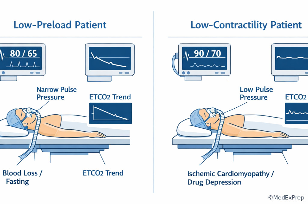
- Hypovolemia is a common, testable cause of low preload in the OR.
- High intrathoracic pressure decreases venous return.
- Volatile anesthetics and many induction agents can reduce contractility and/or SVR.
- Neuraxial blockade often causes vasodilation first; preload falls because venous capacitance increases.
- A sudden fall in ETCO2 can be a clue to sudden reduction in cardiac output, not just hyperventilation.
- Fixing the wrong determinant may worsen instability.
Exam takeaway: Start every hemodynamic stem by sorting the problem into heart rate, preload, afterload, or contractility before choosing a treatment.
flowchart TD
A[Low perfusion or hypotension] --> B{Which determinant is primary?}
B --> C[Low preload
bleeding fasting PPV neuraxial]
B --> D[Low contractility
ischemia cardiomyopathy drug effect]
B --> E[Abnormal heart rate
bradycardia or tachycardia]
B --> F[Excess afterload
vasoconstriction outflow burden]
C --> G[Reduce venous pooling and restore volume]
D --> H[Support inotropy and treat cause]
E --> I[Correct rate or rhythm]
F --> J[Reduce resistance if appropriate] Exam takeaway: Visualize filling, pumping strength, and outflow resistance as separate levers so you do not treat all hypotension the same way.
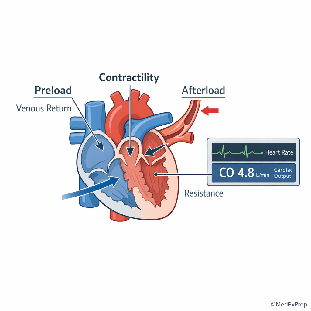
| Variable | Primary effect on CO | Classic perioperative causes | Typical first clue |
|---|---|---|---|
| Preload | Changes stroke volume through filling | Blood loss, fasting, bowel prep, vasodilation, high PEEP | Narrow pulse pressure, low filling, improved response to volume |
| Contractility | Changes stroke volume through pump strength | Myocardial ischemia, cardiomyopathy, anesthetic depression | Poor forward flow despite filling, rising filling pressures if measured |
| Heart rate | Changes CO directly and via filling time | Vagal events, beta-blockade, dysrhythmia, light anesthesia, pain | Bradycardia or tachycardia out of proportion to surgical stimulus |
| Afterload | Excess load can reduce stroke volume | Vasoconstrictors, chronic hypertension, sympathetic surge | High MAP with low forward flow or worsening LV performance |
Core content
1.1.1.3 Foundational model: cardiac output equals heart rate times stroke volume
A Explanation
The first exam decision conflict is whether to think in pressure or in flow. The correct choice is flow first. Cardiac output is the amount of blood delivered per minute, expressed by \( CO = HR \times SV \). That formula looks simple, but the tested skill is choosing which side of the equation has failed. A patient with a MAP of 68 mm Hg may have adequate flow, while another with a MAP of 75 mm Hg may still have poor output if vasoconstriction is artificially maintaining pressure. WHY THIS IS TESTED: stems often give a blood pressure value that tempts you to ignore low flow physiology.
Choose heart rate when the rate abnormality is itself severe enough to limit output: marked bradycardia reduces beats per minute, and marked tachycardia shortens diastolic filling time enough to lower stroke volume. Reject heart rate as the primary explanation when the rate is only mildly abnormal but other clues point to hypovolemia or pump failure. Choose stroke volume when the pattern suggests low filling, weak squeeze, or high ejection resistance.
In anesthesia, this matters because many interventions change more than one determinant at once. Propofol may reduce SVR and contractility; positive-pressure ventilation may reduce preload; laryngoscopy may increase HR and afterload. See Fig 1 for the fastest way to organize these variables before acting.
B Worked example
A 67-year-old man undergoing open colectomy becomes hypotensive 10 minutes after induction. He has hypertension, coronary artery disease, and chronic kidney disease. HR is 58/min, BP is 78/42 mm Hg, ETCO2 has fallen from 34 to 26 mm Hg, and the ventilator settings are unchanged. He received fentanyl, lidocaine, propofol, rocuronium, and a phenylephrine bolus 5 minutes earlier. Hemoglobin from preop labs was 12.3 g/dL. The surgeon has not yet made incision.
Identify the task: determine the most likely determinant of reduced cardiac output. Extract key facts: no surgical blood loss yet, low ETCO2 suggests reduced pulmonary blood flow, bradycardia is present but not extreme, recent induction plus vasoconstrictor can lower contractility and increase afterload, and the patient is likely vasodilated/relatively underfilled after induction and fasting. Apply the rule: when several determinants shift, choose the one best supported by timing and physiology. Eliminate distractors: acute anemia is tempting because hemoglobin affects oxygen delivery, but hemoglobin does not explain the immediate drop. Hyperventilation is tempting because ETCO2 fell, but the ventilator has not changed. Verify: the likely issue is induction-related reduced venous return with possible vasodilation and relative bradycardia, so the safest next anesthesia-focused response is to support preload/rate/pressure while reassessing depth and response to ventilation.
C Exam trap
Exam trap: The common wrong answer is “treat the blood pressure number alone with more vasoconstrictor.” Why it is tempting: MAP is low, and phenylephrine predictably raises arterial pressure. The single clue that eliminates it: a falling ETCO2 with unchanged ventilation points toward reduced flow; pure afterload increase can worsen output if the ventricle is already underfilled or depressed.
D Checkpoint
Question: During induction of anesthesia, which statement best guides the most appropriate interpretation of a sudden fall in ETCO2 from 35 to 24 mm Hg when minute ventilation is unchanged?
- The patient is necessarily hyperventilated, so cardiac output is unchanged.
- The fall may reflect decreased pulmonary blood flow from reduced cardiac output.
- The patient has increased oxygen consumption, causing ETCO2 to fall.
- The patient has improved alveolar ventilation because of stronger spontaneous effort.
- A: Tempting because ETCO2 often tracks ventilation, but wrong because unchanged ventilation means perfusion changes must be considered.
- B: Tempting because low ETCO2 is a low-flow clue; correct because ETCO2 can fall when less CO2 is delivered to the lungs.
- C: Tempting because metabolism influences CO2 production, but increased oxygen consumption would not be the classic immediate cause here.
- D: Tempting because breathing pattern can alter ETCO2, but the patient is mechanically ventilated with unchanged settings.
1.1.1.4 Preload and venous return: when low filling is the real problem
A Explanation
The next decision conflict is choosing between giving volume versus giving pressure support. Preload refers to ventricular filling before contraction, and in practical anesthesia terms it tracks venous return. The most testable rule is this: a ventricle cannot eject what it never receives. Underfilling decreases stroke volume even if contractility is normal. Frank-Starling physiology explains why moderate increases in filling can raise stroke volume, but only up to a point.
Choose low preload when the setting includes fasting, bowel prep, hemorrhage, venodilation from neuraxial block, positive-pressure ventilation, high PEEP, or rapid position change that impairs venous return. Reject low preload when the patient appears congested or when there is evidence of primary pump failure. WHY THIS IS TESTED: many stems are designed so that the examinee confuses venodilation or positive-pressure effects with “vasodilatory shock” alone and chooses the wrong drug first.
Positive-pressure ventilation matters because raising intrathoracic pressure reduces the pressure gradient for venous return. A hypovolemic patient may tolerate spontaneous breathing but become hypotensive after induction and positive-pressure ventilation. Similarly, neuraxial anesthesia often causes venous pooling before major arterial effects become obvious. See Fig 2 for the decision pattern that distinguishes low preload from other causes of low output.
Exam takeaway: When the stem features fasting, blood loss, neuraxial block, or positive-pressure ventilation, think low venous return before assuming primary pump failure.
flowchart LR
A[Low CO suspected] --> B{Perioperative context}
B --> C[Fasting or hemorrhage]
B --> D[PEEP or PPV]
B --> E[Spinal or epidural sympathectomy]
C --> F[Low venous return]
D --> F
E --> F
F --> G[Low stroke volume]
G --> H[Hypotension low ETCO2 weak pulse]
H --> I[Volume ventilation adjustment vasopressor if needed]
B Worked example
A 29-year-old woman presents for urgent postpartum tubal ligation under spinal anesthesia. She had prolonged labor, poor oral intake, and an estimated blood loss of 700 mL after delivery. Her history includes mild asthma and iron-deficiency anemia. Five minutes after the block, she becomes nauseated and pale. HR is 112/min, BP is 72/38 mm Hg, and ETCO2 by facemask sampling declines slightly after sedation. A small wheal from tape adhesive on her shoulder is noted and distracts the team toward allergic reaction.
Identify the task: choose the most likely determinant causing low output. Extract key facts: recent spinal, probable relative hypovolemia, tachycardia, hypotension, nausea, postpartum blood loss. Apply the rule: venodilation from sympathectomy plus depleted intravascular volume lowers preload dramatically. Eliminate distractors: anaphylaxis is tempting because there is skin irritation and hypotension, but a minor local tape reaction is not the classic discriminator; there is no bronchospasm, diffuse urticaria, or abrupt exposure-linked collapse. Primary myocardial depression is less likely in a healthy young patient immediately after neuraxial sympathectomy. Verify: restoring venous return and supporting blood pressure is the safest determinant-based move.
C Exam trap
Exam trap: The common wrong answer is “treat tachycardia first as the cause of low output.” Why it is tempting: learners memorize that tachycardia reduces filling time. The single clue that eliminates it: the stem timing right after spinal anesthesia with recent volume loss points to preload collapse; the tachycardia is compensatory, not primary.
D Checkpoint
Question: A patient becomes hypotensive soon after induction and positive-pressure ventilation. Which additional finding most strongly supports low preload as the primary cause of reduced cardiac output?
- Improvement in blood pressure and ETCO2 after a fluid bolus
- New S3 gallop with pulmonary crackles
- Progressive hypertension after phenylephrine
- Wide pulse pressure with bounding pulses
- A: Tempting because response-to-therapy can reveal mechanism; correct because improved flow after volume supports underfilling.
- B: Tempting because cardiac signs matter, but wrong because congestion suggests pump failure or overload, not isolated low preload.
- C: Tempting because vasopressors raise MAP, but wrong because pressure response alone does not prove preload deficit.
- D: Tempting because pulse pressure gives hemodynamic clues, but wrong because bounding pulses do not fit classic low-volume physiology.
- The most common perioperative cause of reduced preload is relative or absolute hypovolemia.
- True or False: Positive-pressure ventilation can reduce venous return. True.
- Name 3 stem clues favoring low preload: recent volume loss, falling ETCO2, response to fluid.
1.1.1.5 Contractility and afterload: weak pump versus hard-to-eject pump
A Explanation
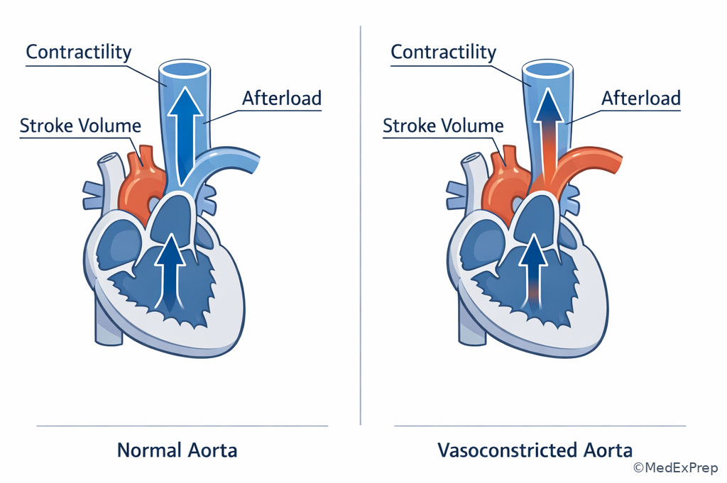
Once preload seems inadequate or already corrected, the next conflict is whether stroke volume remains low because the ventricle is weak or because it is ejecting against excessive resistance. Contractility is intrinsic pump strength at a given preload and afterload. Afterload is the load opposing ventricular ejection; in most CRNA exam stems, think SVR for the left ventricle. These are commonly confused because both can produce low output with hypotension or poor perfusion.
Choose low contractility when the patient has ischemic heart disease, cardiomyopathy, severe myocardial depressant exposure, new wall-motion abnormality if such data are given, or signs of poor forward flow despite adequate filling. Choose excess afterload when pressure is high or artificially increased and the ventricle struggles to eject, particularly in a hypertensive patient or after heavy vasoconstrictor use. Reject excess afterload as the main problem when hypotension occurs immediately after neuraxial anesthesia, because that pattern more strongly favors venodilation and low preload. WHY THIS IS TESTED: the wrong vasopressor or inotrope selection follows from confusing these two states.
In anesthesia, volatile agents, propofol, and ischemia can depress contractility. Phenylephrine can support pressure, but in a failing left ventricle it may worsen stroke volume by increasing afterload. See Fig 3 for a side-by-side sorting diagram.
Exam takeaway: A weak ventricle needs forward-flow support, whereas an overconstricted ventricle may worsen if afterload is raised further.
flowchart TB
A[Low stroke volume] --> B{Primary reason}
B --> C[Low contractility]
B --> D[High afterload]
C --> E[History of CAD cardiomyopathy myocardial depressant effect]
C --> F[Poor forward flow despite filling]
D --> G[Marked vasoconstriction chronic HTN phenylephrine effect]
D --> H[Pressure may rise while flow stays low]
E --> I[Consider inotropy and cause]
G --> J[Reduce unnecessary resistance and reassess] Exam takeaway: A ventricle pumping against a tight arterial system may generate pressure without generating adequate forward flow.
B Worked example
A 74-year-old man with ischemic cardiomyopathy, prior CABG, diabetes, and chronic hypertension undergoes hip fracture repair. After induction, MAP is 61 mm Hg. The CRNA gives repeated phenylephrine boluses, and MAP rises to 78 mm Hg, but ETCO2 falls from 33 to 24 mm Hg and urine output remains minimal. HR is 84/min. Lung exam is unchanged, and there has been no surgical blood loss yet. A tempting clue is his chronic hypertension, which may make a higher MAP seem automatically desirable.
Identify the task: choose why forward flow remains poor. Extract key facts: severe LV disease, pressure improved but ETCO2 worsened, no evidence yet for hemorrhage, HR is reasonable. Apply the rule: pressure recovery with worsening flow clues after repeated alpha-agonist dosing suggests excessive afterload imposed on an already weak ventricle. Eliminate distractors: “just give more phenylephrine to meet a target MAP” is tempting because pressure matters, but it ignores the falling ETCO2. Pure hypovolemia is possible, but there is no blood loss and the deterioration tracks vasoconstrictor administration. Verify: inotropy or a less afterload-intensive strategy fits better than more pure alpha stimulation.
C Exam trap
Exam trap: The common wrong answer is “high blood pressure means cardiac output is adequate.” Why it is tempting: learners equate perfusion pressure with perfusion flow. The single clue that eliminates it: ETCO2 drops as MAP rises, signaling less pulmonary blood flow and therefore worse forward output.
D Checkpoint
Question: Which finding most strongly favors excessive afterload rather than isolated low preload as the reason stroke volume remains depressed?
- Cardiac output clues worsen after repeated phenylephrine despite a higher MAP
- Blood pressure improves after fluid administration
- Hypotension follows bowel prep and prolonged fasting
- Tachycardia improves after pain control
- A: Tempting because phenylephrine is a common fix; correct because worsening flow after alpha stimulation points to too much ejection resistance.
- B: Tempting because improvement matters, but wrong because that pattern supports low preload.
- C: Tempting because it is a classic setup; wrong because it also supports low preload, not high afterload.
- D: Tempting because rate affects output, but wrong because it addresses heart rate rather than distinguishing afterload from preload.
1.1.1.6 Heart rate as a determinant: too slow, too fast, or just a clue
A Explanation
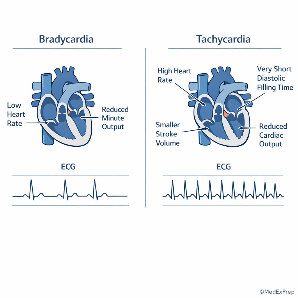
Heart rate is deceptively simple. The conflict is deciding whether the rate is the primary problem or merely a compensatory sign. Severe bradycardia lowers cardiac output because fewer beats occur each minute. Severe tachycardia lowers cardiac output by shortening diastole and reducing ventricular filling, especially in stiff ventricles, coronary disease, or hypovolemia. The testable rule is: rate becomes the target when it clearly prevents adequate filling or adequate beat frequency.
Choose bradycardia as the main determinant when hypotension appears with pronounced slow rate after vagal stimulation, neuraxial block, high opioid dose, beta-blockade, or conduction disease. Choose tachycardia as the main determinant when the rate is so fast that filling time collapses, often in patients with limited diastolic reserve. Reject rate as the primary issue when the rate abnormality is mild and fits compensation for hemorrhage or vasodilation. WHY THIS IS TESTED: stems commonly include a dramatic number that pulls you away from the actual cause.
For anesthesia candidates, the key is not memorizing one “good” heart rate. A patient with hypertrophic physiology or severe diastolic dysfunction may require lower rates for filling; another with complete heart block may need urgent rate support because stroke volume cannot compensate enough.
Exam takeaway: Bradycardia lowers output by too few beats, while severe tachycardia lowers output by stealing filling time.
B Worked example
A 61-year-old woman undergoing shoulder arthroscopy in the sitting position receives fentanyl and a deep volatile anesthetic. She has hypertension, obesity, and remote paroxysmal atrial fibrillation controlled with metoprolol. During positioning, HR drops to 34/min and BP to 58/30 mm Hg. ETCO2 falls from 32 to 20 mm Hg. The surgeon notes the heart rate “looks calm now,” creating a tempting false reassurance.
Identify the task: decide if rate is the primary determinant. Extract key facts: abrupt profound bradycardia, hypotension, low ETCO2, beta-blocker use, sitting position, anesthetic depth. Apply the rule: this is pathologic bradycardia causing low output, not a helpful slowing of the heart. Eliminate distractors: fluid alone might help because positioning can reduce venous return, but a rate of 34/min is too severe to ignore. More anesthetic is tempting if one mistakes this for sympathetic response, but the pulse is already profoundly slow. Verify: the immediate priority is correcting the bradycardia while supporting circulation.
C Exam trap
Exam trap: The common wrong answer is “tachycardia or bradycardia should always be normalized first.” Why it is tempting: the rate is visible and emotionally salient. The single clue that eliminates it: if the stem clearly gives a cause of compensation such as hemorrhage or vasodilation, the rate is often the response, not the primary target.
D Checkpoint
Question: Which scenario most strongly supports heart rate abnormality as the primary determinant of reduced cardiac output?
- HR 118/min after acute blood loss with narrow pulse pressure
- HR 36/min with abrupt hypotension after vagal stimulation during eye surgery
- HR 104/min after spinal anesthesia in a fasting patient
- HR 92/min with persistent low ETCO2 after repeated phenylephrine
- A: Tempting because tachycardia can reduce filling, but wrong because here it is more likely compensating for low preload.
- B: Tempting because the trigger is classic; correct because profound bradycardia itself is sufficient to reduce output.
- C: Tempting because spinal can affect rate, but wrong because mild tachycardia in this setup is more likely compensatory.
- D: Tempting because low flow is present, but wrong because this pattern points more toward excessive afterload/weak pump than rate.
- True or False: Mild tachycardia in hemorrhage is usually the primary determinant to treat first. False.
- The single best clue that bradycardia is causing low output is abrupt severe slowing with hypotension and low ETCO2.
- Name the mechanism by which severe tachycardia lowers stroke volume: reduced diastolic filling time.
1.1.1.7 Applying determinants in anesthesia: induction, ventilation, neuraxial block, and vasoactive choices
A Explanation
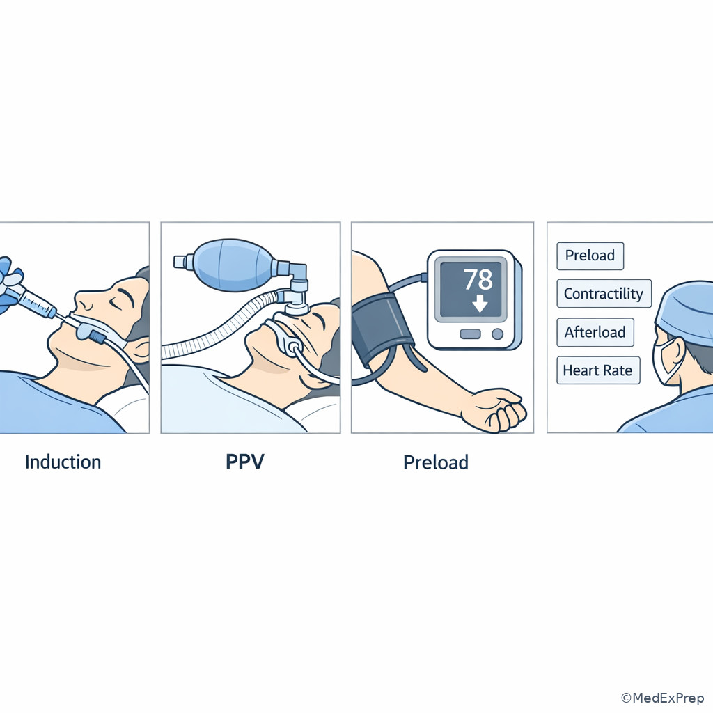
The final foundational step is integration: the exam rarely asks about determinants in isolation. Instead, it asks which anesthesia event changed the determinant and what the safest response is. Induction often reduces preload, afterload, and contractility simultaneously. Positive-pressure ventilation mainly reduces venous return, especially in the volume-depleted patient. Neuraxial anesthesia often causes venous pooling and reduced SVR. Vasoactive drugs shift the balance differently: some increase vascular tone, some increase rate/contractility, and some do both.
The decision conflict is choosing between a “number fix” and a “mechanism fix.” If a patient becomes hypotensive after spinal anesthesia with nausea and tachycardia, choose restoration of venous return and vascular tone; reject isolated treatment of the heart rate. If a patient with severe LV dysfunction has low ETCO2 after repeated alpha-agonist boluses, choose a strategy that supports forward flow rather than further increasing afterload. WHY THIS IS TESTED: anesthesia stems are deliberately dynamic; the correct answer depends on the trend after your last intervention.
Forward reference: in the next topic on Oxygen Transport Variables, you will connect these determinant-based changes in flow to \( DO_2 \), where cardiac output multiplies arterial oxygen content. See Fig 5 for a procedure-room algorithm tying common events to the determinant most likely affected.
Exam takeaway: Match the hemodynamic change to the anesthesia event that caused it before selecting a rescue drug or fluid strategy.
flowchart TD
A[Perioperative hypotension or low flow] --> B{What just happened?}
B --> C[Induction drugs]
B --> D[Positive-pressure ventilation or high PEEP]
B --> E[Spinal epidural sympathectomy]
B --> F[Repeated alpha agonist doses]
B --> G[Vagal stimulus or conduction problem]
C --> H[Low preload plus lower SVR and possible lower contractility]
D --> I[Reduced venous return]
E --> J[Venous pooling and lower SVR]
F --> K[Higher afterload]
G --> L[Bradycardia]
H --> M[Choose determinant-matched support]
I --> M
J --> M
K --> M
L --> M Exam takeaway: Timing relative to induction, ventilation, and positioning often reveals the determinant before advanced monitoring does.
B Worked example
A 58-year-old man with obesity, untreated OSA, hypertension, and an EF of 30% undergoes laparoscopic cholecystectomy. After induction and initiation of positive-pressure ventilation with pneumoperitoneum, BP drops to 82/46 mm Hg. HR is 88/min. ETCO2 falls from 35 to 28 mm Hg. The abdomen is insufflated, PEEP has been increased, and the patient received phenylephrine twice with only transient improvement. A distractor is the presence of obesity and OSA, which might make airway causes seem primary despite stable oxygen saturation.
Identify the task: select the dominant determinant affected by the sequence of events. Extract key facts: low EF, PPV, pneumoperitoneum, increased intrathoracic pressure, transient response to alpha support, falling ETCO2. Apply the rule: reduced venous return from pressure effects plus a ventricle with poor contractile reserve can sharply lower output. Eliminate distractors: isolated bronchospasm is less likely because oxygenation and airway pressures are not presented as the main change. Pure vasodilation is less likely now that pneumoperitoneum and alpha boluses are in play. Verify: determinant-matched correction means reducing reversible preload impediments, supporting circulation carefully, and reassessing whether afterload-heavy therapy is worsening forward flow.
C Exam trap
Exam trap: The common wrong answer is “repeat the last intervention because it partially worked once.” Why it is tempting: transient improvement feels like confirmation. The single clue that eliminates it: persistent or worsening low-flow markers such as falling ETCO2 after repeated doses tell you the mechanism was not fully matched.
D Checkpoint
Question: After induction, PPV, and a rise in PEEP, a fasting patient becomes hypotensive with a lower ETCO2. Which is the most appropriate initial interpretation?
- Reduced venous return is likely contributing to low cardiac output
- High oxygen consumption is lowering ETCO2 and causing hypotension
- Primary anemia is the most likely cause of the abrupt change
- The lower ETCO2 proves the patient is too deeply paralyzed
- A: Tempting because the timing fits physiology; correct because PPV and PEEP can impair preload, especially after fasting.
- B: Tempting because metabolism matters, but wrong because this does not explain the immediate event linked to ventilation changes.
- C: Tempting because anemia affects oxygen delivery, but wrong because abrupt post-induction change points elsewhere unless bleeding occurred.
- D: Tempting because depth changes ventilation mechanics, but wrong because paralysis does not by itself explain this perfusion pattern.
Exam Traps & Differentiators
Common traps & distractors: The exam rewards determinant matching, not memorized reflexes. Wrong answers are usually tempting because they correct a number, but not the mechanism.
1.1.1.8 Most common wrong answer and why
Most common wrong answer: give a pure vasoconstrictor for every hypotensive patient. This is tempting because MAP visibly rises fast and exam takers feel safer with a higher pressure. The problem is that pressure and flow are not interchangeable. In underfilled or poorly contracting ventricles, excessive afterload may worsen output even if the cuff reading improves.
1.1.1.9 Looks similar but isn't
| Pattern | Looks like | Actually think | Single clue that separates them |
|---|---|---|---|
| Hypotension after spinal with tachycardia | Primary arrhythmia problem | Low preload from venous pooling | Timing immediately after sympathectomy |
| Low ETCO2 after repeated phenylephrine | Improved circulation because MAP rose | Reduced forward flow from excessive afterload | ETCO2 fell as pressure rose |
| Tachycardia during bleeding | Need to slow the heart first | Compensatory response to low preload | Volume loss context with narrow pulse pressure |
| Hypotension after PPV starts | Only anesthetic overdose | Reduced venous return is contributing | Change begins with PPV/PEEP transition |
| Normal MAP with poor urine output and low ETCO2 | Adequate perfusion | Low cardiac output hidden by vascular tone | Pressure preserved but flow markers worsened |
1.1.1.10 If the stem says ___, think ___
- If the stem says “after induction and positive-pressure ventilation”, think reduced venous return until proven otherwise.
- If the stem says “ETCO2 falls without a ventilator change”, think reduced pulmonary blood flow/low cardiac output.
- If the stem says “pressure rose but perfusion markers worsened after phenylephrine”, think excess afterload or unresolved low flow.
- If the stem says “profound bradycardia after vagal stimulus or neuraxial event”, think heart rate is now the primary determinant.
- If the stem says “fasting, bowel prep, blood loss, neuraxial block”, think low preload before primary pump failure.
- If the stem says “ischemic cardiomyopathy with poor flow despite filling”, think contractility problem.
| Related topic pair | Why they are confusable | How to separate them |
|---|---|---|
| Cardiac Output Determinants vs Oxygen Transport Variables | Both can present as tissue hypoperfusion | This lesson asks why flow is low; the next asks how oxygen delivery depends on both flow and arterial oxygen content |
| Cardiac Output Determinants vs Ventilation Perfusion Relationships | Both can produce low ETCO2 or hypoxemia clues | Low output lowers ETCO2 by reducing perfusion; V/Q problems alter gas exchange patterns and the ETCO2-PaCO2 relationship |
| Low preload vs low SVR | Both can cause hypotension after neuraxial anesthesia | Low preload shows underfilling clues; low SVR emphasizes vasodilation, but clinically they often coexist |
Exam takeaway: Compare context and trend clues rather than relying on a single blood pressure value.
Tables
| Summary/reference | Increase in variable usually does what to CO? | When that rule fails | Exam use |
|---|---|---|---|
| Preload | Usually increases CO up to the Starling limit | Failing or overdistended ventricle | Do not assume more fluid always helps |
| Afterload | Usually decreases CO if excessive | Profound vasodilation may require more tone | Pressure support is not always flow support |
| Contractility | Increased contractility increases SV and CO | Severe underfilling limits effect | Weak pump stems require cause-directed support |
| Heart rate | Moderate increases may raise CO | Severe tachycardia shortens filling time | Decide when rate is cause versus compensation |
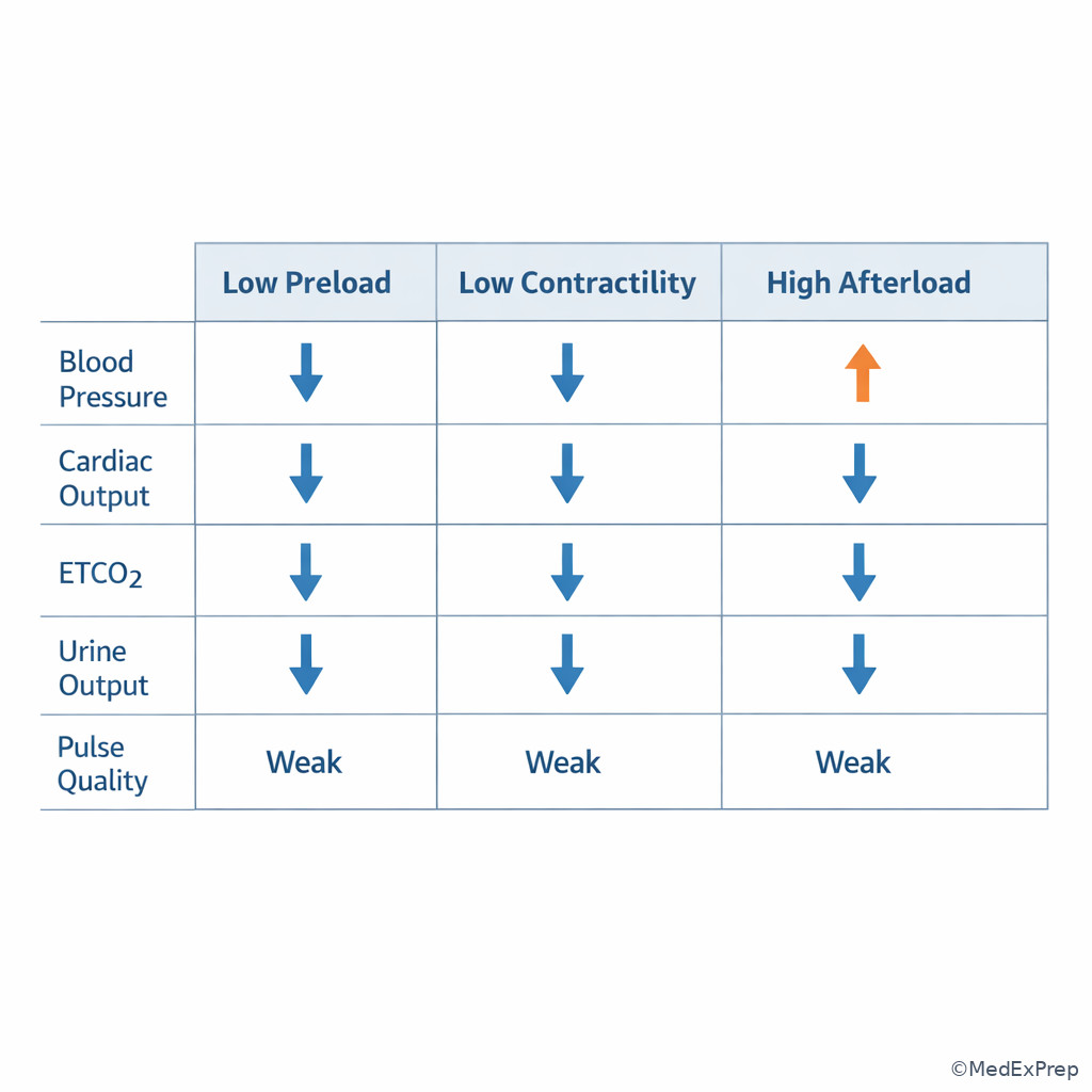
| Comparison | Low preload | Low contractility | Excess afterload |
|---|---|---|---|
| Typical setup | Fasting, bleeding, PPV, neuraxial block | CAD, cardiomyopathy, depressant drugs | Alpha agonists, severe HTN, vasoconstricted state |
| Response to fluid | Often improves | Limited or may worsen congestion | Minimal unless underfilling coexists |
| Pressure-flow relationship | Low pressure and low flow | Low flow often despite adequate filling | Pressure may be high or rise while flow stays poor |
| ETCO2 clue | May improve with volume restoration | Low from poor forward flow | Can worsen after extra vasoconstriction |
| Wrong reflex answer | Pure rate control | Give only volume | Give more alpha agonist |
| Differential/classification | Primary determinant disturbed | Classic board-style clue | First reasoning move |
|---|---|---|---|
| Spinal anesthesia hypotension | Preload and SVR | Nausea, pallor, timing after block | Think venous pooling before primary cardiac failure |
| Induction hypotension in fasting patient | Preload with possible lower contractility/SVR | Occurs after induction and PPV | Sort underfilling from anesthetic depression |
| Profound vagal event | Heart rate | Abrupt bradycardia and hypotension | Treat rate while supporting circulation |
| Cardiomyopathy with low ETCO2 despite adequate pressure | Contractility | Known poor EF, weak forward flow clues | Avoid assuming MAP equals adequate CO |
| Worsening ETCO2 after repeated phenylephrine | Afterload | Pressure improves while flow markers worsen | Reconsider pure alpha strategy |
Exam takeaway: The exam often separates students who can distinguish flow from pressure from those who cannot.
Algorithm / Approach
Exam takeaway: In perioperative hypotension, identify the trigger, sort the determinant, then choose determinant-matched support and verify with flow markers.
flowchart TD
A[Hypotension low ETCO2 weak pulse low urine output] --> B[Check immediate safety: oxygenation ventilation rhythm]
B --> C{What changed just before deterioration?}
C --> D[Induction PPV PEEP position block]
C --> E[Bleeding or volume loss]
C --> F[Vagal event or dysrhythmia]
C --> G[Repeated vasoconstrictor or severe HTN]
C --> H[Known weak ventricle ischemia depressant drugs]
D --> I[Think preload first]
E --> I
F --> J[Think heart rate first]
G --> K[Think afterload first]
H --> L[Think contractility first]
I --> M[Restore venous return and reassess]
J --> N[Correct pathologic rate and reassess]
K --> O[Avoid unnecessary resistance increase]
L --> P[Support forward flow and treat cause]
M --> Q[Verify MAP ETCO2 pulse quality urine output]
N --> Q
O --> Q
P --> Q
Use this algorithm exactly the way exam writers expect: identify the task, extract the trigger, select the determinant, choose the safest next step, and verify the expected effect. See Fig 6.
Rapid Review
- Cardiac output -> think flow per minute, not blood pressure alone
- Stroke volume -> separated by preload, afterload, contractility
- Falling ETCO2 with unchanged ventilation -> suspect reduced pulmonary blood flow
- Induction plus PPV -> low preload until proven otherwise
- Spinal anesthesia hypotension -> venous pooling is usually central to the mechanism
- Repeated phenylephrine with worsening ETCO2 -> pressure up, flow down
- Profound bradycardia -> heart rate becomes the determinant, not just a monitor finding
- Mild tachycardia after blood loss -> compensation, not the primary target
- Known poor EF with poor flow despite filling -> contractility problem
- High PEEP in a dry patient -> impaired venous return
- Normal MAP with low urine output and low ETCO2 -> do not assume adequate CO
- Response to fluid -> strong clue toward underfilling, not weak squeeze
- Afterload -> too much can reduce stroke volume even when pressure looks better
- Upcoming oxygen delivery lesson -> low CO will directly reduce \( DO_2 \)
Self-check quiz
1.1.1.11 Question 1
A 54-year-old fasting patient receives induction agents and positive-pressure ventilation for elective hernia repair. He has hypertension and mild CKD. Within minutes, BP falls to 80/46 mm Hg, HR is 96/min, and ETCO2 falls from 34 to 28 mm Hg without a ventilator change. Which determinant most likely requires initial attention?
- Preload
- Afterload
- Contractility
- Heart rate
1.1.1.12 Question 2
A 63-year-old patient with glaucoma develops abrupt HR 32/min and BP 56/30 mm Hg during ocular traction under general anesthesia. He has CAD and takes metoprolol. Which determinant is most likely the primary cause of reduced cardiac output?
- Preload
- Heart rate
- Afterload
- Contractility
1.1.1.13 Question 3
A 72-year-old woman with ischemic cardiomyopathy, diabetes, and chronic hypertension has MAP 60 mm Hg after induction for hip arthroplasty. After repeated phenylephrine, MAP rises to 78 mm Hg, but ETCO2 falls from 32 to 24 mm Hg and peripheral perfusion remains poor. Which interpretation is most appropriate?
- The higher MAP confirms cardiac output has improved
- Excess afterload may be worsening forward flow
- Minute ventilation must have suddenly increased
- Bradycardia is the main determinant of instability
1.1.1.14 Question 4
A 30-year-old postpartum patient with recent blood loss, prolonged labor, and poor oral intake becomes nauseated and hypotensive after spinal anesthesia for tubal ligation. HR is 116/min, BP is 74/40 mm Hg. Which is the most appropriate initial interpretation?
- Tachycardia is the primary cause of low output and should be slowed first
- Low preload from venous pooling and volume depletion is likely central
- Primary myocardial depression is most likely in this otherwise healthy patient
- The blood pressure change proves isolated high afterload
1.1.1.15 Question 5
A 66-year-old man with obesity, OSA, and EF 30% becomes hypotensive after induction, PEEP increase, and pneumoperitoneum for laparoscopy. HR is 88/min, BP is 82/46 mm Hg, ETCO2 falls from 35 to 28 mm Hg, and oxygen saturation remains stable. Which determinant is most likely reduced first by the most recent events?
- Preload
- Heart rate
- Contractility
- Oxygen consumption
1.1.1.16 Question 6
A 77-year-old man with severe aortic stenosis, CAD, and chronic hypertension becomes hypotensive after induction. HR is 118/min, BP is 78/44 mm Hg, ETCO2 is 27 mm Hg. He had bowel prep and poor intake for colon surgery. Which detail most strongly argues that the tachycardia is compensatory rather than the primary determinant to correct first?
- The presence of hypertension in the medical history
- The preoperative volume-depleting bowel preparation and poor intake
- The age older than 75 years
- The ETCO2 value is below 30 mm Hg
1.1.1.17 Question 7
A 69-year-old woman with cardiomyopathy, CKD, and peripheral vascular disease becomes hypotensive after induction. A fluid bolus produces minimal change, but repeated pure alpha-agonist boluses increase MAP while ETCO2 continues to fall. Which is the most appropriate next hemodynamic interpretation?
- The ventricle is likely underfilled and needs more fluid alone
- Excess afterload in a weak ventricle is impairing stroke volume
- The heart rate must be the dominant problem even if normal
- The low ETCO2 excludes a cardiac output problem
1.1.1.18 Question 8
A 58-year-old patient with obesity, GERD, and controlled hypertension develops BP 64/34 mm Hg and HR 38/min immediately after peritoneal traction during abdominal surgery. ETCO2 falls from 33 to 21 mm Hg. A colleague suggests the patient is simply underfilled from fasting and should receive only fluid. Which is the best next interpretation?
- Low preload is the sole determinant because the patient was NPO
- Heart rate is now the primary determinant because profound bradycardia is causing low output
- High afterload is the most likely cause because ETCO2 is falling
- Contractility failure is proven by the presence of GERD and obesity
Answer key
1.1.1.19 Question 1: A. Preload
A: Tempting because induction and positive-pressure ventilation commonly reduce venous return in a fasting patient; correct because the stem links the event timing to underfilling and a low-flow clue (falling ETCO2). Single discriminating clue: hypotension begins after induction plus PPV in an NPO patient. Source: Barash et al.; Miller.
B: Tempting because induction agents can alter vascular tone, but incorrect here because the falling ETCO2 with PPV timing points more specifically to reduced venous return than isolated afterload change. Single clue: unchanged ventilation with lower ETCO2.
C: Tempting because induction drugs may depress the myocardium, but incorrect because there is no history or clue prioritizing weak pump over the immediate PPV/fasting setup. Single clue: the most recent trigger is PPV in a volume-depleted patient.
D: Tempting because heart rate affects output, but incorrect because HR 96/min is compatible with compensation rather than a primary rate problem. Single clue: no profound bradycardia or extreme tachycardia.
1.1.1.20 Question 2: B. Heart rate
A: Tempting because underfilling can cause hypotension in anesthetized patients, but incorrect because the abrupt profound bradycardia during ocular traction is a classic vagal event. Single clue: HR 32/min after eye manipulation.
B: Tempting because rate problems are obvious on the monitor; correct because severe bradycardia directly lowers cardiac output and is sufficient to explain the collapse. Single clue: abrupt profound bradycardia with hypotension.
C: Tempting because blood pressure is low, but incorrect because no evidence suggests excessive afterload. Single clue: the trigger is vagal, not vasoconstrictive.
D: Tempting because CAD can impair pump function, but incorrect because the sudden event is better explained by bradycardia than by acute contractility failure. Single clue: timing with ocular traction.
1.1.1.21 Question 3: B. Excess afterload may be worsening forward flow
A: Tempting because higher MAP feels reassuring, but incorrect because pressure does not guarantee improved flow; the ETCO2 has fallen further. Single clue: ETCO2 worsens after phenylephrine.
B: Tempting because phenylephrine increases SVR; correct because in a patient with ischemic cardiomyopathy, added afterload can reduce stroke volume despite a better cuff pressure. Single clue: repeated alpha-agonist doses with falling ETCO2.
C: Tempting because ETCO2 tracks ventilation, but incorrect because no ventilator change is provided. Single clue: the only intervention emphasized is vasoconstrictor administration.
D: Tempting because rhythm matters in low-output states, but incorrect because no bradycardia is present. Single clue: the HR is not slow.
1.1.1.22 Question 4: B. Low preload from venous pooling and volume depletion is likely central
A: Tempting because tachycardia can reduce filling, but incorrect because here it is more likely a compensatory response to sympathectomy plus volume depletion. Single clue: immediate post-spinal timing with recent blood loss and poor intake.
B: Tempting because spinal anesthesia commonly redistributes venous volume; correct because neuraxial sympathectomy plus depleted intravascular volume sharply reduces preload. Single clue: postpartum blood loss and recent spinal block.
C: Tempting because hypotension can always reflect poor contractility, but incorrect because the healthy patient and timing strongly favor a preload problem. Single clue: no evidence of primary myocardial disease.
D: Tempting because hemodynamic variables are intertwined, but incorrect because isolated high afterload does not fit hypotension after spinal anesthesia. Single clue: sympathectomy produces vasodilation, not excess resistance.
1.1.1.23 Question 5: A. Preload
A: Tempting because PPV, higher PEEP, and pneumoperitoneum all impede venous return; correct because these recent changes reduce preload first, especially in a patient with limited cardiac reserve. Single clue: deterioration follows PEEP increase and insufflation.
B: Tempting because HR contributes to output, but incorrect because HR is stable and not pathologic. Single clue: HR 88/min.
C: Tempting because EF is low and contractility is relevant, but incorrect as the question asks which determinant is reduced first by the most recent events; those events primarily impair venous return. Single clue: “most recent events” are pressure-related ventilation changes.
D: Tempting because metabolism affects physiology, but incorrect because oxygen consumption is not one of the four determinant options and does not explain the trigger-linked hemodynamic pattern. Single clue: stable oxygen saturation with hemodynamic timing.
1.1.1.24 Question 6: B. The preoperative volume-depleting bowel preparation and poor intake
A: Tempting because chronic hypertension changes hemodynamic targets, but incorrect because it does not specifically prove tachycardia is compensatory. Single clue: history alone is nonspecific.
B: Tempting because bowel prep often matters; correct because preoperative volume depletion makes tachycardia more likely to be a response to low preload than the primary problem. Single clue: bowel prep and poor intake.
C: Tempting because older adults tolerate tachycardia poorly, but incorrect because age does not distinguish primary from compensatory tachycardia. Single clue: age is supportive context, not the discriminator.
D: Tempting because low ETCO2 suggests low flow, but incorrect because it does not by itself tell you whether tachycardia is cause or compensation. Single clue: ETCO2 shows low flow, not mechanism.
1.1.1.25 Question 7: B. Excess afterload in a weak ventricle is impairing stroke volume
A: Tempting because fluid is a common first-line reaction to hypotension, but incorrect because minimal response to fluid and worsening flow after alpha support argue against isolated underfilling. Single clue: fluid produced little change.
B: Tempting because cardiomyopathy plus alpha agonists create a classic mismatch; correct because a weak ventricle may lose forward flow when afterload is pushed higher. Single clue: MAP up, ETCO2 down after pure alpha boluses.
C: Tempting because rate always matters, but incorrect because a normal rate is not the dominant issue here. Single clue: no pathologic rate abnormality.
D: Tempting because ETCO2 is often treated as a ventilation number, but incorrect because low ETCO2 is fully compatible with low cardiac output when ventilation is unchanged. Single clue: low ETCO2 during hemodynamic decline.
1.1.1.26 Question 8: B. Heart rate is now the primary determinant because profound bradycardia is causing low output
A: Tempting because fasting lowers preload and fluid may help, but incorrect because profound bradycardia after peritoneal traction is immediately dangerous and is the dominant determinant. Single clue: HR 38/min with abrupt trigger.
B: Tempting because traction can produce a vagal response; correct because severe bradycardia directly reduces cardiac output and explains the abrupt fall in ETCO2 and BP. Single clue: sudden profound bradycardia after surgical traction.
C: Tempting because ETCO2 is falling, but incorrect because nothing suggests high afterload; the trigger is vagal. Single clue: peritoneal traction with bradycardia.
D: Tempting because comorbidities can distract, but incorrect because GERD and obesity do not prove contractility failure. Single clue: no pump-failure evidence, strong vagal trigger instead.
Keep going with the full CRNA NCE Prep Course (CRNA NCE)
You just studied the way members do, in the same lesson interface. Full access unlocks every lesson, the question bank and flashcards for a full year.
One payment · 1 year of access · No auto-renewal