1.1.1 - Antigen-Antibody Reaction Basics
Antigen-Antibody Reaction Basics
- Immunohematology Foundations: Serology principles -> antigen-antibody interaction mechanisms
- Immunohematology Foundations: Serology principles -> lattice formation and agglutination interpretation
- Pretransfusion Testing: Compatibility testing decisions -> recognize conditions required for visible serologic reactions
- Laboratory Troubleshooting: Serologic interpretation -> distinguish true reaction strength from technical or biologic variation
- Explain what must be present for a detectable antigen-antibody reaction in blood bank testing.
- Distinguish sensitization from visible agglutination and decide when each matters.
- Predict how antibody class, antigen site spacing, and zeta potential affect test results.
- Interpret why IgM and IgG behave differently across routine serologic phases.
- Apply a basic blood bank reasoning framework to early serology questions and vignettes.
Why it matters
- Most BB serology questions reduce to one core decision: did antibody bind, did it bridge cells, or did testing fail to reveal the binding?
- Understanding this topic prevents classic exam errors such as confusing sensitization with agglutination, or assuming a negative immediate-spin result excludes clinically significant antibody.
- These principles directly support later topics in antibody screening, AHG testing, crossmatch interpretation, and transfusion reaction workups.
- When a result pattern looks odd, this foundation tells you whether to suspect antibody class, phase of testing, antigen density, or technical conditions first.
Key Terms & Must-Know Facts
1.1.1.1 Key terms
- Antigen: Red cell structure capable of reacting with a specific antibody.
- Antibody: Immunoglobulin directed against a corresponding antigen.
- Paratope: Antigen-binding site on the antibody.
- Epitope: Specific antigenic determinant recognized by the antibody.
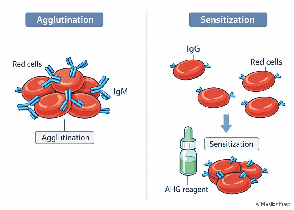
- Sensitization: Antibody coating of red cells without visible agglutination.
- Agglutination: Visible clumping caused by bridging between cells.
- Hemolysis: Red cell destruction; in vitro may be a positive reaction depending on method and controls.
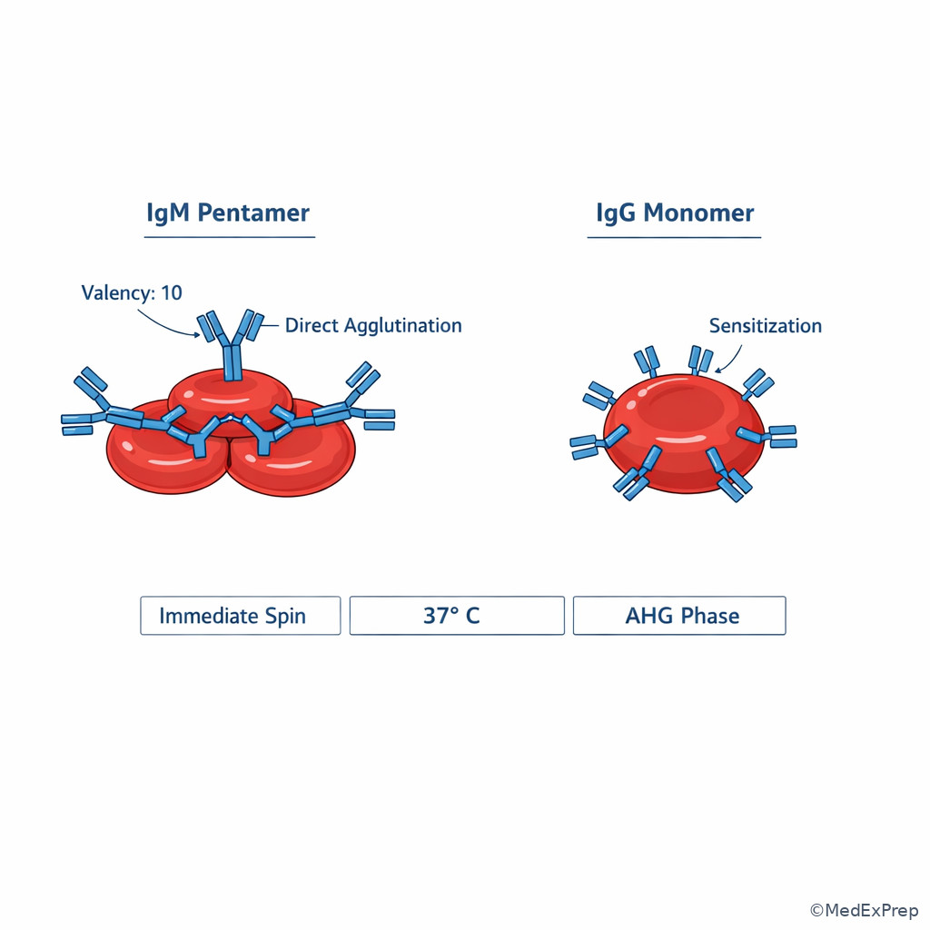
- IgM: Pentameric antibody that often reacts best at colder temperatures and can directly agglutinate cells.
- IgG: Monomeric antibody that commonly sensitizes cells and is detected at AHG phase.
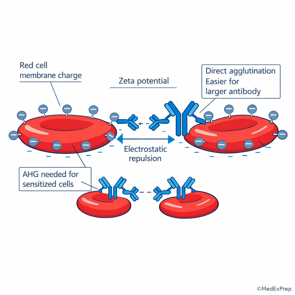
- Zeta potential: Net electrostatic repulsion between red cells that limits spontaneous cell-to-cell approach.
- Lattice formation: Multivalent antigen-antibody network required for visible agglutination.
- Affinity: Strength of binding between one antibody site and one epitope.
- Avidity: Overall binding strength produced by multiple interactions.
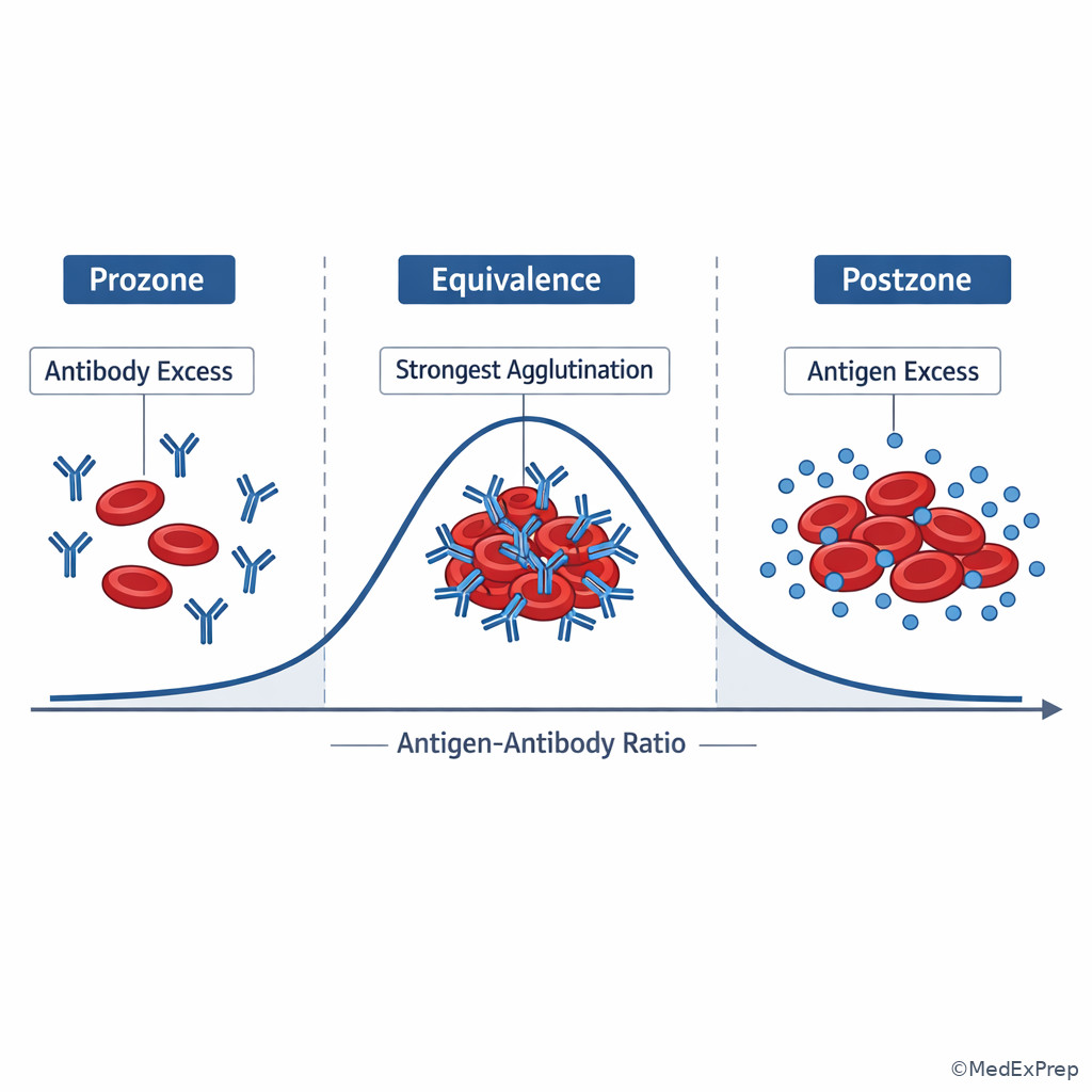
- Zone of equivalence: Relative antigen-antibody proportions most favorable for lattice formation.
- Prozone/postzone: Excess antibody or excess antigen that weakens visible reaction formation.
1.1.1.2 Must-know facts
- Binding alone is not enough; visible agglutination requires crosslinking and favorable testing conditions.
- IgM usually causes direct agglutination more easily than IgG because of its larger structure and higher valency.
- Many clinically significant red cell antibodies are IgG and are best detected at 37 C/AHG rather than immediate spin.
- A negative immediate-spin result does not rule out an IgG alloantibody.
- Red cells naturally repel each other because of zeta potential; enhancement media reduce this barrier in later testing. See Fig 1.
- Sensitized cells may look nonreactive until antihuman globulin bridges attached IgG molecules.
- Visible reaction strength depends on antigen density, antibody concentration, temperature, pH, ionic conditions, and incubation time.
- ABO antibodies are classically strong, naturally occurring, and often reactive at immediate spin because many are IgM.
- Reaction grade reflects observed strength, not automatically clinical significance.
| Term | What it means in BB questions | Why it matters |
|---|---|---|
| Binding | Antibody attaches to antigen | May be invisible unless bridging or AHG reveals it |
| Sensitization | IgG-coated cells without clumping | Common setup for positive AHG testing |
| Agglutination | Visible cell clumping | Usually means direct bridging occurred |
| Hemolysis | Cell destruction in test system | Can indicate a positive reaction depending on context |
| Zeta potential | Cell repulsion barrier | Explains why IgG often needs AHG or enhancement |
| Lattice | Network of linked cells/antibodies | Required for visible agglutination |
Core content
1.1.1.3 Foundational case: Why one antibody gives visible agglutination and another only sensitizes cells
A. Explanation
A newly hired transfusion service technologist observes that one patient sample shows immediate visible clumping, while another remains smooth until the AHG phase. The decision conflict is simple but heavily tested: should you conclude the second sample lacks antibody, or should you recognize that antibody may be present but only coating the cells? The single rule that resolves this is that visible agglutination requires effective bridging between red cells; antibody binding alone may produce only sensitization.
In red cell serology, the key sequence is antigen present -> antibody binds -> if enough crosslinking occurs under favorable conditions, a lattice forms -> visible agglutination appears. Large multivalent antibodies such as many IgM antibodies can bridge the gap between red cells directly. IgG antibodies, however, are smaller and often attach to red cells without spanning the intercellular distance created by zeta potential. That produces sensitized cells rather than obvious clumps. An AHG reagent can then bridge the cell-bound IgG molecules and convert invisible sensitization into visible agglutination. See Fig 1.
WHY THIS IS TESTED: Exam items frequently present a negative immediate-spin reaction and ask whether antibody is absent or merely undetectable at that phase. The tested pattern is choosing reject absence of antibody when the likely issue is antibody class and detection phase.
Exam takeaway: A negative visible reaction can still mean red cells are sensitized, especially with IgG that requires AHG detection.
flowchart LR
A[Antigen on red cell] --> B[Antibody binds]
B --> C{Can it bridge cells directly?}
C -->|Yes| D[Visible agglutination]
C -->|No| E[Sensitization only]
E --> F[AHG reagent added]
F --> G[Visible agglutination at AHG] Exam takeaway: Start by deciding whether the issue is no binding, invisible binding, or visible bridging.
Exam takeaway: Direct clumping and AHG-revealed sensitization are different outcomes of antigen-antibody interaction.
B. Worked example
A 63-year-old man with myelodysplastic syndrome, chronic kidney disease, and a history of multiple transfusions has a pretransfusion workup. His antibody screen is negative at immediate spin but 2+ positive at AHG. ABO typing is straightforward, and his bilirubin is mildly elevated from chronic disease, which is distracting but not central. A new learner is tempted to call the immediate-spin phase “nonreactive therefore clinically insignificant.”
Reasoning chain: Identify task -> explain phase-dependent reactivity. Extract key facts -> multiply transfused patient, negative immediate spin, positive AHG. Apply rule -> many clinically significant alloantibodies are IgG and may sensitize rather than directly agglutinate. Eliminate distractors -> this is not explained by ABO antibody behavior because those are often immediate-spin reactive; not by “no antibody present” because AHG is positive; not by autoagglutination because the pattern given is phase-specific rather than panreactive across all steps. Verify -> most appropriate interpretation is a likely IgG antibody detectable at AHG phase.
C. Exam trap
D. Checkpoint
Question: A patient’s screen cells show no agglutination at immediate spin but react at AHG. Which interpretation is most appropriate?
- The antibody is absent because visible agglutination did not occur initially.
- The antibody likely sensitized the cells without direct agglutination until AHG was added.
- The reagent cells lacked antigens, so the AHG phase created a false-positive result.
- The reaction pattern proves the antibody is IgM.
Answer: B
- A: Tempting because learners focus on what they can see at the earliest phase; wrong because invisible sensitization can still be present.
- B: Correct because IgG commonly coats red cells without directly bridging them until AHG reagent reveals the sensitization.
- C: Tempting because reagent issues can cause odd patterns; wrong because lack of antigen would not selectively create a true AHG reaction pattern.
- D: Tempting because antibody class is being tested; wrong because IgM more often reacts directly at immediate spin, not only at AHG.
1.1.1.4 Foundational case: Why IgM and IgG behave differently in blood bank testing
A. Explanation
A candidate sees “IgM” and “IgG” in an answer set and must choose between them without memorizing random lists. The decision conflict is whether to classify an antibody by what phase it reacts in or by how its structure changes visible serology. The single rule is that antibody structure and valency influence its ability to bridge red cells directly.
IgM is pentameric, with multiple binding sites that make it efficient at forming lattices and causing direct agglutination. In routine blood bank teaching, this is why ABO antibodies are strongly associated with immediate-spin reactivity. IgG is monomeric and smaller; it commonly binds at 37 C but does not by itself overcome the red cell spacing caused by zeta potential. Therefore IgG often requires AHG detection. This is a structural explanation, not merely a memorized phase list. When the exam asks you to choose between “cold-reactive direct agglutinator” and “warm-reactive sensitizer,” it is really testing whether you connect antibody size, valency, and testing phase. See Fig 2.
WHY THIS IS TESTED: Later questions on ABO antibodies, Rh/Kell/Duffy/Kidd antibodies, and immediate-spin versus AHG crossmatch all depend on this mechanism.
Exam takeaway: On exams, IgM usually explains immediate-spin direct agglutination, while IgG usually explains 37 C/AHG sensitization patterns.
flowchart TD
A[Antibody class] --> B{IgM or IgG?}
B -->|IgM| C[Higher valency]
C --> D[More direct cell bridging]
D --> E[Often immediate-spin agglutination]
B -->|IgG| F[Monomer]
F --> G[Coats red cells at 37 C]
G --> H[Often requires AHG for detection] Exam takeaway: Start with antibody class when deciding which testing phase is most likely to detect the reaction.
Exam takeaway: Structural differences between IgM and IgG explain common blood bank phase patterns.
B. Worked example
A 27-year-old postpartum patient with iron deficiency anemia and a recent cesarean delivery has a positive antibody screen at AHG only. She has no recent transfusion history but received prenatal testing. A tempting clue is that she has mild hypothermia after surgery, which might push a learner toward a cold-reactive antibody. However, the strongest exam clue is the AHG-only pattern.
Reasoning chain: Identify task -> infer likely antibody behavior class, not the exact specificity. Extract key facts -> warm-phase/AHG detection, no immediate-spin reactivity, postpartum immune exposure background. Apply rule -> IgG antibodies are the classic explanation for sensitization detectable at AHG. Eliminate distractors -> IgM is less likely because it more often gives direct agglutination earlier; hypothermia is noise because the lab phase pattern matters more. Verify -> the likely serologic behavior is IgG-mediated.
C. Exam trap
D. Checkpoint
Question: Which antibody property best explains why many IgG antibodies are detected at AHG rather than immediate spin?
- IgG lacks specificity for red cell antigens.
- IgG is too small to directly bridge red cells efficiently in the presence of zeta potential.
- IgG reacts only below room temperature.
- IgG destroys AHG reagent before agglutination can occur.
Answer: B
- A: Tempting because the question asks about detection limits; wrong because IgG can be highly specific.
- B: Correct because the key issue is geometry and cell spacing, not lack of binding.
- C: Tempting if cold-reactive concepts are mixed up with immunoglobulin class; wrong because many IgG antibodies are warm reactive.
- D: Tempting because AHG is named in the stem; wrong because AHG is used to detect cell-bound IgG, not destroyed by it in this manner.
- The antibody class most associated with direct immediate-spin agglutination is IgM.
- True or False: IgG binding always causes visible clumping without AHG. False.
- Name the key barrier that keeps red cells apart: zeta potential.
1.1.1.5 Application case: Why zeta potential, antigen density, and spacing determine visible reaction strength
A. Explanation
A reagent panel shows weak or variable agglutination. The decision conflict is whether to blame “weak antibody” or to recognize that reaction strength depends on more than concentration. The single rule is that visible strength reflects how easily antibodies form lattices under the physical conditions of the test system.
Red cells carry a negative surface charge that creates electrostatic repulsion, called zeta potential. This keeps cells separated and makes direct bridging difficult, especially for smaller antibodies. Antigen density also matters: cells with more accessible target sites allow more antibody binding and more effective lattice formation. When antigens are sparse or poorly positioned, even a specific antibody may give weak reactions. Affinity and avidity also contribute. A high-affinity interaction may bind well, but visible agglutination still depends on whether enough linked interactions form between multiple cells. That is why reaction grade is informative but never interpreted alone. See Fig 3.
For BB exams, choose-vs-reject logic matters: choose “physical or test-system limitation” when the pattern is weak but consistent with known antibody behavior; reject “no antibody” unless the setup truly excludes binding. The upcoming topics on enhancement media and serologic variables will show how laboratories deliberately reduce ionic shielding or alter conditions to improve lattice formation.
WHY THIS IS TESTED: Weak reactions are common distractors. The exam wants you to know that weak does not automatically mean insignificant, absent, or technical failure.
Exam takeaway: Weak agglutination can result from spacing and antigen density issues even when the antibody is real.
flowchart TD
A[Observed weak reaction] --> B{Why weak?}
B --> C[Low antigen density]
B --> D[Poor lattice formation]
B --> E[Zeta potential keeps cells apart]
B --> F[Low effective antibody concentration]
C --> G[Reduced visible agglutination]
D --> G
E --> G
F --> G Exam takeaway: When strength is lower than expected, first ask whether binding conditions support lattice formation.
Exam takeaway: Red cell repulsion explains why some antibodies bind without producing immediate visible agglutination.
B. Worked example
A 71-year-old woman with metastatic colon cancer, recent chemotherapy, and two prior transfusions has a weakly positive screen at AHG with one screening cell and negative immediate-spin testing. A slightly elevated plasma protein level raises concern for nonspecific interference, and that distracting clue makes rouleaux feel appealing. However, the given pattern is one of weak phase-specific reactivity, not broad nonspecific clumping.
Reasoning chain: Identify task -> explain why a true antibody may appear weak. Extract key facts -> AHG-only reactivity, transfusion history, selective cell positivity. Apply rule -> weak reaction may reflect limited antigen density, lower antibody concentration, or less efficient lattice formation. Eliminate distractors -> rouleaux usually affects broader interpretation and has different serologic behavior; “no antibody” is unsafe because there is reproducible AHG reactivity. Verify -> a real alloantibody remains possible despite weak strength.
C. Exam trap
D. Checkpoint
Question: Which factor best explains why a specific IgG antibody may bind red cells yet fail to produce strong direct agglutination?
- Zeta potential keeps red cells separated and limits direct bridging.
- IgG can react only with plasma proteins, not red cell antigens.
- Antigen density has no effect on agglutination strength.
- Visible agglutination occurs whenever one antibody molecule binds one antigen.
Answer: A
- A: Correct because electrostatic repulsion is a central physical reason direct bridging is limited.
- B: Tempting if one confuses reagent interactions with antibody specificity; wrong because IgG commonly targets red cell antigens.
- C: Tempting because some students overfocus on class alone; wrong because antigen density strongly affects lattice formation.
- D: Tempting because binding seems sufficient at first glance; wrong because visible agglutination requires multivalent lattice formation.
1.1.1.6 Application case: Why antigen-antibody ratio and test conditions matter before later media are even considered
A. Explanation
A technologist gets inconsistent reactions in parallel tubes. The decision conflict is whether to interpret this as a biologic antibody difference or as a setup problem. The single rule is that the best visible reaction occurs near an optimal relative proportion of antigen and antibody. Too much antibody can produce prozone; too much antigen can produce postzone; both reduce lattice visibility.
Although advanced discussion belongs in later topics, the BB exam often expects the basic concept now: serologic reactions depend on more than “antibody present or absent.” A test system with marked excess antibody may saturate antigen sites without effective cell-to-cell bridging. A test system with excess antigen may spread available antibody too thinly to build a strong lattice. In practical blood bank terms, this means a weak or unexpectedly negative result is not always biologically negative. It may reflect proportions, incubation conditions, or system setup. See Fig 4.
This is especially useful when answering “best explanation” questions. Choose a ratio/conditions explanation when the stem suggests unexpected weakness despite plausible antibody presence. Reject disease-based speculation if the question is really about serologic mechanics. The next lesson on Reaction Phases and Media will build directly on this by showing how saline, LISS, PEG, and incubation alter expression of these same principles.
WHY THIS IS TESTED: The exam likes negative or weak reactions that are not truly negative. You must recognize that reaction failure may be technical or conditional rather than immunologically absent.
Exam takeaway: False-weak or false-negative reactions can occur when antigen and antibody proportions are far from equivalence.
flowchart LR
A[Excess antibody] --> B[Prozone]
B --> E[Poor lattice visibility]
C[Optimal proportion] --> D[Equivalence]
D --> F[Best visible reaction]
G[Excess antigen] --> H[Postzone]
H --> E
Exam takeaway: Before assuming no antibody, check whether the test system allowed lattice formation.
Exam takeaway: The strongest visible serologic reaction occurs when antigen and antibody are present in favorable proportion.
B. Worked example
A 35-year-old patient with sickle cell disease, recent emergency transfusion, and high immunologic exposure history has repeat testing because one tube in a manual workup is unexpectedly negative while a parallel setup is weakly positive. The patient also has hypergammaglobulinemia, a distracting clue that could steer a candidate toward nonspecific causes. However, the question asks for the best basic serologic explanation for inconsistent visible reaction.
Reasoning chain: Identify task -> explain tube-to-tube reaction variation. Extract key facts -> same clinical sample, inconsistent visible strength, manual setup context. Apply rule -> differences in test conditions or antigen-antibody proportion can change lattice formation. Eliminate distractors -> a true antibody does not vanish biologically from one tube to the next without some test-system explanation; “clinically insignificant” does not explain inconsistency. Verify -> the most appropriate explanation is suboptimal reaction conditions, including possible zone effects.
C. Exam trap
D. Checkpoint
Question: A reaction expected to be positive appears unexpectedly weak. Which principle is most important to remember first?
- Any true antibody must always give the same visible strength regardless of test setup.
- Visible agglutination is optimal only when antigen and antibody are present in favorable proportion.
- Only hemolysis, not agglutination, reflects antigen-antibody interaction.
- Zone effects occur only in donor infectious disease testing, not blood bank serology.
Answer: B
- A: Tempting because reproducibility is important; wrong because setup differences can affect visible strength.
- B: Correct because lattice formation depends on relative proportions as well as antibody presence.
- C: Tempting if one overgeneralizes complement concepts; wrong because agglutination is a core positive serologic endpoint.
- D: Tempting because zone terminology appears in multiple lab areas; wrong because the concept applies directly to serology.
- The region with best visible lattice formation is the zone of equivalence.
- True or False: Antibody excess can weaken visible agglutination. True.
- Name the two classic zone problems: prozone and postzone.
1.1.1.7 Integration case: Using basic reaction principles to interpret early blood bank scenarios safely
A. Explanation
A board-style item rarely asks only for definitions. Instead, it gives a patient with recent transfusion, pregnancy history, or a positive screen and asks for the most appropriate interpretation. The decision conflict is whether to chase specificity too early or first classify the reaction pattern correctly. The single rule is that the first safe interpretation step is to decide whether the pattern suggests direct agglutination, sensitization, or a condition-limited false-weak result.
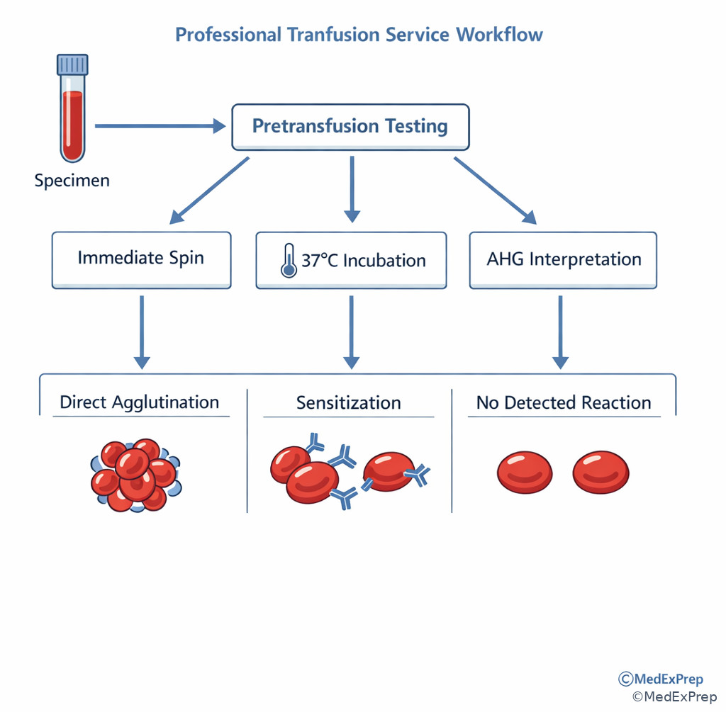
For BB-level reasoning, follow a fixed framework: identify the task, extract phase and strength, connect likely mechanism, then decide the safest next interpretation. If the reaction is immediate-spin positive, think first about antibodies capable of direct agglutination and about ABO relevance. If the reaction appears only after incubation/AHG, think sensitizing antibodies, especially clinically significant IgG alloantibodies. If the reaction is weaker than expected or inconsistent, think of physical/test-system limitations before concluding absence. See Fig 5. This framework prevents overcalling, undercalling, and wrong-phase mistakes.
WHY THIS IS TESTED: BB exams reward organized interpretation. Many distractors are technically possible but wrong because they skip the first classification step.
Exam takeaway: Start with reaction phase and visibility pattern before naming likely antibody categories.
flowchart TD
A[Serologic result appears] --> B{Visible at immediate spin?}
B -->|Yes| C[Think direct agglutination pattern]
C --> D[Often IgM / ABO-type behavior]
B -->|No| E{Positive at AHG?}
E -->|Yes| F[Think sensitization pattern]
F --> G[Often IgG / clinically significant alloantibody behavior]
E -->|No| H[Consider no binding or suboptimal conditions] Exam takeaway: Classify the reaction mechanism before advancing to specificity or compatibility decisions.
Exam takeaway: Early serologic interpretation depends on classifying phase-specific reaction behavior before unit selection.
B. Worked example
A 54-year-old woman with systemic lupus erythematosus, prior pregnancies, and a remote transfusion history needs red cells. Her screen shows no immediate-spin agglutination, 1+ reactivity at 37 C, and 2+ at AHG. She also has a positive ANA, a distracting laboratory clue that may make the case feel autoimmune and therefore nonspecific. The question asks for the most appropriate first-pass interpretation.
Reasoning chain: Identify task -> classify mechanism before naming specificity. Extract key facts -> phase increases with incubation/AHG, no direct immediate-spin agglutination, immune exposure history. Apply rule -> this pattern fits a sensitizing warm-reactive IgG alloantibody more than a cold direct-agglutinating IgM antibody. Eliminate distractors -> ANA is not the deciding blood bank clue; “no antibody” is disproved by AHG reactivity; ABO-like behavior is less likely because immediate spin is negative. Verify -> most appropriate interpretation is probable IgG-mediated sensitization requiring further antibody identification.
C. Exam trap
D. Checkpoint
Question: In a first-pass interpretation of serologic results, which clue should carry the greatest weight initially?
- The patient’s admitting diagnosis, because clinical disease determines antibody class.
- The reaction phase pattern, because it indicates whether direct agglutination or sensitization is more likely.
- The patient’s age alone, because older patients usually produce IgM antibodies.
- The strength at one phase only, because stronger reactions always identify specificity.
Answer: B
- A: Tempting because clinical context matters; wrong because serologic phase pattern is the initial laboratory discriminator.
- B: Correct because phase pattern provides the earliest mechanism-based interpretation.
- C: Tempting because age is often included in stems; wrong because age alone does not determine antibody class.
- D: Tempting because strength feels decisive; wrong because one strength value without phase context is incomplete.
1.1.1.8 Integration case: Separating true reaction principles from common early misconceptions
A. Explanation
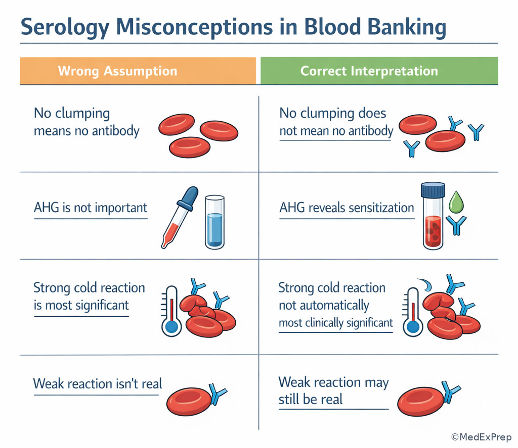
A learner says, “If there is antibody, there should be clumping.” Another says, “If there is no immediate-spin reaction, it is not important.” These statements sound efficient but are unsafe. The decision conflict is choosing between oversimplified exam shortcuts and the actual governing rule. The single rule is that blood bank serology measures a detectable expression of antigen-antibody interaction, not merely the existence of antibody in isolation.
Several misconceptions fall away once you keep that rule in mind. First, no visible clumping does not equal no binding. Second, strong clumping does not automatically mean clinical significance; many cold-reactive antibodies can agglutinate strongly while being less important for red cell survival than certain warm-reactive IgG antibodies. Third, a weak or variable reaction is not automatically an artifact. Fourth, the laboratory professional’s role is to interpret and escalate safely within protocol, not to diagnose the patient’s overall disease state. These distinctions matter because BB exam distractors often include medically tempting but out-of-scope actions or overconfident interpretations. See Fig 6.
WHY THIS IS TESTED: Foundational lessons are often assessed through misconceptions, not straight definitions. The exam asks which assumption to reject.
Exam takeaway: Do not equate visible clumping with all antibody activity or early negativity with true absence.
flowchart TD
A[Misconception] --> B{Claim}
B --> C[No clumping means no antibody]
B --> D[Strongest reaction means most significant]
B --> E[Immediate-spin negative means safe to ignore]
C --> F[Incorrect: sensitization may be invisible]
D --> G[Incorrect: phase and specificity matter]
E --> H[Incorrect: IgG may appear later at AHG] Exam takeaway: Correct answers in BB often come from rejecting oversimplified rules and following phase-based serologic logic.
Exam takeaway: The biggest beginner mistakes come from overinterpreting visible strength and underinterpreting phase-specific reactivity.
B. Worked example
A 46-year-old trauma patient with diabetes, prior pregnancy, and emergency-release transfusion 3 months ago has a current antibody screen that is negative at immediate spin and weakly positive at AHG. The patient also has elevated lactate from sepsis, which is clinically important but not the key serologic clue. A novice suggests issuing blood based on the negative immediate-spin phase because “there is no visible reaction.”
Reasoning chain: Identify task -> reject an unsafe simplification. Extract key facts -> history of immune exposure, AHG reactivity, phase-specific pattern. Apply rule -> antibody activity may be invisible until AHG, so immediate-spin negativity cannot clear the case. Eliminate distractors -> lactate does not resolve serologic interpretation; trauma urgency does not authorize the laboratorian to ignore compatibility findings outside policy. Verify -> the correct laboratory action is to continue compatibility workup/escalation according to SOP rather than treating early negativity as absence.
C. Exam trap
D. Checkpoint
Question: Which statement is most appropriate for a BB candidate interpreting an early serology pattern?
- A strong immediate-spin agglutinin is always more clinically significant than an AHG-reactive antibody.
- If no clumping is seen initially, the specimen can be treated as antibody negative.
- Visible reaction endpoint, phase, and mechanism must all be considered before judging significance.
- The laboratorian should independently authorize emergency issue whenever a reaction pattern is difficult.
Answer: C
- A: Tempting because strength is easy to see; wrong because clinical significance depends on more than visual strength.
- B: Tempting because early negative results feel reassuring; wrong because sensitization may still be present.
- C: Correct because safe BB interpretation integrates endpoint, phase, and mechanism before conclusions.
- D: Tempting in urgent scenarios; wrong because release decisions must follow policy and escalation boundaries.
- True or False: Visible agglutination is required to prove antibody binding occurred. False.
- The phase that often reveals IgG-coated cells is AHG.
- The safest first interpretation step is to classify the reaction mechanism.
Exam Traps & Differentiators
| Looks similar | Actually means | Discriminator |
|---|---|---|
| No visible clumping | Could be no binding or sensitization only | AHG phase may reveal coated cells |
| Strong immediate-spin reaction | Often direct agglutination pattern | Think IgM/ABO-type behavior first, not automatically highest clinical significance |
| Weak AHG reaction | Could still be real alloantibody | Phase-appropriate, reproducible weak reactions matter |
| Inconsistent tube strength | Could reflect test conditions | Consider zone/setup effects before dismissing |
| Strong reaction overall | Observed serologic endpoint | Strength alone does not define specificity or clinical importance |
| If the stem says... | Think... | Reject... |
|---|---|---|
| “Negative at immediate spin, positive at AHG” | Sensitization pattern, often IgG | “No antibody present” |
| “Direct visible clumping without AHG” | Direct bridging is occurring | “AHG is required for all antibodies” |
| “Weak but reproducible reactivity” | Possible true antibody with limited lattice formation | “Weak means irrelevant” |
| “Unexpectedly negative despite strong suspicion” | Consider condition/setup issues | “Biologically impossible” |
| “Urgent transfusion scenario with AHG positivity” | Follow policy and escalate safely | Out-of-scope independent release decisions |
Related future trap: in Reaction Phases and Media, a common error is choosing the wrong phase for the antibody behavior. Another related future trap from Factors Affecting Serologic Results is blaming the antibody when the real issue is temperature, ionic strength, incubation, or centrifugation.
Tables
| Key fact/value | High-yield meaning | Exam use |
|---|---|---|
| IgM | Large, multivalent, direct bridge easier | Think immediate-spin/direct agglutination pattern first |
| IgG | Smaller, often sensitizes cells | Think 37 C/AHG detection first |
| Sensitization | Antibody attached, no visible clumping yet | Explains AHG-only positivity |
| Zeta potential | Red cell repulsion | Explains why direct bridging may fail |
| Equivalence zone | Best relative antigen-antibody proportion | Explains strongest visible reaction |
| Prozone | Antibody excess | Can cause false-weak/false-negative appearance |
| Postzone | Antigen excess | Also can reduce visible reaction |
| Feature | IgM-type pattern | IgG-type pattern |
|---|---|---|
| Usual structural clue | Pentameric, high valency | Monomeric |
| Direct agglutination tendency | Higher | Lower |
| Common test-phase association | Immediate spin/colder reactivity | 37 C/AHG |
| Typical BB example theme | ABO-style behavior | Clinically significant alloantibody behavior |
| Need for AHG | Often not required for visibility | Often required for detection |
| Observed pattern | Best first interpretation | Common wrong inference | Why wrong |
|---|---|---|---|
| Immediate-spin positive | Direct agglutination likely | Must be clinically most significant antibody | Strength/phase do not alone define significance |
| AHG-only positive | Sensitization detected by AHG | No reaction occurred earlier so antibody absent | Binding may have been invisible without AHG |
| Weak reproducible positive | Potential real antibody with limited lattice formation | Probably meaningless artifact | Weak does not equal irrelevant |
| Unexpected negative | Review conditions/ratio before exclusion | No antigen or antibody can be present | Zone or setup factors may reduce visibility |
| Urgent scenario with AHG reactivity | Continue protocol/escalate | Laboratorian should ignore reaction and issue anyway | Outside scope and unsafe |
Algorithm / Approach
Rapid Review
- Binding vs agglutination -> antibody can bind without visible clumping
- AHG-only pattern -> think sensitization first
- IgM -> more efficient direct bridge across red cells
- IgG -> commonly detected after incubation/AHG
- Zeta potential -> main physical reason direct IgG agglutination is limited
- Reaction strength -> not equal to clinical significance
- Weak reproducible reaction -> do not dismiss as noise by default
- Immediate-spin negative -> does not exclude clinically significant IgG
- Equivalence zone -> best visible lattice formation
- Prozone -> antibody excess can hide a reaction
- Postzone -> antigen excess can also weaken visibility
- ABO-style antibodies -> classic immediate-spin/direct agglutination association
- First-pass interpretation -> classify phase/mechanism before naming specificity
- Laboratory scope -> interpret, document, and escalate; do not bypass policy
Self-check quiz
1.1.1.9 Question 1
A screening cell is nonreactive at immediate spin but becomes positive at AHG. Which interpretation is most appropriate?
- No antibody bound to the cell.
- The red cells were sensitized and required AHG for visible detection.
- The reaction proves the antibody is IgM.
- The test is invalid because agglutination must occur before AHG.
1.1.1.10 Question 2
A serology question asks which antibody class most readily causes direct visible agglutination of red cells in routine testing. Which is the best answer?
- IgA
- IgD
- IgG
- IgM
1.1.1.11 Question 3
A 68-year-old patient with myeloma, prior transfusions, and chronic anemia has a weak but reproducible AHG-positive screen with one reagent cell. Immediate-spin testing is negative. Total protein is elevated, which makes nonspecific interference tempting. Which principle best explains why the antibody may still be real?
- Any weak reaction is clinically insignificant unless hemolysis is present.
- Visible reaction strength depends on lattice formation, not only on whether antibody exists.
- AHG positivity usually means reagent contamination.
- Immediate-spin negativity excludes alloantibody in transfused patients.
1.1.1.12 Question 4
A 29-year-old postpartum patient with prior pregnancies, no recent transfusion, and a positive screen at AHG only is being reviewed in the blood bank. She is mildly hypothermic after surgery, a distracting clue. Which mechanism best fits the serologic pattern?
- A warm-reactive IgG bound red cells without direct bridging until AHG was added.
- An IgM antibody reacted strongly at immediate spin but the technologist missed the clumps.
- No antibody is present because only the final phase is positive.
- The patient’s body temperature directly proves a cold autoantibody.
1.1.1.13 Question 5
A 44-year-old man with sickle cell disease, recent transfusion, and an antibody history has inconsistent reaction strength in parallel manual tubes during a workup. One tube is weakly positive and another is negative. Which explanation is most appropriate first?
- A true antibody cannot vary between tubes, so the positive tube must be ignored.
- Differences in test conditions or antigen-antibody proportions may alter visible lattice formation.
- Only infectious disease assays show zone effects.
- If one tube is negative, the patient is serologically cleared for immediate issue.
1.1.1.14 Question 6
A 57-year-old woman with ovarian cancer, multiple prior transfusions, and a current need for red cells has no immediate-spin reactivity, 1+ at 37 C, and 2+ at AHG. A new employee suggests that the weak 37 C reaction means the antibody is probably unimportant. Which is the most appropriate blood bank interpretation?
- The pattern most strongly suggests a sensitizing IgG antibody and requires continued compatibility workup.
- The absence of immediate-spin agglutination excludes clinically significant antibody.
- The weak 37 C phase proves the result is random noise unrelated to antibody binding.
- The laboratorian should independently authorize release because the reaction is not strong at every phase.
1.1.1.15 Question 7
A 38-year-old trauma patient with diabetes, prior pregnancy, and emergency-release transfusion 2 months ago now has a negative immediate-spin screen and a positive AHG screen. The surgeon is waiting, and lactate is elevated. Which action requires immediate clarification within laboratory scope?
- Interpreting the pattern as possible IgG-mediated sensitization and following SOP for further workup/escalation.
- Treating the screen as definitively negative because the earliest phase had no clumping.
- Documenting the phase-specific result in the workup record.
- Notifying appropriate supervisory/clinical personnel if compatibility is delayed by significant findings.
1.1.1.16 Question 8
A 72-year-old man with chronic lymphocytic leukemia, recent transfusion, and a positive antibody history has an unexpectedly weak reaction in a setup where antibody is strongly suspected. The stem asks for the best basic serologic explanation, not the exact specificity. Which answer is best?
- Because one antibody molecule bound one antigen, strong visible agglutination should always occur.
- The reaction may appear weak if lattice formation is limited by spacing, antigen density, or suboptimal antigen-antibody proportion.
- Any true antibody in blood bank testing must hemolyze cells to be considered present.
- Visible strength alone determines whether the antibody is clinically significant.
Answer key
1.1.1.17 Question 1 Answer: B
A: Tempting because no clumping was seen early, which can feel like no reaction occurred. Incorrect for this scenario because AHG positivity shows antibody-coated cells were present; the single discriminator is the positive AHG phase. Authoritative support: AABB teaching on AHG detection of sensitized cells and standard immunohematology texts emphasize that IgG sensitization may be invisible until AHG.
B: Tempting because it requires mechanism-based thinking rather than simple observation. Correct because AHG reveals sensitization when direct agglutination did not occur; the discriminator is negative immediate spin with later positive AHG. Source support: AABB Technical Manual; Harmening.
C: Tempting because antibody class is related to phase behavior. Incorrect because IgM more commonly causes direct visible agglutination earlier rather than AHG-only reactivity; the discriminator is AHG-only positivity.
D: Tempting if one misunderstands AHG as a confirmatory add-on only after visible clumps. Incorrect because AHG is specifically intended to detect cell-bound IgG or complement when direct agglutination is absent; the discriminator is the function of AHG testing itself.
1.1.1.18 Question 2 Answer: D
A: Tempting because it is an immunoglobulin class from the same category. Incorrect because IgA is not the classic answer for direct red cell agglutination in routine BB serology; the discriminator is routine visible agglutination behavior.
B: Tempting because it is another immunoglobulin option that sounds technical. Incorrect because IgD is not the standard blood bank answer for direct visible agglutination; the discriminator is blood bank relevance of class behavior.
C: Tempting because many clinically significant antibodies are IgG. Incorrect because IgG more often sensitizes cells and requires AHG for visible detection; the discriminator is direct visible agglutination.
D: Correct because IgM’s pentameric structure and high valency favor direct bridging between red cells; the discriminator is most readily causes direct visible agglutination. Source support: Harmening; Roback et al.
1.1.1.19 Question 3 Answer: B
A: Tempting because weak reactions seem less convincing. Incorrect because weak but reproducible AHG reactivity can still represent a clinically significant antibody; the discriminator is reproducible phase-appropriate positivity. Sources: AABB Technical Manual; Harmening.
B: Correct because visible reaction strength depends on lattice formation, zeta potential, antigen density, and related conditions, not simply on binary antibody presence; the discriminator is weak but reproducible AHG positivity with immediate-spin negativity.
C: Tempting because contamination is a plausible lab concern. Incorrect for this scenario because the stem is testing serologic principles, not random reagent failure; the discriminator is consistent phase-specific pattern.
D: Tempting because immediate-spin is often overinterpreted. Incorrect because clinically significant IgG alloantibodies may be immediate-spin negative but AHG positive; the discriminator is transfused patient with AHG reactivity.
1.1.1.20 Question 4 Answer: A
A: Correct because an AHG-only pattern classically supports warm-reactive IgG sensitization rather than direct immediate-spin bridging; the discriminator is positive at AHG only. Sources: AABB Technical Manual; Harmening.
B: Tempting because IgM is associated with direct agglutination. Incorrect because the observed pattern is not immediate-spin positive; the discriminator is absence of early clumping.
C: Tempting if one equates visible negativity at earlier phases with no antibody. Incorrect because AHG positivity directly contradicts absence; the discriminator is final phase positivity.
D: Tempting because the patient is hypothermic after surgery. Incorrect because the key clue is the laboratory phase pattern, not bedside temperature noise; the discriminator is AHG-only serologic behavior.
1.1.1.21 Question 5 Answer: B
A: Tempting because reproducibility matters in lab medicine. Incorrect because visible strength can vary when setup conditions differ; the discriminator is parallel manual tube variation. Sources: standard serology principles in Harmening and AABB materials.
B: Correct because test conditions and antigen-antibody proportions affect lattice formation and can explain one weakly positive and one negative tube; the discriminator is inconsistent visible strength in manual setups.
C: Tempting because zone terminology appears in multiple testing areas. Incorrect because zone effects are directly applicable to serology; the discriminator is basic antigen-antibody reaction mechanics.
D: Tempting in a patient needing blood quickly. Incorrect because one negative tube does not clear a patient when another tube is positive and history is significant; the discriminator is discordant results with antibody history.
1.1.1.22 Question 6 Answer: A
A: Correct because a pattern that strengthens from 37 C to AHG without immediate-spin reactivity is most consistent with sensitizing IgG behavior and requires continued compatibility workup within SOP; the discriminator is 37 C/AHG phase progression. Source support: AABB Technical Manual; Roback et al.
B: Tempting because immediate-spin negativity feels reassuring. Incorrect because clinically significant IgG antibodies are often not detected at immediate spin; the discriminator is later phase positivity.
C: Tempting because the 37 C reaction is only 1+. Incorrect because weak warm-phase reactivity can still be meaningful and becomes more convincing when AHG is 2+; the discriminator is strength increase at AHG.
D: Tempting because the patient needs blood and the reactions are not maximal at every phase. Incorrect because independent release outside policy is beyond laboratory scope and unsafe; the discriminator is need for continued workup with significant serologic findings.
1.1.1.23 Question 7 Answer: B
A: Tempting because it sounds cautious and technical. Incorrect as the choice requiring clarification because it is actually appropriate: the pattern may represent IgG-mediated sensitization, and further workup/escalation is correct; the discriminator is alignment with BB scope and mechanism. Sources: AABB standards-informed practice and Technical Manual.
B: Correct because treating the screen as definitively negative based only on lack of immediate-spin clumping ignores AHG positivity and is unsafe; the discriminator is positive AHG screen despite negative immediate spin.
C: Tempting because documentation can seem secondary in urgent cases. Incorrect as the answer requiring clarification because documenting phase-specific results is appropriate and required; the discriminator is traceable phase interpretation.
D: Tempting because some candidates may think notification is outside lab role. Incorrect as the answer requiring clarification because notifying appropriate personnel when significant findings delay compatibility is within scope and expected; the discriminator is safety escalation within SOP.
1.1.1.24 Question 8 Answer: B
A: Tempting because one may think any correct antibody-antigen match should be enough for a strong reaction. Incorrect because visible agglutination requires lattice formation across multiple cells, not just one binding event; the discriminator is question asks about weak visible expression despite suspected antibody. Sources: Harmening; AABB Technical Manual.
B: Correct because spacing, antigen density, and antigen-antibody proportion all affect lattice formation and therefore visible strength; the discriminator is best basic serologic explanation for unexpectedly weak reaction.
C: Tempting because hemolysis is a recognized positive endpoint in some settings. Incorrect because antibody presence does not require hemolysis; agglutination and sensitization are sufficient serologic concepts here; the discriminator is basic reaction mechanics, not complement-only outcomes.
D: Tempting because clinical significance is a frequent blood bank concern. Incorrect because visible strength alone does not determine clinical importance; the discriminator is the stem asks for reaction weakness explanation, not significance ranking.
Keep going with the full ASCP BB/SBB Prep Course (BB)
You just studied the way members do, in the same lesson interface. Full access unlocks every lesson, the question bank and flashcards for a full year.
One payment · 1 year of access · No auto-renewal