1.1.1 - Tarsal And Metatarsal Anatomy
Why it matters
- Board questions often hide straightforward anatomy inside injury stems; one landmark or tendon attachment is the single clue that separates the right bone from a close distractor.
- Tarsal and metatarsal relationships are the scaffold for later topics on joints, gait, fractures, ligament complexes, and neurovascular pathways.
- Lower-extremity imaging, stress injury patterns, and tendon dysfunction all depend on precise localization of the involved bone surface, process, or base.
- APMLE Part I favors structure-function logic: if you know what articulates, what attaches, and what bears load, you can answer questions even when the stem adds distracting details.
Key Terms & Must-Know Facts
1.1.1.1 Key terms
- Talus — tarsal bone transmitting body weight from tibia to foot; no muscular attachments.
- Calcaneus — largest tarsal; heel bone; insertion site of Achilles tendon.
- Navicular — medial midfoot tarsal between talus and cuneiforms; has navicular tuberosity.
- Cuboid — lateral midfoot tarsal anterior to calcaneus; contains groove for fibularis longus tendon.
- Medial, intermediate, lateral cuneiforms — distal row medial midfoot tarsals articulating with first three metatarsals.
- Metatarsal base — proximal end articulating with tarsals and adjacent metatarsals.
- Metatarsal shaft — long central portion.
- Metatarsal neck — narrowed region proximal to the head.
- Metatarsal head — distal rounded articular surface with proximal phalanx.
- Tuberosity of fifth metatarsal — lateral proximal prominence; fibularis brevis insertion.
- Sustentaculum tali — medial calcaneal projection supporting talus.
- Trochlea of talus — superior articular surface for tibia at ankle.
- Tarsus — hindfoot and midfoot bones excluding metatarsals and phalanges.
- Lisfranc region — tarsometatarsal complex, especially base of second metatarsal and cuneiform relationships.
1.1.1.2 Must-know facts
- The talus articulates with tibia, fibula, calcaneus, and navicular.
- The talus is unique because it has no direct muscular insertions.
- The calcaneus is the largest tarsal bone and forms the heel.
- The navicular is medial; the cuboid is lateral.
- The first metatarsal is shortest and thickest; the second is commonly the longest.
- The base of the fifth metatarsal has a palpable tuberosity on the lateral foot.
- The second metatarsal base is recessed between cuneiforms, contributing to midfoot stability.
- The cuneiforms articulate distally with metatarsals 1–3; the cuboid articulates distally with metatarsals 4–5.
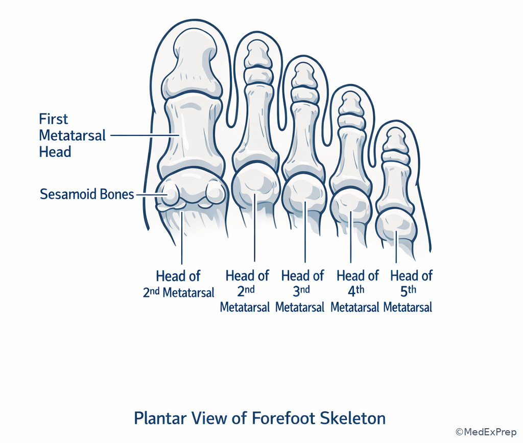
- Sesamoids are typically found under the head of the first metatarsal.
| Bone | Region | High-yield landmark | Most tested discriminator |
|---|---|---|---|
| Talus | Hindfoot | Trochlea, neck, head | No muscular attachments |
| Calcaneus | Hindfoot | Sustentaculum tali, tuberosity | Achilles insertion; heel bone |
| Navicular | Midfoot | Navicular tuberosity | Medial bone between talus and cuneiforms |
| Cuboid | Midfoot | Fibularis longus groove | Lateral bone anterior to calcaneus |
| Medial cuneiform | Midfoot | Largest cuneiform | Articulates with first metatarsal |
| Intermediate cuneiform | Midfoot | Smallest cuneiform | Articulates with second metatarsal |
| Lateral cuneiform | Midfoot | Lateral member of cuneiform trio | Articulates with third metatarsal |
| Fifth metatarsal | Forefoot | Base tuberosity | Palpable lateral prominence; fibularis brevis insertion |
Exam takeaway: Divide the foot into hindfoot, midfoot, and forefoot first, then localize the landmark within that region.
flowchart LR
A[Hindfoot] --> B[Talus]
A --> C[Calcaneus]
D[Midfoot] --> E[Navicular]
D --> F[Cuboid]
D --> G[Medial cuneiform]
D --> H[Intermediate cuneiform]
D --> I[Lateral cuneiform]
J[Forefoot] --> K[Metatarsals 1 to 5]
K --> L[Phalanges]
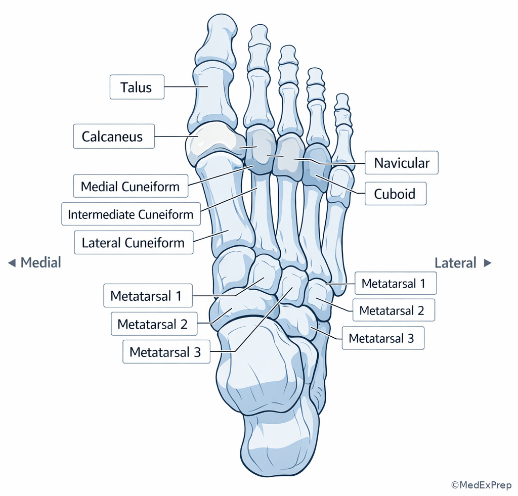
Exam takeaway: Use dorsal spatial relationships to avoid mixing up the medial navicular-cuneiform chain with the lateral calcaneocuboid chain.
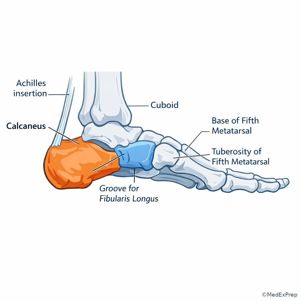
Exam takeaway: Lateral foot questions usually turn on the calcaneocuboid relationship or the fifth metatarsal tuberosity.
Core content
1.1.1.3 Foundational map: hindfoot, midfoot, and forefoot
A Explanation
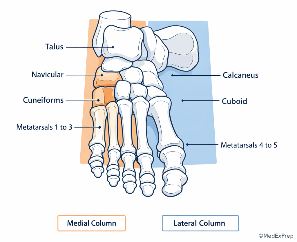
The first decision conflict on anatomy questions is simple but high yield: which region of the foot is the stem describing? If you do not localize first, you will confuse bones that share similar words like base, head, tuberosity, and facet. The resolving rule is this: \(hindfoot = talus and calcaneus\); \(midfoot = navicular\), cuboid, and cuneiforms; \(forefoot = metatarsals and phalanges\). That one map eliminates many distractors before you even think about detailed landmarks. See Fig 1 and Fig 2.
WHY THIS IS TESTED: board stems commonly present a tenderness location, radiograph clue, or tendon insertion and ask for the involved bone. The exam rewards candidates who organize the skeleton spatially rather than memorizing isolated facts.
B Worked example
A 22-year-old man twists his foot while playing basketball. He has obesity and mild asthma. He reports pain over the lateral midfoot, but the stem also mentions bruising over the toes and a prior ankle sprain last year. A radiograph question asks which bone lies anterior to the calcaneus and posterior to the fourth and fifth metatarsals. The tempting wrong clue is the history of ankle sprain, which may push you toward the talus. Reasoning chain: identify task — name the bone from location; extract key facts — lateral midfoot, anterior to calcaneus, linked to fourth and fifth metatarsals; apply rule — lateral midfoot tarsal in that position is the cuboid; eliminate distractors — navicular is medial, cuneiforms articulate with metatarsals 1–3, talus is proximal/hindfoot; verify — cuboid is part of the calcaneocuboid chain on the lateral side.
C Exam trap
Common wrong answer: Navicular.
Why it is tempting: Students remember “midfoot bone between hindfoot and metatarsals” and forget to separate medial from lateral anatomy.
Single clue that eliminates it: The stem says lateral or links the bone to the fourth and fifth metatarsals — that points to cuboid, not navicular.
D Checkpoint
Question: A 19-year-old woman with hypermobility and a prior stress fracture has focal tenderness over the medial midfoot after running. Imaging localizes the abnormality to the bone between the talar head and the three cuneiforms. Which structure is most likely involved?
- Navicular
- Cuboid
- Calcaneus
- Intermediate cuneiform
Answer: A. Navicular.
- A is tempting and correct because “between the talar head and the cuneiforms” is the defining positional rule for the navicular.
- B is tempting but wrong because it is also a midfoot tarsal; however, the cuboid is lateral and relates to calcaneus plus metatarsals 4–5, not the talar head.
- C is tempting but wrong because hindfoot pain after running can suggest calcaneal stress, but the described articulation pattern is not calcaneal.
- D is tempting but wrong because the intermediate cuneiform is near the area, but it lies distal to the navicular rather than between the talus and the cuneiform set.
1.1.1.4 Tarsal bones: landmarks, articulations, and unique signatures
A Explanation
The next decision conflict is which specific tarsal bone fits the clue? The rule that resolves this is to pair each bone with its most unique signature. Talus: no muscular attachments and a superior trochlea for the tibia. Calcaneus: largest tarsal, heel, Achilles insertion, sustentaculum tali. Navicular: medial midfoot connector between talus and cuneiforms, with a tuberosity. Cuboid: lateral midfoot, groove for fibularis longus. Cuneiforms: three wedge-shaped bones distal to navicular and proximal to metatarsals 1–3. See Fig 2.
WHY THIS IS TESTED: these unique identifiers are classic single-clue discriminators. On exams, one landmark is often enough. If the stem mentions a tendon groove on the plantar-lateral midfoot, choose cuboid. If it mentions a bone receiving body weight from the tibia without muscle attachment, choose talus.
Exam takeaway: One high-yield landmark usually identifies the tarsal bone faster than memorizing long lists of attachments.
flowchart TD
A[Landmark clue] --> B{Best match}
B -->|No muscular attachments| C[Talus]
B -->|Achilles insertion or heel tuberosity| D[Calcaneus]
B -->|Medial tuberosity between talus and cuneiforms| E[Navicular]
B -->|Groove for fibularis longus| F[Cuboid]
B -->|Distal to navicular and proximal to MT 1 to 3| G[Cuneiforms] Exam takeaway: Medial foot stems often hinge on distinguishing navicular tuberosity from calcaneal sustentaculum tali.
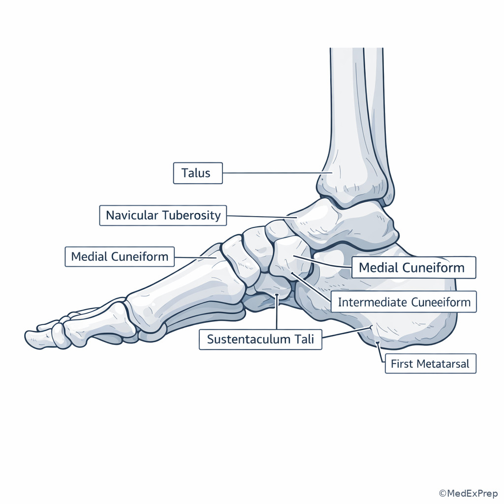
B Worked example
A 14-year-old boy with obesity and mild pes planus develops prominence and pain along the medial midfoot after soccer. He also has eczema and a mildly elevated alkaline phosphatase from growth, which is distracting but age-appropriate. The stem asks for the bone bearing the palpable tuberosity where a tendon inserts medially. Reasoning chain: identify task — locate a medial prominence; extract key facts — medial midfoot, tuberosity, tendon insertion, not heel; apply rule — navicular tuberosity is the key landmark; eliminate distractors — sustentaculum tali is calcaneal and more posterior/inferior, medial cuneiform lacks the classic named tuberosity clue, talus has no muscular attachment; verify — the navicular is the medial midfoot connector and common board favorite.
C Exam trap
Common wrong answer: Talus for any “important ankle-area bone” clue.
Why it is tempting: Students overgeneralize the talus because it is central to weight transfer and ankle questions.
Single clue that eliminates it: Any clue involving a direct tendon insertion excludes the talus, because the talus has no muscular attachments.
D Checkpoint
Question: A 31-year-old woman with hypothyroidism and chronic lateral foot pain has imaging focused on a plantar groove that transmits the tendon of fibularis longus. Which bone is the best match?
- Navicular
- Cuboid
- Calcaneus
- Lateral cuneiform
Answer: B. Cuboid.
- A is tempting but wrong because it is also a midfoot tarsal; however, its signature clue is medial tuberosity rather than a plantar tendon groove.
- B is tempting and correct because the groove for fibularis longus on the plantar-lateral aspect is the cuboid’s classic identifying feature.
- C is tempting but wrong because the calcaneus is also lateral/posterior and tendon-rich, but the fibularis longus groove is specifically cuboid.
- D is tempting but wrong because the lateral cuneiform is in the distal midfoot, yet it lacks the hallmark tendon-groove association in standard board anatomy.
- The only tarsal with no muscular attachments is the talus.
- True or False: The cuboid is medial to the navicular. False.
- Name the 3 bones distal to the navicular on the medial side: medial, intermediate, and lateral cuneiforms.
1.1.1.5 Metatarsal anatomy: base, shaft, neck, head, and numbering logic
A Explanation
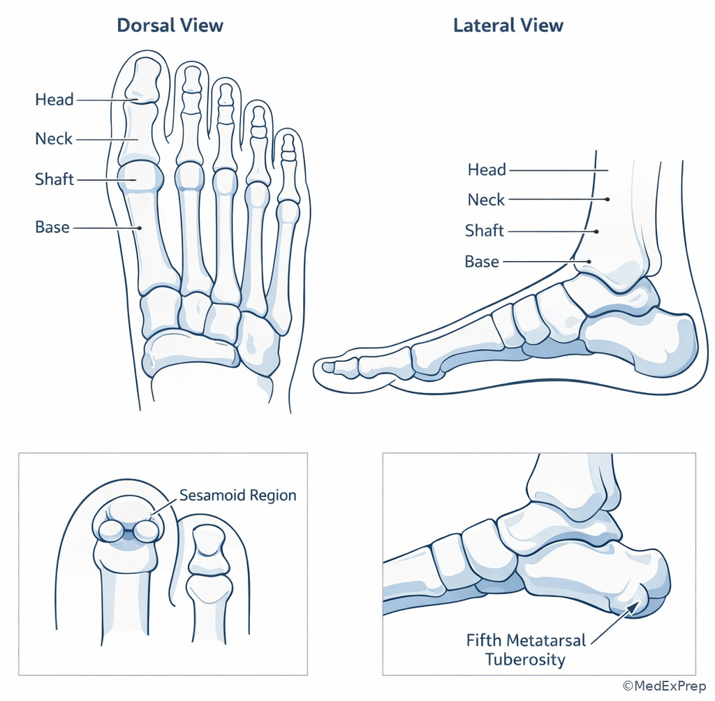
The decision conflict here is which part of which metatarsal does the stem describe? The resolving rule is to separate number from segment. Metatarsals are numbered medial to lateral: first through fifth. Each has a base proximally, shaft, neck, and head distally. The first metatarsal is thick and built for load transmission; the second is commonly longest and sits recessed between cuneiforms; the fifth has a prominent tuberosity at its base. See Fig 4.
WHY THIS IS TESTED: fracture, stress reaction, tendon insertion, and gait questions often depend on whether the abnormality is at the base, neck, or head. The wrong answer is commonly the correct metatarsal but the wrong segment, or the correct segment on the wrong metatarsal.
Exam takeaway: Separate which metatarsal is involved from which segment of that metatarsal is involved.
| Metatarsal | Key feature | Articular pattern | Board-style clue |
|---|---|---|---|
| 1st | Shortest, thickest | With medial cuneiform | Weight-bearing; sesamoids under head |
| 2nd | Commonly longest | With intermediate cuneiform; recessed | Midfoot stability/Lisfranc clue |
| 3rd | Central ray | With lateral cuneiform | Often confused with second in central foot stems |
| 4th | Lateral forefoot | With cuboid | Shares lateral articulation pattern with fifth |
| 5th | Proximal tuberosity | With cuboid | Lateral base prominence; fibularis brevis insertion |
Exam takeaway: Distal metatarsal-head questions often become easier when you remember the unique sesamoid relationship under the first head.
B Worked example
A 27-year-old woman training for a half marathon has tenderness over the distal forefoot. She has iron deficiency and a history of irregular menses; the stem also mentions a recent blister on the heel, which is distracting because it is unrelated to the painful site. Radiographs are negative, and the question asks which segment lies just proximal to the rounded articular surface that contacts the proximal phalanx. Reasoning chain: identify task — name a metatarsal segment; extract key facts — “just proximal to the rounded articular surface” means near the head; apply rule — the narrowed segment proximal to the head is the neck; eliminate distractors — base is proximal, shaft is longer central portion, tuberosity is specific to certain proximal regions; verify — head articulates distally, neck is immediately proximal.
C Exam trap
Common wrong answer: Shaft instead of neck.
Why it is tempting: Both are central portions of a long bone, and stems often use broad language like “distal metatarsal pain.”
Single clue that eliminates it: If the stem specifies the narrowed region immediately proximal to the head, that is the neck, not the shaft.
D Checkpoint
Question: A 20-year-old soccer player with mild cavus feet and a prior ankle sprain has tenderness over a palpable prominence at the proximal lateral foot. Which structure is most likely involved?
- Head of the first metatarsal
- Base tuberosity of the fifth metatarsal
- Neck of the second metatarsal
- Base of the first metatarsal
Answer: B. Base tuberosity of the fifth metatarsal.
- A is tempting but wrong because the first metatarsal head is prominent and clinically important, but it is distal and medial rather than proximal and lateral.
- B is tempting and correct because the palpable proximal lateral foot prominence is the tuberosity at the base of the fifth metatarsal.
- C is tempting but wrong because central forefoot stress symptoms commonly involve the second metatarsal, yet the location given is not distal-central.
- D is tempting but wrong because the first metatarsal base is proximal, but it lies medially and articulates with the medial cuneiform.
1.1.1.6 Medial vs lateral column logic and articulation patterns
A Explanation
Now move from naming bones to recognizing the column pattern of the foot. The decision conflict is whether the stem describes the medial chain or the lateral chain. The resolving rule: the medial column runs talus → navicular → cuneiforms → metatarsals 1–3; the lateral column runs calcaneus → cuboid → metatarsals 4–5. The second metatarsal is especially important because its base fits between cuneiforms, creating a recessed keystone configuration that stabilizes the tarsometatarsal region. See Fig 5.
WHY THIS IS TESTED: articulation patterns explain why certain injuries localize where they do and why some distractors are almost right. If a stem mentions a lateral plantar tendon groove, think cuboid-lateral column. If it mentions a recessed second metatarsal base, think medial/central cuneiform relationship and midfoot stability.
Exam takeaway: Column logic is the fastest way to solve articulation questions and reject near-neighbor distractors.
flowchart LR
A[Medial column] --> B[Talus]
B --> C[Navicular]
C --> D[Cuneiforms]
D --> E[Metatarsals 1 to 3] F[Lateral column] --> G[Calcaneus]
G --> H[Cuboid]
H --> I[Metatarsals 4 to 5] J[Central stability clue] --> K[2nd metatarsal base recessed between cuneiforms]
| Pattern | Medial column | Lateral column | Discriminator |
|---|---|---|---|
| Proximal tarsal relationship | Talus-associated chain | Calcaneus-associated chain | Talus is medial-superior; calcaneus dominates heel/lateral column |
| Midfoot connector | Navicular | Cuboid | Navicular is medial; cuboid has fibularis longus groove |
| Distal tarsals | Cuneiforms | No cuneiforms | Metatarsals 1–3 vs 4–5 |
| Metatarsal partners | 1st, 2nd, 3rd | 4th, 5th | Second metatarsal base keystone is a central clue |
| Typical exam landmark | Navicular tuberosity | 5th metatarsal tuberosity / cuboid groove | Medial prominence vs lateral prominence |
Exam takeaway: Visualizing the foot as two columns reduces confusion on articulation and stability questions.
B Worked example
A 35-year-old man with diabetes and obesity presents in a stem describing midfoot instability after a twisting injury. The question notes pain near the base of the second metatarsal and mentions ecchymosis over the heel as a distracting detail. It asks which neighboring tarsal bone most directly explains the recessed “keystone” relationship. Reasoning chain: identify task — determine the proximal articular partner pattern; extract key facts — second metatarsal base, recessed, midfoot stability; apply rule — second metatarsal relates to the cuneiform complex, especially the intermediate cuneiform region; eliminate distractors — cuboid matches lateral metatarsals 4–5, navicular is proximal to cuneiforms not directly the distal partner; verify — this is a cuneiform-based central stabilization clue.
C Exam trap
Common wrong answer: Cuboid for any midfoot-stability question.
Why it is tempting: The cuboid is memorable and frequently tested, so students overapply it whenever the stem says “midfoot.”
Single clue that eliminates it: If the stem centers on the second metatarsal base, think cuneiform complex, not cuboid.
D Checkpoint
Question: A 24-year-old dancer with ligamentous laxity and recent midfoot pain has imaging showing a bone chain running from talus to navicular to distal tarsals before reaching metatarsals 1–3. Which distal tarsal group is involved?
- Cuneiforms
- Cuboid
- Calcaneus
- Sesamoids
Answer: A. Cuneiforms.
- A is tempting and correct because the medial column progresses from talus to navicular to the three cuneiforms before metatarsals 1–3.
- B is tempting but wrong because cuboid is a distal tarsal too, but it belongs to the lateral chain with metatarsals 4–5.
- C is tempting but wrong because calcaneus is proximal hindfoot and belongs to the lateral/posterior chain rather than the described medial sequence.
- D is tempting but wrong because sesamoids are associated with the first metatarsal head, not the proximal medial tarsal chain.
- The lateral midfoot bone distal to calcaneus is the cuboid.
- True or False: Metatarsals 4 and 5 articulate proximally with the cuboid. True.
- Name the central stability clue in the tarsometatarsal region: recessed base of the second metatarsal between cuneiforms.
1.1.1.7 High-yield localization in board-style stems
A Explanation
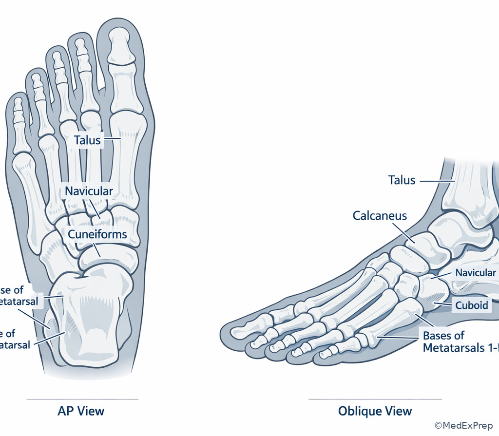
The final integration skill is choosing between look-alike anatomy answers when the stem adds noise. The decision conflict is not “Do I know anatomy?” but “Which single clue rules out the tempting alternative?” The best method is: locate the region, identify the landmark, confirm the articular neighbors, then reject the most tempting distractor. See Fig 3 and Fig 5. On APMLE-style questions, distractions may include age, sports history, diabetes, prior sprain, or unrelated bruising. Those details matter only if they localize the structure. Otherwise, ignore them.
WHY THIS IS TESTED: basic science board questions reward disciplined reasoning over trivia. A candidate who uses a structured localization approach will outperform someone who memorizes lists without hierarchy.
Exam takeaway: Many anatomy stems are really palpation-location questions translated into bone-identification language.
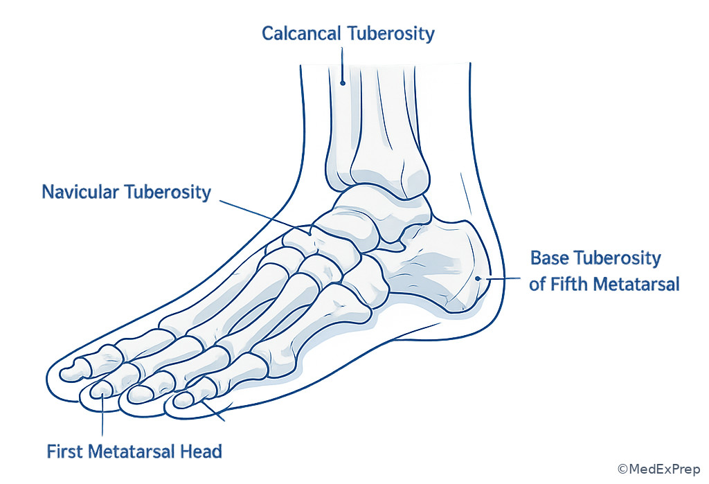
Exam takeaway: Oblique and AP relationships help distinguish overlapping midfoot structures in image-based questions.
B Worked example
A 42-year-old woman with rheumatoid arthritis and chronic steroid exposure develops lateral foot pain after stepping off a curb. She also reports morning hand stiffness, which is distracting because it does not help localize the foot structure. The question asks which bone provides a prominent proximal lateral attachment site for fibularis brevis. Reasoning chain: identify task — choose the bony insertion site; extract key facts — proximal, lateral, prominent tuberosity, fibularis brevis; apply rule — base tuberosity of the fifth metatarsal; eliminate distractors — cuboid has fibularis longus groove, navicular is medial, calcaneus is posterior with Achilles insertion; verify — lateral proximal prominence on palpation matches fifth metatarsal tuberosity. See Fig 6.
C Exam trap
Common wrong answer: Cuboid when the stem names a fibularis tendon.
Why it is tempting: Students remember the cuboid-fibularis longus association and apply it to every lateral tendon clue.
Single clue that eliminates it: Fibularis brevis inserts on the tuberosity of the fifth metatarsal; the cuboid is the tendon groove for fibularis longus.
D Checkpoint
Question: A 29-year-old runner with vitamin D deficiency and prior shin splints reports tenderness at a medial bony prominence in the midfoot. Which option best matches the anatomic structure?
- Navicular tuberosity
- Sustentaculum tali
- Base of the fifth metatarsal
- Cuboid groove
Answer: A. Navicular tuberosity.
- A is tempting and correct because a palpable medial midfoot prominence classically indicates the navicular tuberosity.
- B is tempting but wrong because it is also medial, but it is on the calcaneus and lies more posterior/inferior, supporting the talus.
- C is tempting but wrong because it is a palpable prominence, but it is lateral rather than medial.
- D is tempting but wrong because it is lateral midfoot and a groove rather than a palpable medial tuberosity.
Exam Traps & Differentiators
Most common wrong answer and why: navicular vs cuboid. This pair is tempting because both are midfoot bones between hindfoot and metatarsal regions. The discriminator is medial vs lateral: navicular is medial and linked to talus/cuneiforms; cuboid is lateral and linked to calcaneus/metatarsals 4–5.
Looks similar but isn't: navicular tuberosity vs sustentaculum tali. Both may appear as medial prominences. The navicular is in the midfoot and lies distal to the talar head; the sustentaculum tali is a calcaneal shelf more posterior and supports the talus.
Cross-topic trap: upcoming Ankle And Subtalar Joints questions may mention talus and calcaneus together. In this lesson, the goal is the bone map; in the next lesson, the tested idea becomes the articulation and motion.
| Confusable pair | Why they look similar | Single discriminator | Think this instead |
|---|---|---|---|
| Navicular vs Cuboid | Both are midfoot tarsals | Medial talar-head relation vs lateral calcaneal relation | \(Medial = navicular\); \(lateral = cuboid\) |
| Talus vs Calcaneus | Both are hindfoot bones | No muscular attachments vs Achilles insertion/heel | Weight-transfer dome = talus; \(heel = calcaneus\) |
| 5th metatarsal tuberosity vs Cuboid | Both are lateral foot structures | Prominent proximal lateral base vs tarsal tendon groove | Palpable lateral bump = 5th metatarsal tuberosity |
| Navicular tuberosity vs Sustentaculum tali | Both can be described as medial prominences | Midfoot bone distal to talus vs calcaneal shelf under talus | \(Anterior-medial = navicular\); posterior-medial support = sustentaculum |
| Metatarsal neck vs Shaft | Both are long-bone segments | Neck lies immediately proximal to head | Distal narrowing = neck |
- If the stem says “between talus and cuneiforms,” think navicular.
- If the stem says “groove for fibularis longus,” think cuboid.
- If the stem says “no muscular attachment,” think talus.
- If the stem says “Achilles insertion” or “heel bone,” think calcaneus.
- If the stem says “proximal lateral tuberosity,” think base of the fifth metatarsal.
- If the stem says “recessed second metatarsal base,” think cuneiform-based midfoot stability/Lisfranc region.
Algorithm / Approach
Rapid Review
- Talus → only tarsal with no muscular attachments.
- Calcaneus → largest tarsal and Achilles insertion site.
- Navicular → medial midfoot bridge between talus and cuneiforms.
- Cuboid → lateral midfoot bone with fibularis longus groove.
- Medial cuneiform → distal to navicular, proximal to first metatarsal.
- Intermediate cuneiform → smallest cuneiform; second metatarsal partner.
- Lateral cuneiform → proximal partner for third metatarsal.
- First metatarsal → shortest and thickest ray.
- Second metatarsal → commonly longest; recessed base supports midfoot stability.
- Fifth metatarsal → proximal lateral tuberosity on the base.
- Metatarsal neck → narrowed segment just proximal to head.
- Metatarsal head → distal articular end with proximal phalanx.
- Medial column → talus-navicular-cuneiforms-metatarsals 1–3.
- Lateral column → calcaneus-cuboid-metatarsals 4–5.
Self-check quiz
1. A 21-year-old man with a prior inversion injury and mild obesity has a board-style anatomy question asking which tarsal bone lacks direct muscular attachments. Which is the single best answer?
- Talus
- Calcaneus
- Navicular
- Cuboid
2. A 17-year-old runner with pes planus and medial foot tenderness has focal pain over the bone between the talar head and the three cuneiforms. Which bone is most likely involved?
- Navicular
- Cuboid
- Talus
- Medial cuneiform
3. A 28-year-old woman with vitamin D deficiency and a history of metatarsalgia reports pain at a palpable prominence on the proximal lateral foot after missing a step. The stem asks which structure best explains this location. Which is the most appropriate interpretation?
- Head of the first metatarsal
- Base tuberosity of the fifth metatarsal
- Navicular tuberosity
- Sustentaculum tali
4. A 25-year-old soccer player with recurrent midfoot pain and prior ankle sprains has imaging that highlights a plantar groove for tendon passage in the lateral midfoot. Which bone is the single best answer?
- Navicular
- Calcaneus
- Cuboid
- Lateral cuneiform
5. A 34-year-old man with diabetes and obesity has a twisting midfoot injury. The stem emphasizes the recessed base of the second metatarsal as a key stabilizing feature. Which proximal tarsal group best explains this relationship?
- Cuneiforms
- Cuboid
- Navicular
- Calcaneus
6. A 30-year-old woman with rheumatoid arthritis and chronic steroid use reports medial foot pain. Examination localizes tenderness to a palpable prominence distal to the talar head. A tempting note in the stem mentions discomfort behind the medial malleolus. Which structure is most likely involved?
- Navicular tuberosity
- Sustentaculum tali
- Medial cuneiform base
- Talus neck
7. A 23-year-old dancer with ligamentous laxity and a prior stress reaction has pain at the distal forefoot. The stem asks for the metatarsal segment immediately proximal to the rounded articular surface that contacts a proximal phalanx. Which is the best answer?
- Base
- Shaft
- Neck
- Tuberosity
8. A 26-year-old basketball player with cavus feet has lateral midfoot pain. Imaging localizes the abnormality to the bone anterior to the calcaneus and proximal to the bases of the fourth and fifth metatarsals. The report also notes unrelated mild first-MTP degenerative change. Which bone is most likely involved?
- Navicular
- Cuboid
- Lateral cuneiform
- Fifth metatarsal
Answer key
1. Correct answer: A. Talus.
- A. Talus — Tempting because it is the major ankle-region tarsal, and correct because the talus uniquely has no muscular attachments. Single stem clue: “lacks direct muscular attachments.” Source: Moore, Dalley, and Agur; Drake et al.
- B. Calcaneus — Tempting because it is another hindfoot bone, but incorrect because it has major tendon attachment including Achilles insertion. Single discriminating clue: any muscular/tendinous insertion excludes talus and fits calcaneus. Source: Moore, Dalley, and Agur.
- C. Navicular — Tempting because it is highly tested in medial foot anatomy, but incorrect because it is not the unique no-muscle-attachment tarsal. Single discriminating clue: navicular is identified by location between talus and cuneiforms, not by lack of muscular insertion. Source: Drake et al.
- D. Cuboid — Tempting because it is a classic landmark bone with a tendon groove, but that same tendon relationship proves it is not correct here. Single discriminating clue: groove for fibularis longus means tendon interaction exists. Source: Moore, Dalley, and Agur.
2. Correct answer: A. Navicular.
- A. Navicular — Tempting because it sits in the exact region described, and correct because it lies between the talar head proximally and cuneiforms distally. Single stem clue: “between the talar head and the three cuneiforms.” Source: Drake et al.; Moore, Dalley, and Agur.
- B. Cuboid — Tempting because it is also a midfoot tarsal, but incorrect because it is lateral and articulates with calcaneus plus metatarsals 4–5. Single discriminating clue: talar-head relation points medial, not lateral. Source: Moore, Dalley, and Agur.
- C. Talus — Tempting because the talar head appears in the stem, but incorrect because the questioned bone is distal to the talus, not the talus itself. Single discriminating clue: “between” identifies the intermediate bone. Source: Drake et al.
- D. Medial cuneiform — Tempting because the cuneiforms are adjacent, but incorrect because they lie distal to the navicular rather than between talus and cuneiform set. Single discriminating clue: cuneiforms are the distal partners, not the middle bridge. Source: Moore, Dalley, and Agur.
3. Correct answer: B. Base tuberosity of the fifth metatarsal.
- A. Head of the first metatarsal — Tempting because it is a palpable landmark, but incorrect because it is distal and medial rather than proximal and lateral. Single discriminating clue: “proximal lateral foot.” Source: Moore, Dalley, and Agur.
- B. Base tuberosity of the fifth metatarsal — Tempting and correct because it creates the classic palpable prominence on the proximal lateral foot. Single stem clue: “palpable prominence on the proximal lateral foot.” Source: Drake et al.
- C. Navicular tuberosity — Tempting because it is also a palpable tuberosity, but incorrect because it is medial midfoot, not lateral. Single discriminating clue: side of the foot. Source: Moore, Dalley, and Agur.
- D. Sustentaculum tali — Tempting because it is a notable bony projection, but incorrect because it is on the medial calcaneus, more posterior and inferior. Single discriminating clue: the described landmark is lateral. Source: Drake et al.
4. Correct answer: C. Cuboid.
- A. Navicular — Tempting because it is a midfoot tarsal, but incorrect because its classic landmark is a medial tuberosity rather than a lateral plantar tendon groove. Single discriminating clue: “plantar groove” in the lateral midfoot. Source: Moore, Dalley, and Agur.
- B. Calcaneus — Tempting because it neighbors the cuboid and contains important plantar structures, but incorrect because the characteristic fibularis longus groove is on the cuboid. Single discriminating clue: tendon passage across the lateral midfoot rather than posterior heel. Source: Drake et al.
- C. Cuboid — Tempting and correct because the cuboid has the classic groove for the tendon of fibularis longus on its plantar aspect. Single stem clue: “plantar groove for tendon passage in the lateral midfoot.” Source: Moore, Dalley, and Agur.
- D. Lateral cuneiform — Tempting because it is a distal midfoot tarsal, but incorrect because it lacks the hallmark tendon-groove association. Single discriminating clue: groove, not just position. Source: Drake et al.
5. Correct answer: A. Cuneiforms.
- A. Cuneiforms — Tempting and correct because the recessed base of the second metatarsal fits into the cuneiform complex and supports central midfoot stability. Single stem clue: “recessed base of the second metatarsal.” Source: Drake et al.; Moore, Dalley, and Agur.
- B. Cuboid — Tempting because it is a midfoot articulation partner for lateral metatarsals, but incorrect because it mainly serves metatarsals 4–5 rather than the second ray keystone relation. Single discriminating clue: second metatarsal, not fourth/fifth. Source: Moore, Dalley, and Agur.
- C. Navicular — Tempting because it is central in the medial midfoot, but incorrect because it lies proximal to the cuneiforms and is not the immediate distal tarsal group receiving the second metatarsal base. Single discriminating clue: proximal partner group of the second metatarsal. Source: Drake et al.
- D. Calcaneus — Tempting because twisting injuries involve the hindfoot too, but incorrect because the calcaneus does not explain the recessed second metatarsal base configuration. Single discriminating clue: midfoot central stability, not heel anatomy. Source: Moore, Dalley, and Agur.
6. Correct answer: A. Navicular tuberosity.
- A. Navicular tuberosity — Tempting and correct because it is a palpable medial prominence distal to the talar head in the midfoot. Single stem clue: “palpable prominence distal to the talar head.” Source: Moore, Dalley, and Agur.
- B. Sustentaculum tali — Tempting because it is also medial and the note about discomfort behind the medial malleolus nudges you posteriorly, but incorrect because sustentaculum tali is on the calcaneus and sits more posterior/inferior. Single discriminating clue: distal to the talar head, not beneath it. Source: Drake et al.
- C. Medial cuneiform base — Tempting because it is in the medial midfoot, but incorrect because the classic palpable tuberosity clue points to navicular, not cuneiform. Single discriminating clue: named prominence. Source: Moore, Dalley, and Agur.
- D. Talus neck — Tempting because the talus is adjacent and the stem mentions the talar head, but incorrect because the prominence is distal to the talus rather than part of it. Single discriminating clue: position relative to talar head. Source: Drake et al.
7. Correct answer: C. Neck.
- A. Base — Tempting because it is a standard long-bone segment, but incorrect because the base is proximal, not immediately adjacent to the distal articular head. Single discriminating clue: “immediately proximal to the rounded articular surface.” Source: Moore, Dalley, and Agur.
- B. Shaft — Tempting because many distal stress questions involve shaft-versus-neck confusion, but incorrect because the shaft is the longer central region, not the short narrowed region just before the head. Single discriminating clue: immediate adjacency to head. Source: Drake et al.
- C. Neck — Tempting and correct because the neck is the narrowed segment immediately proximal to the metatarsal head. Single stem clue: “immediately proximal to the rounded articular surface.” Source: Moore, Dalley, and Agur.
- D. Tuberosity — Tempting because some metatarsals have named tuberosities, but incorrect because tuberosity refers to a projection, classically at the base of the fifth metatarsal, not a universal distal segment. Single discriminating clue: this is a segment question, not a prominence question. Source: Drake et al.
8. Correct answer: B. Cuboid.
- A. Navicular — Tempting because it is another midfoot tarsal, but incorrect because it lies medially and relates to talus and cuneiforms rather than calcaneus with metatarsals 4–5. Single discriminating clue: “anterior to the calcaneus and proximal to the bases of the fourth and fifth metatarsals.” Source: Moore, Dalley, and Agur.
- B. Cuboid — Tempting and correct because the cuboid sits exactly anterior to the calcaneus and proximal to the fourth and fifth metatarsals in the lateral column. Single stem clue: lateral column articulation pattern. Source: Drake et al.
- C. Lateral cuneiform — Tempting because it is a distal tarsal and lies laterally within the cuneiform trio, but incorrect because it does not articulate with the calcaneus or the bases of metatarsals 4 and 5 as described. Single discriminating clue: relationship to calcaneus. Source: Moore, Dalley, and Agur.
- D. Fifth metatarsal — Tempting because it is in the lateral foot and the stem mentions metatarsal bases, but incorrect because the questioned structure is proximal to those bases, making it a tarsal rather than a metatarsal. Single discriminating clue: “proximal to the bases.” Source: Drake et al.
Keep going with the full APMLE Part I Prep Course (APMLE I)
You just studied the way members do, in the same lesson interface. Full access unlocks every lesson, the question bank and flashcards for a full year.
One payment · 1 year of access · No auto-renewal