1.1.1 - Cross-Sectional Vascular Anatomy
Cross-Sectional Vascular Anatomy
What you'll learn
- FOUNDATIONS: Imaging Anatomy Basics -> Cross-sectional vessel identification — distinguish major arteries and veins on CT and MR by position, branching pattern, and relationship to organs.
- FOUNDATIONS: Imaging Anatomy Basics -> Procedural route planning — choose the safest access or target pathway based on vascular course and adjacent structures.
- VASCULAR_INTERVENTION: Anatomy and imaging findings -> Territory recognition — map vascular territories relevant to embolization, revascularization, and complication avoidance.
- PERIPROCEDURAL_CARE: Safety planning -> Non-target risk recognition — identify branches and variants that increase risk of non-target puncture, embolization, or ischemia.
- TRAUMA_EMERGENCY_IR: Imaging interpretation -> Rapid vessel localization — localize likely bleeding source or access target from cross-sectional anatomy under time pressure.
- FOUNDATIONS → Imaging Anatomy Basics → Cross-sectional vessel identification
- FOUNDATIONS → Imaging Anatomy Basics → Anatomic relationships for image-guided planning
- VASCULAR_INTERVENTION → Anatomy and imaging findings → Arterial territory recognition
- VENOUS_AND_DIALYSIS_ACCESS → Anatomy and access strategy → Central and peripheral venous pathway recognition
- PERIPROCEDURAL_CARE → Procedure planning → Avoidance of non-target injury based on anatomy
Why it matters
- IR questions often hide the real task inside anatomy: the best next step depends on knowing which vessel you are seeing before you choose a wire, catheter, puncture site, or embolic target.
- Cross-sectional vascular anatomy determines whether a route is technically feasible, whether a branch is expendable, and whether a planned intervention risks non-target ischemia.
- Many board distractors exploit side confusion, branch mislabeling, or failure to recognize a variant that changes access strategy.
- In emergency IR, rapid vessel localization on CTA can separate urgent embolization from a futile or unsafe procedure.
Key Terms & Must-Know Facts
1.1.1.1 Key terms
- Aorta — central arterial conduit; branch order and caliber guide orientation.
- IVC — thin-walled right-sided retroperitoneal venous structure receiving renal, hepatic, and iliac inflow.
- Celiac axis — first major anterior abdominal aortic branch supplying foregut territory.
- SMA — major anterior branch supplying midgut; key landmark below celiac axis.
- IMA — inferior anterior branch supplying hindgut; smaller and lower than SMA.
- Portal vein — formed by SMV and splenic vein; enters liver at porta hepatis.
- Hepatic artery proper — arterial supply to liver; runs with bile duct and portal vein in portal triad.
- Renal hilum — classic venous-anterior, arterial-middle, pelvis-posterior relationship, with variation.
- External iliac artery — continues to common femoral below inguinal ligament.
- Internal iliac artery — pelvic branch with hemorrhage and embolization relevance.
- Common femoral artery — preferred access target over profunda or superficial femoral puncture.
- Jugular-carotid relationship — internal jugular usually lateral to carotid, but position varies with level and head rotation.
- Anatomic variant — common deviation from textbook branching that changes planning but is not pathologic.
1.1.1.2 Must-know facts
- The IVC is usually right of the aorta; loss of this rule should trigger careful re-orientation before labeling structures.
- The left renal vein crosses anterior to the aorta and posterior to the SMA in normal anatomy.
- The right renal artery often courses posterior to the IVC, a favorite board anatomy clue.
- The portal vein is posterior to the common bile duct and hepatic artery at the porta hepatis.
- The SMA lies left of the SMV in standard axial orientation; reversal suggests malrotation or labeling error.
- Femoral access safety depends on inguinal ligament level, not just vessel size.
- Internal iliac branches matter in pelvic embolization because non-target embolization can affect gluteal, vesical, uterine, or rectal territories.
- Aortic branch order is testable: celiac, SMA, renal arteries, IMA.
| Landmark | Usual Relationship | Why IR Cares |
|---|---|---|
| Aorta vs IVC | Aorta left, IVC right | Core orientation for abdominal CTA reading |
| SMA vs SMV | SMA left of SMV | Mesenteric planning, malrotation check |
| Left renal vein | Anterior to aorta, posterior to SMA | Renal venography, nutcracker recognition |
| Right renal artery | Posterior to IVC | Catheter selection, CTA interpretation |
| Portal triad | Portal vein posterior, duct lateral, artery medial/anterior variable | Transhepatic procedures, embolization planning |
| Common femoral artery | Over femoral head, above bifurcation | Safe arterial access |
Core content
Exam takeaway: Use fixed right-left and anterior-posterior relationships first, then branch order, to avoid mislabeling vessels on axial imaging.
flowchart TD
A[Start with axial CT slice] --> B[Find vertebral body]
B --> C[Identify aorta left of midline]
B --> D[Identify IVC right of aorta]
C --> E[Look anteriorly for celiac then SMA]
D --> F[Look for renal veins entering IVC]
E --> G[Use branch order to confirm level]
F --> G
G --> H[Then assign target vessel or access route]
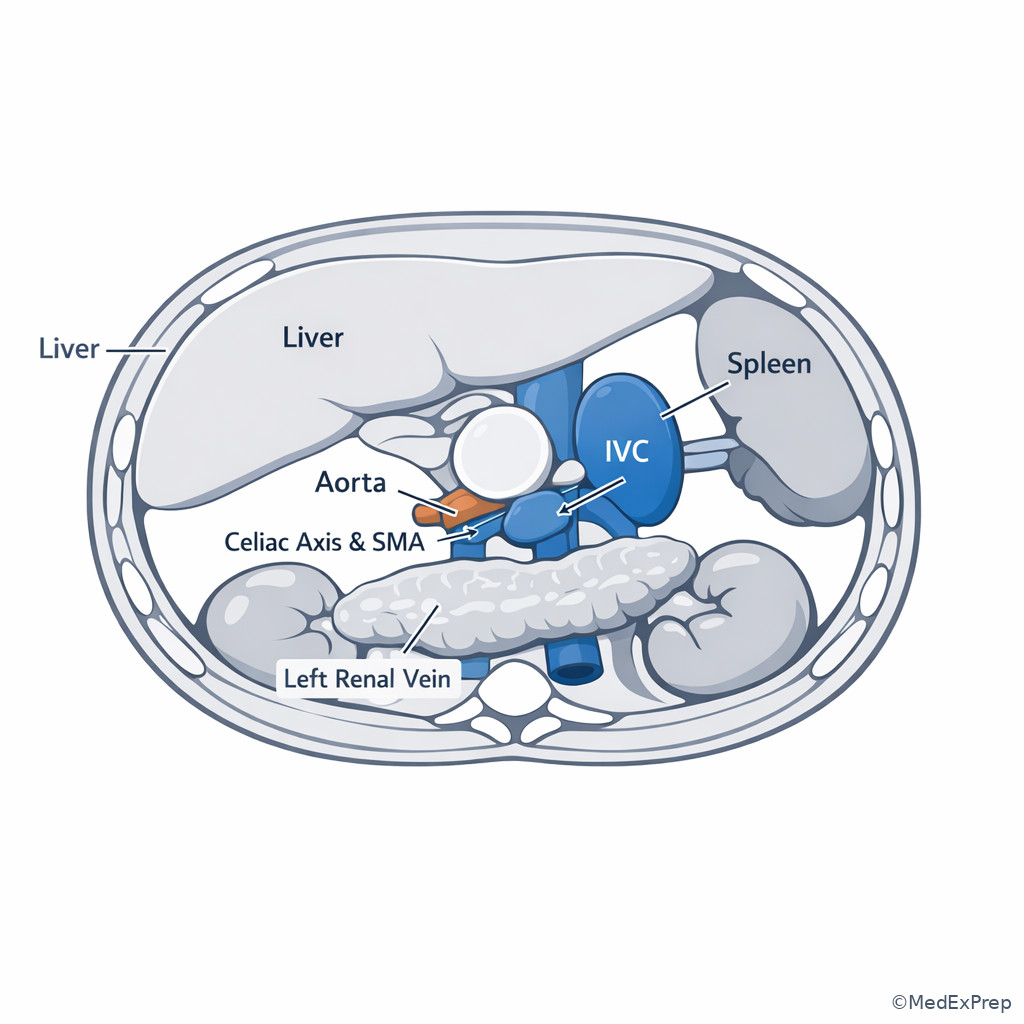
Exam takeaway: Anchor yourself with aorta, IVC, SMA, SMV, renal vessels, pancreas, and portal vein before answering procedural planning questions.
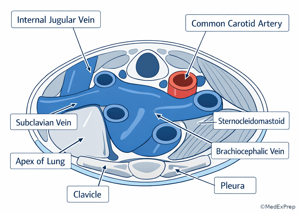
Exam takeaway: Recognize internal jugular, subclavian, brachiocephalic veins, and carotid relationship before planning central venous access.
Exam takeaway: Internal iliac branches create the classic pelvic embolization distractors, so branch territory matters more than memorizing every small vessel.
flowchart TD
A[Common iliac] --> B[External iliac]
A --> C[Internal iliac]
C --> D[Anterior division]
C --> E[Posterior division]
D --> F[Uterine/Vesical/Obturator/Internal pudendal]
E --> G[Iliolumbar/Lateral sacral/Superior gluteal]
B --> H[Common femoral after inguinal ligament]
1.1.1.3 Foundational orientation: how to label vessels before you decide anything
A Explanation
The first exam conflict is simple but decisive: are you looking at the right vessel, or are you about to make a procedural decision based on a mislabeled structure? The single rule that resolves many anatomy questions is to start with stable landmarks before variable branches. On axial imaging, identify the vertebral body, then the aorta, then the IVC, then major anterior aortic branches. Only after this should you label mesenteric, renal, portal, or pelvic vessels. See Fig 1 and Fig 2.
WHY THIS IS TESTED: board stems often present a CTA slice and ask for the bleeding source, the target vessel, or the safest catheter path. The error is usually not ignorance of therapy; it is failure to orient properly. Choose the interpretation supported by fixed relationships: aorta usually left of the IVC; SMA usually left of the SMV; left renal vein crosses anterior to the aorta; right renal artery often runs posterior to the IVC. Reject answers that depend on memorized branch names if the global orientation is wrong.
For IR planning, anatomy is functional. If you misidentify the SMA for the SMV, you may choose the wrong sheath side or underestimate risk to bowel branches. If you mistake a portal venous structure for an arterial one, you may misread tumor vascularity or hemorrhage source. The best strategy is a sequence: orient the patient, identify paired anchors, assign level, confirm branch order, then decide the procedure.
B Worked example
A 67-year-old man with hypertension, atrial fibrillation on apixaban, chronic pancreatitis, and prior laparoscopic cholecystectomy undergoes CTA for melena. A trainee points to a round enhancing structure anterior to the aorta and right of midline and calls it the SMA origin. Another reader notes the structure is larger, thin-walled, and contiguous with venous tributaries. A nearby artery arises separately from the aorta just left of this structure. Mild pancreatic calcifications distract from the task.
Reasoning chain: Identify task: label the vessel correctly before planning mesenteric angiography. Extract key facts: the questioned structure is right of the expected arterial position, appears larger and venous, and is associated with tributaries. Apply rule: the SMA is usually left of the SMV on axial imaging. Eliminate distractors: pancreatic inflammatory change does not redefine vessel orientation; prior cholecystectomy is irrelevant. Verify: the structure is the SMV, and the adjacent left-sided artery is the SMA. The best planning implication is that the operator should not interpret this venous structure as the culprit arterial target.
C Exam trap
D Checkpoint
Question: On axial abdominal CTA, which relationship most reliably supports correct vessel labeling when you must rapidly distinguish mesenteric artery from mesenteric vein?
- The SMA is usually right of the SMV.
- The SMA is usually left of the SMV.
- The portal vein is usually anterior to the common bile duct.
- The right renal artery is usually anterior to the IVC.
Answer: B
- A: Tempting because some learners remember the pair but reverse the sides; wrong because standard axial anatomy places the artery left of the vein.
- B: Correct because the SMA-left-of-SMV relationship is a high-yield orientation rule for abdominal cross-sectional imaging.
- C: Tempting because it references a real porta hepatis relationship; wrong because the portal vein is posterior, not anterior.
- D: Tempting because the right renal artery has a distinctive course; wrong because it often passes posterior to the IVC.
1.1.1.4 Thoracic inlet, neck, and central venous pathways for access planning
A Explanation
In the neck and thoracic inlet, the decision conflict is which venous path is straightforward and compressible versus which path increases risk to artery, pleura, or mediastinum? The key resolving rule is to understand not just vessel names, but their cross-sectional relationship to surrounding danger structures. Internal jugular access is favored in many settings because the vein is typically superficial and lateral to the carotid, whereas subclavian access may reduce infection or preserve comfort in selected circumstances but introduces pleural and noncompressible bleeding risk. For the IR exam, anatomy questions here are really access strategy questions.
The brachiocephalic veins join to form the SVC, with the left brachiocephalic crossing obliquely across the mediastinum. This longer course matters when central venous stenosis, ports, dialysis catheters, or pacer leads are in play. Right-sided approaches usually provide a straighter line to the SVC and right atrium; left-sided approaches may be necessary but can be more angulated. Reject any answer that treats all central venous routes as equivalent. Vessel course influences wire behavior, catheter tip position, and complication profile.
See Fig 3 for a central venous pathway summary. This anatomy will directly connect to the next lesson on ultrasound access landmarks, where static cross-sectional relationships become real-time puncture decisions.
B Worked example
A 59-year-old woman with metastatic colon cancer, prior left chest port, CKD stage 3, and COPD requires urgent tunneled central venous access for therapy after infected port removal. Chest CT shows chronic narrowing of the left brachiocephalic vein and a small right pleural effusion. The intern favors left-sided access because of the effusion on the right and because the patient is left-handed. A carotid bulb prominence on one image is a distracting clue.
Reasoning chain: Identify task: choose the best anatomic access side. Extract key facts: prior left chest device, left brachiocephalic narrowing, need for durable central access, right-sided central pathway usually straighter. Apply rule: pre-existing left central venous stenosis makes left-sided course less favorable despite nonvascular distractions. Eliminate distractors: handedness does not outweigh anatomy; a small right pleural effusion does not automatically mandate left access. Verify: the best route is usually right-sided if ultrasound confirms a safe neck vein, because it avoids the diseased left central pathway.
C Exam trap
D Checkpoint
Question: Which anatomic statement best supports a right-sided central venous approach when all else is equal?
- The right brachiocephalic vein typically has a shorter, straighter route to the SVC.
- The right internal jugular vein is always anterior to the carotid artery.
- The subclavian vein is fully compressible against bone if arterial injury occurs.
- The left brachiocephalic vein never crosses the midline.
Answer: A
- A: Correct because the right-sided central venous pathway is usually more direct, which matters for line passage and tip positioning.
- B: Tempting because learners know the jugular is adjacent to the carotid; wrong because the relationship varies and “always” is unsafe.
- C: Tempting because compression is a hemostatic concept; wrong because subclavian bleeding is less compressible due to location beneath the clavicle.
- D: Tempting because many learners underappreciate the long left-sided course; wrong because the left brachiocephalic vein does cross the mediastinum.
- The vein usually lateral to the carotid in the neck is the internal jugular vein.
- True or False: The left brachiocephalic vein has a longer mediastinal course than the right. True.
- Name the 3 danger structures to remember in lower neck venous access: carotid artery, pleura/apex of lung, clavicle/noncompressible thoracic inlet.
Exam takeaway: A straighter right-sided route often wins unless thrombosis, stenosis, devices, or local anatomy forces an alternative.
flowchart LR
A[Need central access] --> B{Right neck vein patent?}
B -->|Yes| C[Prefer right IJV route]
B -->|No| D[Assess left neck/upper extremity central veins]
D --> E{Left brachiocephalic/SVC stenosis?}
E -->|No| F[Use left route with awareness of longer course]
E -->|Yes| G[Consider alternate access strategy]
C --> H[Confirm tip path to SVC/RA]
F --> H
G --> H Exam takeaway: The exam often tests spatial relationships—vein, artery, pleura, and clavicle—rather than memorized access preferences alone.
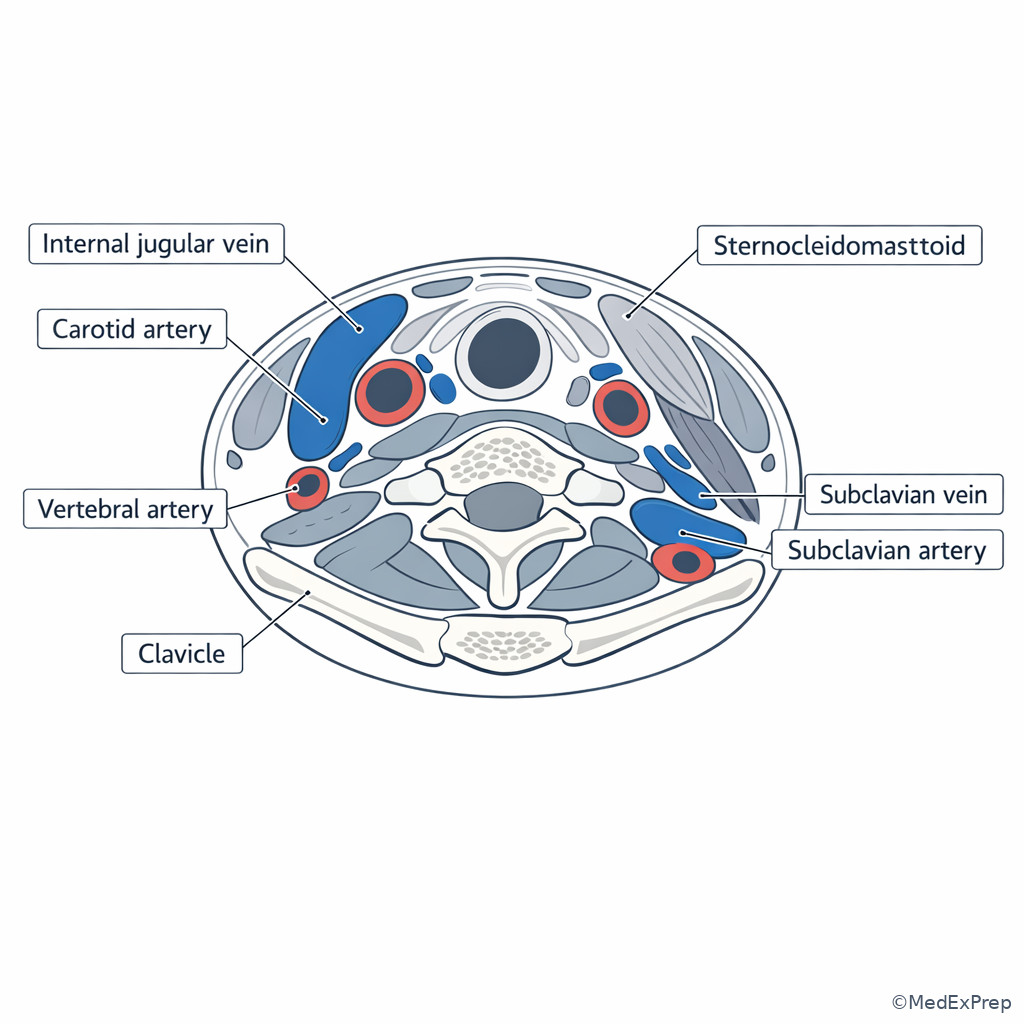
1.1.1.5 Abdominal arterial anatomy: branch order, organ territory, and procedural implications
A Explanation
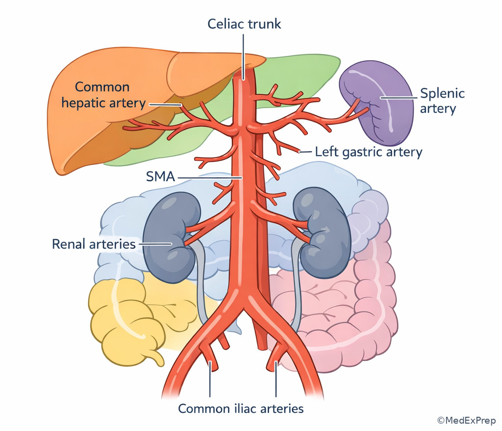
The central conflict in abdominal arterial anatomy is which branch supplies the organ or bleeding bed you care about, and which similar-looking branch would lead you to a wrong territory? The rule that resolves this is branch order plus downstream territory. In standard abdominal aortic anatomy, the celiac axis arises first, followed by the SMA, renal arteries, and IMA. The celiac supports foregut structures through the common hepatic, splenic, and left gastric branches. The SMA serves the midgut. The IMA supplies hindgut territory. On the exam, the safest answer is the one that respects both origin and organ distribution.
For IR, this matters in embolization, trauma, oncology, and GI bleeding. A liver lesion does not imply the portal vein is the therapeutic inflow target for arterial therapy; it points you toward hepatic arterial anatomy. A splenic injury after trauma raises splenic artery territory questions, often through the celiac trunk. A lower GI bleed can push you toward IMA or SMA depending on location, but collateral pathways can complicate the picture. The operator must know the textbook map first, then recognize variant or collateral adjustments.
See Fig 4 for abdominal arterial branch order and territory logic. This is not just memorization: it determines catheter selection, expected collateral recruitment, and non-target risk.
B Worked example
A 73-year-old woman with coronary artery disease, prior colectomy for diverticulitis, and CKD presents with brisk hematochezia. CTA shows active contrast extravasation near the splenic flexure. The resident suggests immediate IMA selection because “lower GI bleed equals IMA.” There is also mild chronic celiac stenosis, a distracting finding that may pull attention away from the actual territory question.
Reasoning chain: Identify task: predict the likely arterial territory to interrogate first. Extract key facts: bleeding is at the splenic flexure, a watershed region with variable supply, prior colectomy may alter local collateral patterns, and the stem asks territory logic rather than final operative care. Apply rule: splenic flexure commonly lies at the border of SMA and IMA territories, so a rigid “lower GI equals IMA” shortcut is unsafe. Eliminate distractors: celiac stenosis does not explain colonic bleeding. Verify: the operator should think in terms of watershed overlap and may need selective evaluation of both SMA and IMA depending on CTA appearance and post-surgical anatomy.
C Exam trap
D Checkpoint
Question: A target lesion in the pancreatic head most strongly points first to which major arterial territory?
- IMA territory
- Celiac axis territory
- External iliac territory
- Internal iliac territory
Answer: B
- A: Tempting because bowel and pancreas are both abdominal viscera; wrong because the IMA supplies hindgut structures, not foregut pancreatic head inflow.
- B: Correct because foregut structures including much of the hepatopancreatobiliary region arise from celiac-derived pathways, though pancreatic supply can be shared with SMA branches.
- C: Tempting if one is thinking only about access route from the groin; wrong because access vessel is not target territory.
- D: Tempting because pelvic branches are heavily tested in embolization; wrong because they do not supply the pancreatic head.
- The first three major anterior abdominal aortic branches in order are celiac axis, SMA, IMA if you ignore renal arteries; with full common test order think celiac, SMA, renal, IMA.
- True or False: A liver-directed arterial therapy is planned from the portal vein because the liver receives most of its blood flow from portal inflow. False.
- Name the watershed colonic region commonly tested in mesenteric territory questions: splenic flexure.
Exam takeaway: When a stem names an organ, convert it into arterial territory before choosing a catheter target or embolization plan.
Exam takeaway: Territory-based thinking helps you choose the correct mesenteric interrogation pathway and avoid oversimplified “upper vs lower GI” shortcuts.
flowchart TD
A[GI bleed or ischemia question] --> B{Location known?}
B -->|Foregut| C[Celiac territory]
B -->|Midgut| D[SMA territory]
B -->|Hindgut| E[IMA territory]
B -->|Watershed or postoperative| F[Consider overlap/collaterals]
C --> G[Target angiography accordingly]
D --> G
E --> G
F --> G
1.1.1.6 Venous anatomy: portal, systemic, renal, and pelvic venous relationships
A Explanation
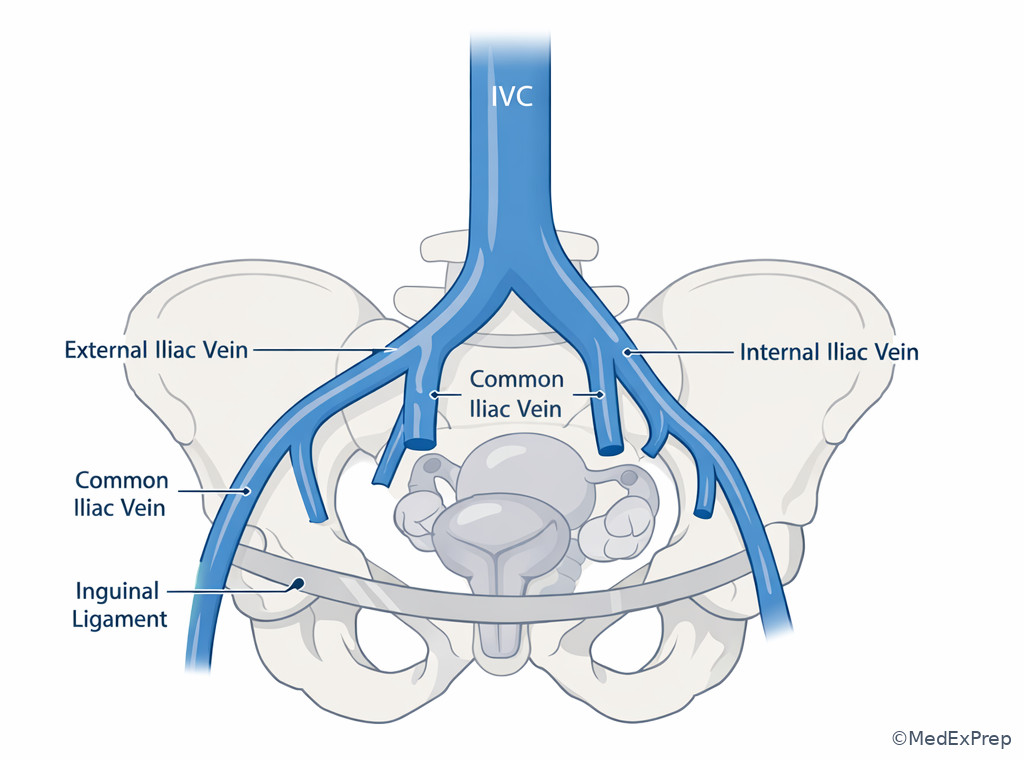
The key conflict in venous anatomy is whether the structure you are following represents systemic venous return, portal venous inflow, or a local tributary that changes procedural feasibility. The rule that resolves this is to trace the vessel to its confluence. The portal vein is formed by the SMV and splenic vein and courses into the liver at the porta hepatis. The IVC receives hepatic veins superiorly and renal veins lower in the retroperitoneum. Pelvic veins converge to the common iliac veins and then the IVC. For IR, confluent anatomy determines whether you are planning transhepatic access, portal intervention, renal venography, or systemic venous recanalization.
At the renal level, the left renal vein is especially testable. It crosses anterior to the aorta and posterior to the SMA before draining into the IVC. The right renal vein is shorter. This asymmetry explains why left renal vein compression syndromes are discussed and why left-sided venous anatomy is more distinctive on boards. Another high-yield relationship is that the right renal artery often courses posterior to the IVC, which helps differentiate artery from vein on axial imaging.
At the porta hepatis, the portal vein lies posterior to the bile duct and hepatic artery. In a transhepatic procedure question, this helps you reject a distractor that mislabels a structure based on size or enhancement alone. See Fig 5.
B Worked example
A 46-year-old man with cirrhosis, obesity, and prior pancreatitis undergoes contrast CT before a planned portal venous intervention. A rounded enhancing structure at the porta hepatis is labeled by a junior trainee as the proper hepatic artery because it is prominent and central. Another smaller vessel is seen more anteriorly. There is mild biliary ductal dilatation, which distracts from the anatomy problem.
Reasoning chain: Identify task: determine which structure is the portal vein. Extract key facts: central large hilar vessel, posterior location, confluence with mesenteric-splenic venous anatomy on adjacent images. Apply rule: in the portal triad, the portal vein is posterior and typically largest. Eliminate distractors: a prominent hepatic artery may enhance briskly but does not occupy the classic posterior hilar position; biliary dilation does not redefine vascular anatomy. Verify: the posterior central structure is the portal vein and therefore the key procedural landmark.
C Exam trap
D Checkpoint
Question: Which venous relationship is most useful for recognizing the left renal vein on axial CT?
- It crosses posterior to the aorta.
- It crosses anterior to the aorta and posterior to the SMA.
- It drains directly into the portal vein.
- It courses posterior to the IVC.
Answer: B
- A: Tempting because retroaortic variants exist; wrong because standard anatomy is anterior to the aorta.
- B: Correct because this classic crossing relationship is one of the highest-yield renal venous landmarks.
- C: Tempting if one confuses renal and mesenteric venous confluences; wrong because the left renal vein drains into the IVC.
- D: Tempting because the right renal artery has a posterior-IVC course; wrong because that relationship describes an artery, not the left renal vein.
Exam takeaway: Trace venous confluences and classic crossings to separate portal from systemic anatomy on procedural planning scans.
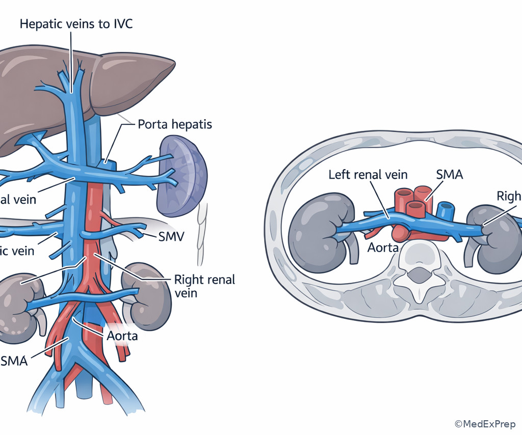
Exam takeaway: Common iliac and pelvic venous anatomy becomes crucial when femoral access or iliocaval recanalization is part of the plan.
1.1.1.7 Pelvic and groin vascular anatomy: access safety, branch recognition, and bleeding control relevance
A Explanation
The exam conflict in the pelvis and groin is where to puncture or embolize safely versus where a seemingly similar vessel location creates avoidable complications. The rule that resolves this is to pair surface access logic with branch anatomy. For groin arterial access, the common femoral artery over the femoral head and above the bifurcation is the preferred target because it balances compressibility and avoids retroperitoneal or branch puncture risk. Too high risks external iliac puncture and retroperitoneal hemorrhage; too low risks profunda or superficial femoral puncture with pseudoaneurysm, AV fistula, or failed closure-device performance.
In the pelvis, the internal iliac artery is the major procedural branch system for hemorrhage control and embolization. The exam rarely requires every branch in exhaustive detail, but it commonly tests territory logic: superior gluteal from posterior division; uterine, vesical, obturator, internal pudendal from anterior division. This matters because non-target embolization can produce buttock ischemic pain, bladder injury, sexual dysfunction, or uterine effects depending on territory. See Fig 6.
When a stem mixes trauma or postpartum hemorrhage with pelvic branch anatomy, choose the answer that respects likely territorial supply and avoids oversimplified “embolize the internal iliac trunk and be done” thinking. Superselective anatomy is safer when feasible.
B Worked example
A 71-year-old man with diabetes, severe PAD, obesity, and prior right groin endarterectomy needs urgent mesenteric angiography. CTA shows heavy calcification at the right common femoral bifurcation and a high bifurcation on the left. The fellow proposes a low left puncture because the vessel looks less calcified distally. A small inguinal hernia is a distracting but nondecisive detail.
Reasoning chain: Identify task: choose safest arterial access site. Extract key facts: right groin is surgically altered, left bifurcation is high, low puncture would land in branch vessels, and common femoral access remains the safety target. Apply rule: avoid low puncture below the common femoral artery. Eliminate distractors: lower calcification distally does not justify branch puncture; the hernia does not define vessel level. Verify: the operator should seek a site that preserves common femoral access principles or select an alternative route rather than accept an unsafe distal puncture.
C Exam trap
D Checkpoint
Question: The most appropriate arterial puncture target for routine femoral access is which of the following?
- External iliac artery above the inguinal ligament
- Common femoral artery over the femoral head and above the bifurcation
- Profunda femoris artery just distal to the bifurcation
- Superficial femoral artery below the femoral head
Answer: B
- A: Tempting because it is a large vessel; wrong because puncture above the inguinal ligament increases retroperitoneal bleeding risk.
- B: Correct because it balances size, compressibility, and closure safety.
- C: Tempting because the profunda can appear sizable; wrong because branch-vessel puncture is not the preferred routine target.
- D: Tempting because it may look superficial on imaging; wrong because puncture below the common femoral segment increases access complications.
- The preferred routine groin arterial target is the common femoral artery.
- True or False: A puncture above the inguinal ligament is safer because the vessel is larger. False.
- Name 3 anterior division internal iliac branches commonly tested: uterine, vesical, obturator (internal pudendal also high yield).
Exam takeaway: Safe groin puncture and pelvic embolization both depend on branch-level anatomy, not just vessel visibility.
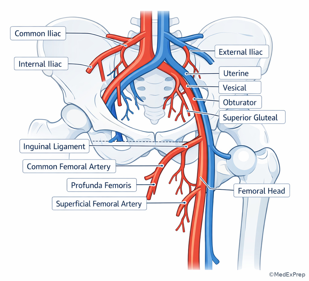
Exam takeaway: The safest access decision comes from puncture level, not convenience, size alone, or least calcified segment alone.
flowchart TD
A[Plan femoral arterial access] --> B{Above inguinal ligament?}
B -->|Yes| C[Reject: retroperitoneal bleed risk]
B -->|No| D{Below CFA bifurcation?}
D -->|Yes| E[Reject: branch puncture risk]
D -->|No| F[Target common femoral artery]
F --> G[Confirm over femoral head]
G --> H[Proceed if anatomy acceptable] Exam Traps & Differentiators
Most common wrong answer and why: The most common wrong move is identifying a vessel by size or brightness alone. This is tempting because contrast timing makes one structure visually dominant. The correct move is to identify the vessel by relationship and confluence, then use enhancement as a secondary clue.
| Looks similar | But isn't | Single discriminator | IR consequence if missed |
|---|---|---|---|
| SMA | SMV | SMA usually left of SMV | Wrong mesenteric target or route planning |
| Portal vein | Proper hepatic artery | Portal vein is posterior at porta hepatis | Misplanned transhepatic or embolization approach |
| Left renal vein | Retroaortic structure or lymphatic soft tissue | Crosses anterior to aorta and posterior to SMA | Renal venous mislabeling |
| Common femoral artery | Superficial femoral artery | Above bifurcation and over femoral head | Access complication risk |
| Right IJV route | Left central route | Right side usually straighter to SVC | Harder wire passage and tip issues |
| If the stem says... | Think... |
|---|---|
| “Posterior hilar vessel” | Portal vein |
| “Structure anterior to aorta, posterior to SMA” | Left renal vein |
| “Large round vessel right of aorta” | IVC until proven otherwise |
| “Need straight central venous course” | Right internal jugular/right brachiocephalic route |
| “Pelvic hemorrhage with need for selective embolization” | Internal iliac branch territory, not just trunk anatomy |
| “Access question with vessel over femoral head” | Common femoral target |
Related traps from neighboring topics: In Ultrasound Access Landmarks, the look-alike pair will shift from static anatomy to compressible vein versus pulsatile artery. In Preprocedural Imaging Interpretation, the trap becomes not just naming the vessel but recognizing when calcification, thrombus, collaterals, bowel, pleura, or postoperative change alters the route.
Tables
| Reference anatomy | High-yield fact | Classic exam use |
|---|---|---|
| Aorta | Usually left of IVC | Abdominal orientation |
| IVC | Receives renal and hepatic veins | Systemic venous planning |
| Celiac axis | Foregut territory | Hepatic/splenic/pancreatic target logic |
| SMA | Left of SMV | Mesenteric branch identification |
| Left renal vein | Anterior to aorta, posterior to SMA | Renal venous identification |
| Portal vein | Posterior at porta hepatis | Transhepatic planning |
| Right brachiocephalic route | Shorter and straighter to SVC | Central access strategy |
| Common femoral artery | Preferred over femoral head and above bifurcation | Safe arterial access |
| Comparison | Arterial structure | Venous structure | Discriminator |
|---|---|---|---|
| Mesenteric pair | SMA | SMV | SMA left of SMV |
| Porta hepatis pair | Hepatic artery | Portal vein | Portal vein posterior and usually larger |
| Renal hilar pair | Right renal artery | Right renal vein | Artery often posterior to IVC; vein enters IVC directly |
| Groin pair | Common femoral artery | Common femoral vein | Arterial access target chosen by puncture level and compressibility logic |
| Differential / classification problem | Option 1 | Option 2 | Single finding that separates them |
|---|---|---|---|
| Upper abdominal midline vessel | Aorta | IVC | Aorta usually left and thicker-walled; IVC right-sided venous confluence |
| Hilar liver vessel | Portal vein | Hepatic artery | Portal vein posterior in portal triad |
| Central venous route choice | Right-sided | Left-sided | Right path usually straighter unless pathology dictates otherwise |
| Groin puncture level | Common femoral | SFA/profunda or external iliac | Above bifurcation and below inguinal ligament wins |
| Mesenteric bleeding territory | SMA | IMA | Location and watershed context determine target |
Algorithm / Approach
Exam takeaway: Most anatomy questions can be solved by deciding whether the stem is testing orientation, territory, venous confluence, or access level.
flowchart TD
A[Cross-sectional vascular question] --> B{Main task?}
B -->|Orientation| C[Use aorta-IVC and SMA-SMV rules]
B -->|Territory| D[Match organ to celiac/SMA/IMA or pelvic branch]
B -->|Venous system| E[Trace confluence to portal or systemic vein]
B -->|Access| F[Choose safest puncture level/path]
C --> G[Make procedural decision]
D --> G
E --> G
F --> G Rapid Review
- Aorta vs IVC → left-sided artery, right-sided vein is the default orientation anchor.
- SMA vs SMV → artery left of vein on standard axial imaging.
- Left renal vein → anterior to aorta, posterior to SMA.
- Right renal artery → often posterior to IVC.
- Portal triad → portal vein posterior at the porta hepatis.
- Right central venous route → usually straighter to SVC than left.
- Celiac territory → foregut, hepatosplenic, proximal pancreatic logic.
- SMA territory → midgut, mesenteric bowel branch logic.
- IMA territory → hindgut, but watershed colon creates overlap.
- Splenic flexure → think watershed, not automatic IMA-only logic.
- Common femoral artery → preferred puncture above bifurcation and below inguinal ligament.
- High puncture → think retroperitoneal bleed risk.
- Low puncture → think SFA/profunda complication and poor closure logic.
- Internal iliac embolization → branch territory matters more than trunk memorization.
Self-check quiz
1. A 62-year-old man undergoing CTA for abdominal pain has a round enhancing vessel just right of the aorta at the renal level. Which structure is most likely being identified?
- IVC
- SMA
- Left renal artery
- IMA
2. A 54-year-old woman with cirrhosis has cross-sectional imaging reviewed before a transhepatic procedure. The posterior major vessel at the porta hepatis is most likely which structure?
- Common bile duct
- Proper hepatic artery
- Portal vein
- Cystic artery
3. A 68-year-old woman with CAD, prior left chest port, and breast cancer needs new central venous access. CT shows severe left brachiocephalic narrowing from prior device-related scarring; the right neck veins appear patent. Which is the most appropriate access strategy?
- Prefer right internal jugular access because the route to the SVC is usually straighter and avoids the diseased left central vein
- Prefer left internal jugular access because prior use suggests that side is proven safe
- Prefer left subclavian access because subclavian puncture is more compressible if bleeding occurs
- Defer all central access because brachiocephalic narrowing on one side contraindicates any upper body access
4. A 73-year-old woman with hematochezia, CKD, and prior partial colectomy has CTA showing active extravasation at the splenic flexure. Which interpretation is most appropriate for angiographic planning?
- Interrogate only the IMA because all lower GI bleeding originates from hindgut supply
- Interrogate only the SMA because the splenic artery shares celiac collaterals with colon
- Consider both SMA and IMA territories because the splenic flexure is a watershed region
- Avoid mesenteric angiography because splenic flexure bleeding is always venous
5. A 70-year-old man with obesity, PAD, and prior contralateral groin surgery requires arterial access for mesenteric angiography. Which puncture target is most appropriate when anatomy permits?
- External iliac artery above the inguinal ligament
- Common femoral artery over the femoral head and above the bifurcation
- Superficial femoral artery below the bifurcation
- Profunda femoris artery to avoid the common femoral bifurcation
6. A 58-year-old man with obesity, hypertension, and hematuria undergoes CT before renal venography. A vessel crosses anterior to the aorta and posterior to the SMA before draining into a large right-sided retroperitoneal vein. Which structure is most likely being traced?
- Left renal vein
- Right renal artery
- Splenic vein
- Left gonadal vein
7. A 65-year-old woman with cirrhosis, portal hypertension, and prior biliary intervention has a multiphase CT reviewed before portal venous planning. The trainee labels the brightest hilar structure on arterial phase as the target portal vein. Which single finding most strongly requires correction of that interpretation?
- The structure is smaller than the bile duct
- The structure lies anterior rather than posterior in the portal triad
- The structure enhances more than the spleen
- The structure is near the gallbladder fossa
8. A 76-year-old man with severe atherosclerosis, prior right femoral endarterectomy, and chronic mesenteric ischemia requires urgent angiography. CTA shows a surgically altered right groin and a high left common femoral bifurcation, with the least calcified left arterial segment located distal to the bifurcation. Which is the most appropriate interpretation?
- Use the least calcified distal left vessel because calcium burden is the only meaningful groin determinant
- Use the left superficial femoral artery because it remains compressible and avoids the bifurcation
- Do not accept a low branch-vessel puncture; seek an alternative site or strategy that preserves common femoral access principles
- Use the external iliac artery because larger caliber outweighs retroperitoneal bleeding risk
Answer key
1.1.1.8 1. Answer: A — IVC
A: Tempting because a large right-sided retroperitoneal vessel at the renal level is a classic IVC appearance; correct because the IVC normally lies right of the aorta and receives renal inflow. Single stem clue: “just right of the aorta at the renal level.” Source: Kandarpa et al.; Standring.
B: Tempting because the SMA is a common tested vessel; incorrect because it is anterior to the aorta and usually left of the SMV, not a large right-sided retroperitoneal structure. Single discriminator: right-sided location. Source: Standring.
C: Tempting because renal vessels are present at this level; incorrect because the left renal artery arises from the aorta and courses laterally, not as a large right-sided venous-appearing structure. Single discriminator: caliber and position relative to aorta. Source: Kandarpa et al.
D: Tempting because the IMA is an abdominal aortic branch; incorrect because the IMA is smaller, lower, and anterior to the aorta rather than a large right-sided vessel. Single discriminator: size and side. Source: Standring.
1.1.1.9 2. Answer: C — Portal vein
A: Tempting because biliary structures travel with hilar vessels; incorrect because the common bile duct is not the posterior major vessel in the portal triad. Single discriminator: posterior hilar position belongs to portal vein. Source: Standring; SIR references.
B: Tempting because arterial enhancement can be striking; incorrect because the proper hepatic artery is more anterior/medial variable and smaller than the main portal vein. Single discriminator: posterior major vessel. Source: Kandarpa et al.
C: Correct because the portal vein is the posterior, dominant hilar vessel and is the key landmark for portal interventions. Single stem clue: “posterior major vessel at the porta hepatis.” Source: Standring.
D: Tempting because the cystic artery is a hepatobiliary branch; incorrect because it is not the principal posterior hilar vessel and is usually too small for this description. Single discriminator: caliber and location. Source: Kandarpa et al.
1.1.1.10 3. Answer: A — Prefer right internal jugular access
A: Correct because central venous access should favor a patent route with a straighter path to the SVC, and the diseased left brachiocephalic vein makes the left route less favorable. Single stem clue: “severe left brachiocephalic narrowing” with patent right neck veins. Source: SIR practice parameters; Kandarpa et al.
B: Tempting because prior successful use of a side can suggest convenience; incorrect because the same side now has significant central venous narrowing that directly affects catheter passage and durability. Single discriminator: current left-sided stenosis. Source: SIR practice guidance.
C: Tempting because subclavian routes may be discussed in line placement; incorrect because subclavian bleeding is less compressible and this option ignores the goal of choosing the better central path. Single discriminator: subclavian noncompressibility and persistent left central narrowing. Source: Kandarpa et al.
D: Tempting because significant venous narrowing raises concern; incorrect because unilateral brachiocephalic narrowing does not contraindicate all upper body access if the contralateral route is patent. Single discriminator: right neck veins appear patent. Source: SIR references.
1.1.1.11 4. Answer: C — Consider both SMA and IMA territories
A: Tempting because lower GI bleeding is often linked to hindgut territory; incorrect because the splenic flexure is a watershed region and prior colectomy may further alter supply. Single discriminator: splenic flexure location. Source: Kandarpa et al.; interventional GI bleed reviews.
B: Tempting because watershed logic suggests overlap; incorrect because saying “only SMA” is still too rigid and the rationale provided is anatomically flawed. Single discriminator: overlap, not exclusivity. Source: standard mesenteric anatomy texts.
C: Correct because angiographic planning for splenic flexure bleeding often requires awareness of both SMA and IMA territories, especially with prior surgery or collateralization. Single stem clue: “active extravasation at the splenic flexure.” Source: Kandarpa et al.; SIR educational material.
D: Tempting because some bleeds are intermittent or occult; incorrect because arterial extravasation on CTA supports mesenteric arterial evaluation rather than “always venous” dismissal. Single discriminator: CTA shows active contrast extravasation. Source: GI hemorrhage angiography literature.
1.1.1.12 5. Answer: B — Common femoral artery over the femoral head and above the bifurcation
A: Tempting because larger caliber can seem safer for access; incorrect because puncture above the inguinal ligament increases retroperitoneal bleeding risk and reduces compressibility. Single discriminator: above-inguinal level. Source: SIR arterial access guidance; Kandarpa et al.
B: Correct because this is the preferred routine arterial puncture zone for compressibility, closure-device performance, and complication reduction. Single stem clue: “when anatomy permits.” Source: SIR quality improvement guidance.
C: Tempting because distal vessels may look easier to reach; incorrect because SFA puncture below bifurcation is a common access complication setup. Single discriminator: below the bifurcation. Source: SIR references.
D: Tempting because avoiding the bifurcation sounds logical; incorrect because the profunda is not the standard routine access target and branch puncture complicates hemostasis. Single discriminator: branch-vessel access. Source: Kandarpa et al.
1.1.1.13 6. Answer: A — Left renal vein
A: Correct because the classic left renal vein course is anterior to the aorta and posterior to the SMA before entering the IVC. Single stem clue: “crosses anterior to the aorta and posterior to the SMA.” Source: Standring.
B: Tempting because the right renal artery has a famous relationship to the IVC; incorrect because the described crossing anterior to the aorta is venous, not the usual right renal arterial course. Single discriminator: anterior-aortic crossing to right-sided large vein. Source: Kandarpa et al.
C: Tempting because the splenic vein is a major abdominal vein; incorrect because it courses along the pancreas and contributes to portal confluence, not across the aorta to the IVC. Single discriminator: drains into a right-sided retroperitoneal vein. Source: Standring.
D: Tempting because gonadal veins relate to renal veins on the left; incorrect because the left gonadal vein drains into the left renal vein and is not itself the large crossing vessel described. Single discriminator: caliber and confluence. Source: anatomic references.
1.1.1.14 7. Answer: B — The structure lies anterior rather than posterior in the portal triad
A: Tempting because size can help identify hilar structures; incorrect because comparative size to the bile duct is less reliable than spatial relationship. Single discriminator: position outweighs size. Source: Standring; Kandarpa et al.
B: Correct because the portal vein is expected posteriorly in the portal triad; a brightly enhancing structure lying anterior is more consistent with artery than portal vein. Single stem clue: arterial-phase brightness plus anterior location. Source: standard hepatobiliary anatomy references.
C: Tempting because enhancement intensity suggests artery; incorrect because comparing to splenic enhancement is indirect and less definitive than anatomic position. Single discriminator: posterior-vs-anterior relationship. Source: imaging anatomy texts.
D: Tempting because nearby gallbladder fossa can orient the liver; incorrect because proximity to the gallbladder fossa does not by itself distinguish portal from arterial hilar structures. Single discriminator: triad spatial arrangement. Source: Kandarpa et al.
1.1.1.15 8. Answer: C — Do not accept a low branch-vessel puncture
A: Tempting because calcium burden is an important technical consideration; incorrect because puncture level is a stronger safety determinant than choosing the least calcified distal segment. Single discriminator: distal segment is below the bifurcation. Source: SIR access guidance.
B: Tempting because the SFA may appear accessible and compressible; incorrect because low SFA puncture is not the preferred routine access site and increases access complications. Single discriminator: branch-vessel location below bifurcation. Source: Kandarpa et al.
C: Correct because the key principle is preserving common femoral access logic; if anatomy does not allow that safely, an alternative site or strategy is preferable to an unsafe low puncture. Single stem clue: “least calcified left arterial segment located distal to the bifurcation.” Source: SIR quality guidance.
D: Tempting because the external iliac is larger; incorrect because puncture above the inguinal ligament raises retroperitoneal bleeding risk and violates standard safe femoral access principles. Single discriminator: external iliac level. Source: SIR references.
Keep going with the full ABR Interventional Radiology Prep Course (IR)
You just studied the way members do, in the same lesson interface. Full access unlocks every lesson, the question bank and flashcards for a full year.
One payment · 1 year of access · No auto-renewal