1.1.1 - Healing Phases And Mediators
Why it matters
- Plastic surgery decisions about closure timing, grafting, flap coverage, and revision all depend on whether the wound is in a healthy healing trajectory or biologically stalled.
- Board stems often hide the diagnosis inside a mediator clue: macrophage-dominant transition, impaired angiogenesis, collagen remodeling failure, or prolonged inflammation.
- Normal healing can look inflamed; infection can also look inflamed. The exam tests whether you can separate expected phase biology from pathology.
- Future topics on closure choice and scar modulation build directly on this lesson. If you do not know the phase, you will miss the timing question.
Key Terms & Must-Know Facts
1.1.1.1 Key terms
- Hemostasis: Immediate vascular constriction and clot formation after injury.
- Inflammatory phase: Early cell-cleanup phase dominated first by neutrophils, then macrophages.
- Proliferative phase: Granulation tissue, angiogenesis, fibroplasia, and re-epithelialization.
- Remodeling phase: Collagen reorganization and scar maturation over months.
- Platelet degranulation: Release of growth factors that initiate repair signaling.
- Macrophage transition: Key switch from inflammation to repair.
- Granulation tissue: Vascular, fibroblast-rich tissue indicating active proliferation.
- Angiogenesis: New capillary growth, largely driven by hypoxia signaling and VEGF.
- Fibroplasia: Fibroblast migration and collagen deposition.
- Myofibroblast: Contractile fibroblast involved in wound contraction.
- Re-epithelialization: Keratinocyte migration over a viable wound bed.
- Type III collagen: Early collagen in immature wound matrix.
- Type I collagen: Stronger collagen that predominates during remodeling.
- MMPs: Matrix metalloproteinases that degrade extracellular matrix; excess impairs healing.
- TIMP: Tissue inhibitor of metalloproteinases; balances matrix breakdown.
1.1.1.2 Must-know facts
- Platelets start the signaling cascade immediately after injury.
- Neutrophils arrive early; macrophages are the pivotal phase-transition cells.
- Macrophages are the major board favorite when the question asks what drives progression from inflammation to proliferation.
- Fibroblasts deposit early collagen, initially type III.
- Tensile strength is low in the first week; dehiscence risk is highest before meaningful collagen maturation.
- Maximum scar strength never returns to unwounded skin.
- Healthy granulation tissue is beefy red, moist, and punctate-bleeding.
- Chronic wounds often remain trapped in prolonged inflammation with excess proteases.
- Hypoxia can stimulate angiogenesis, but severe ischemia prevents healing.
- Infection, necrosis, edema, diabetes, smoking, radiation, and malnutrition delay normal phase progression.
Exam takeaway: Match timing, dominant cell, and visible wound appearance to identify the current healing phase.
flowchart LR
A[Injury] --> B[Hemostasis
minutes to hours
platelets fibrin]
B --> C[Inflammation
hours to days
neutrophils then macrophages]
C --> D[Proliferation
days to weeks
fibroblasts angiogenesis epithelium]
D --> E[Remodeling
weeks to months
collagen reorganization]
Exam takeaway: Visualize the cell sequence so board stems with timing clues become easier to answer.
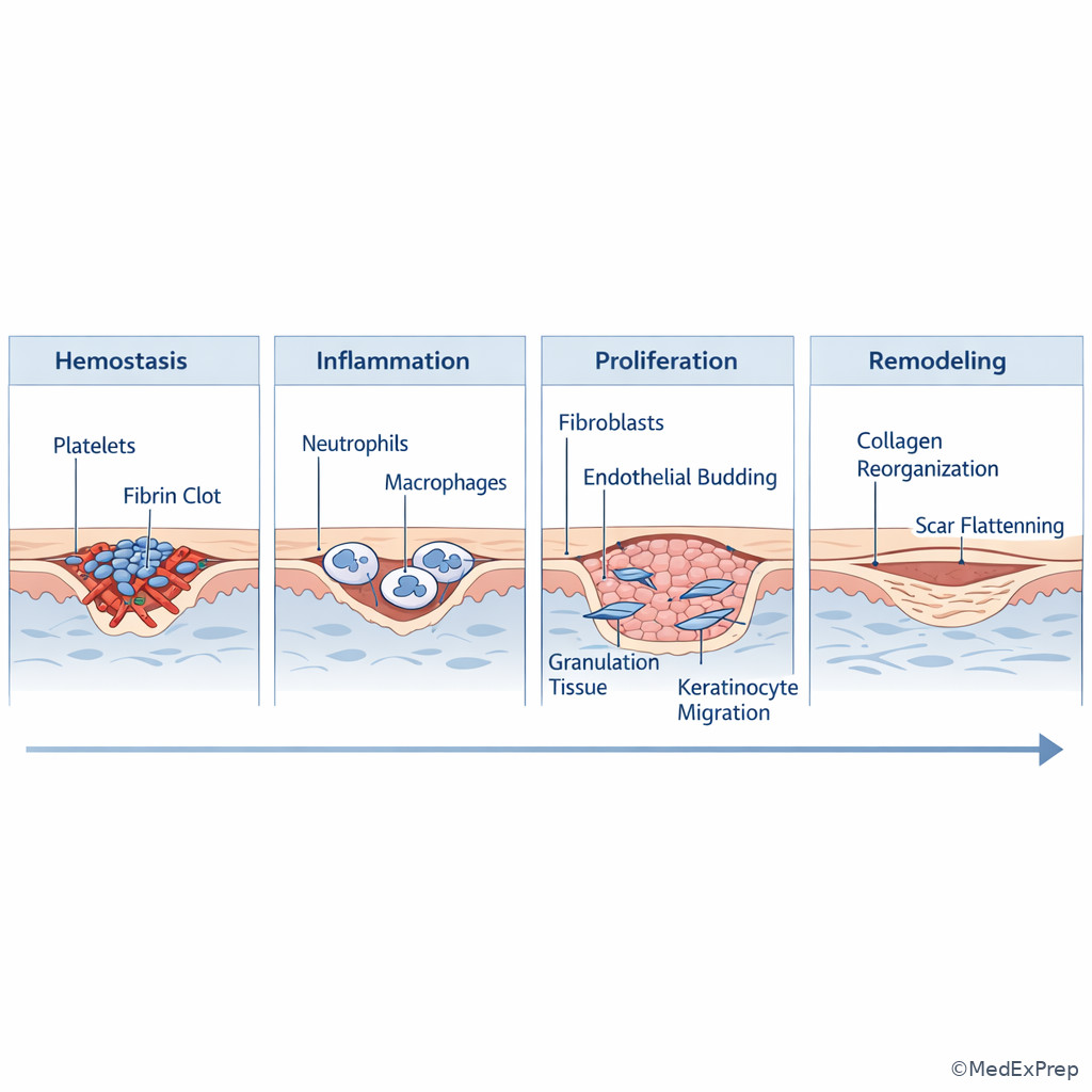
Core content
1.1.1.3 Foundational phase sequence: what phase are you looking at?
A Explanation
The decision conflict is simple but heavily tested: is this wound showing normal phase-appropriate biology or pathology requiring a different next step? The single rule that resolves many stems is to anchor the finding to time since injury or surgery and then ask which cell population should dominate. Immediately after injury, hemostasis controls bleeding and forms a provisional matrix. Over the first 24–48 hours, neutrophils predominate. By about 48–72 hours and beyond, macrophages become central and coordinate transition into repair. Days later, the wound should show granulation, fibroplasia, angiogenesis, and re-epithelialization. Weeks to months later, collagen remodeling and scar maturation dominate. See Fig 1.
WHY THIS IS TESTED: exam stems often give you a wound appearance and postoperative day, then ask for the most likely mediator, cell type, explanation for dehiscence risk, or best timing for closure. Choose “normal inflammation” when erythema is mild, expected, and time-matched; reject infection when the timing and absence of systemic or purulent features favor ordinary inflammatory healing.
Use choose-versus-reject framing. Choose hemostasis when the stem emphasizes immediate clot, vasoconstriction, platelet activation, and fibrin. Choose inflammation when there is edema, leukocyte influx, and debris clearance. Choose proliferation when there is healthy granulation and epithelial edge migration. Choose remodeling when the wound is closed but scar quality is changing. Reject proliferative-phase answers for a fresh postoperative wound, and reject early inflammatory answers for a mature scar question.
B Worked example
A 61-year-old man undergoes lower-extremity soft-tissue debridement after trauma. He has diabetes, chronic tobacco use, and peripheral arterial disease. On postoperative day 2, the wound edge is mildly erythematous and edematous without purulence; temperature is 37.4°C, WBC is 10,800/µL, and a nurse notes “redness concerning for infection.” The dressing shows serosanguinous drainage. Which cell type is most responsible for coordinating the next transition in healing?
Reasoning chain: Identify task: the question is not asking for antibiotics; it is asking for the cell driving phase transition. Extract key facts: day 2, mild erythema/edema, no purulence, no systemic toxicity. Apply rule: day 2–3 is the handoff from neutrophil-heavy inflammation to macrophage-led repair signaling. Eliminate distractors: platelets are too early; fibroblasts dominate later proliferation; keratinocytes matter for re-epithelialization, not the inflammation-to-proliferation switch. Verify: macrophages are the best answer because they clear debris and release growth factors that launch fibroplasia and angiogenesis.
C Exam trap
D Checkpoint
Question: A clean surgical wound on postoperative day 10 is most likely dominated by which process?
- A. Platelet plug formation — Tempting because clotting starts all wounds, but it is wrong because this is an immediate event, not day 10 biology.
- B. Neutrophil chemotaxis — Tempting because acute wounds involve neutrophils, but it is wrong because by day 10 the wound should be moving beyond early leukocyte dominance.
- C. Fibroblast proliferation and angiogenesis — Tempting because granulation and matrix deposition are active in this time window, and it is the correct answer.
- D. Final scar tensile normalization — Tempting because the wound looks externally improved by day 10, but it is wrong because remodeling and maximal strength gains take much longer.
1.1.1.4 Mediators and cells: who does the work, and what are they signaling?
A Explanation
The exam rarely rewards memorizing every cytokine in isolation. Instead, it tests whether you can choose the most likely mediator based on the wound task: coagulation, chemotaxis, angiogenesis, fibroblast activation, epithelial migration, or remodeling. Platelets release PDGF and TGF-β early; these help recruit inflammatory and mesenchymal cells. Neutrophils clear bacteria and debris, but macrophages are the master regulators of transition. Macrophages release PDGF, TGF-β, VEGF, FGF, IL-1, and TNF-related signals that stimulate fibroblasts and endothelial cells. Fibroblasts synthesize matrix and collagen. Endothelial cells respond to VEGF and hypoxia signals with angiogenesis. Keratinocytes drive resurfacing. Myofibroblasts contract the wound. MMPs break down matrix; excessive proteolysis, especially in chronic wounds, prevents stable progression.
WHY THIS IS TESTED: board questions may ask for the best explanation for poor granulation, failure of epithelialization, or excessive chronic inflammation. The resolving rule is to map the wound problem to the biologic job that is failing. Poor angiogenesis points toward VEGF/hypoxia signaling problems. Failure to transition out of inflammation points toward macrophage dysfunction or persistent contamination. Weak matrix and dehiscence relate to fibroblast/collagen problems. Excess destructive protease activity in chronic wounds points toward high MMP burden relative to TIMP.
Choose PDGF/TGF-β when the stem emphasizes recruitment of fibroblasts and matrix deposition. Choose VEGF when the stem stresses new vessel ingrowth. Reject epidermal-only logic when the problem is deeper matrix failure. Reject “collagen maturation” if the wound is still open and granulating; that is not yet the primary issue. See Fig 2.
Exam takeaway: When a stem asks “which factor” or “which cell,” first identify the wound task being tested.
flowchart TD
A[Wound task] --> B{Main problem}
B -->|Immediate clot| C[Platelets
PDGF TGF-beta]
B -->|Debris bacteria cleanup| D[Neutrophils then macrophages]
B -->|Transition to repair| E[Macrophages
growth factors]
B -->|New capillaries| F[VEGF endothelial response]
B -->|Matrix collagen| G[Fibroblasts
type III then I collagen]
B -->|Surface closure| H[Keratinocyte migration]
B -->|Contraction| I[Myofibroblasts]
B -->|Chronic stalled wound| J[Excess MMP inflammatory milieu] B Worked example
A 48-year-old woman with obesity and poorly controlled diabetes undergoes debridement of an abdominal wound after necrotizing soft-tissue infection. Five days later, the wound bed is clean but pale with minimal punctate bleeding and little granulation tissue. Albumin is 2.8 g/dL, and a resident suggests that “keratinocytes are failing.” A wound-care note also mentions moderate edema. Which mediator pathway is most directly associated with the process that should now be increasing to create healthy granulation tissue?
Reasoning chain: Identify task: the stem asks about granulation tissue, not epidermal resurfacing. Extract facts: day 5, clean wound, poor granulation, pale bed, minimal punctate bleeding. Apply rule: granulation requires fibroblasts plus angiogenesis; angiogenesis is strongly linked to VEGF-driven endothelial response. Eliminate distractors: keratinocytes are for re-epithelialization; platelets are early; type I collagen predominance is later remodeling. Verify: the missing biologic feature is new vessel ingrowth, making VEGF the best mechanistic answer.
C Exam trap
D Checkpoint
Question: Which cell is most important in transitioning a wound from inflammatory cleanup to proliferative repair?
- A. Platelet — Tempting because platelets initiate signaling, but they are right at injury onset rather than the key bridge into proliferation.
- B. Neutrophil — Tempting because neutrophils dominate early inflammation, but they are not the main coordinator of the repair transition.
- C. Macrophage — Tempting because it fits both debris clearance and growth-factor release, and it is the correct answer.
- D. Myofibroblast — Tempting because wound contraction matters later, but that is downstream of the transition being asked.
- The most important cell for switching from inflammation to proliferation is macrophage.
- True or False: VEGF is most closely linked to angiogenesis. True.
- Name the 3 key proliferative-phase activities: granulation, angiogenesis, re-epithelialization.
1.1.1.5 Proliferation, collagen, and tensile strength: when is a wound actually strong?
A Explanation
The decision conflict here is whether a wound that looks closed is actually mechanically robust. The single resolving rule: cosmetic closure and biologic strength are not the same thing. During proliferation, fibroblasts deposit collagen, initially rich in type III collagen. Angiogenesis and matrix synthesis improve tissue fill, but tensile strength remains limited early. Remodeling then gradually shifts collagen organization and cross-linking toward stronger scar architecture with increased type I collagen predominance. Even so, healed tissue never regains the full tensile strength of unwounded skin.
WHY THIS IS TESTED: plastic surgery board stems use postoperative timing to ask about dehiscence risk, drain/tension decisions, suture support, staged reconstruction, and why radiated or malnourished tissue fails under load. The key testable pattern is that early external appearance overestimates true mechanical recovery. Choose answers that respect low early tensile strength; reject aggressive assumptions that a sealed epidermal surface means the wound can tolerate major tension.
When asked about collagen type, choose type III for early wound matrix and type I for later mature scar. When asked about contraction, think myofibroblasts. When asked why the wound opened under stress despite appearing acceptable at the skin, think immature collagen and poor matrix organization. See Fig 3.
Exam takeaway: Early closure does not equal mature strength; dehiscence questions hinge on immature collagen and tension.
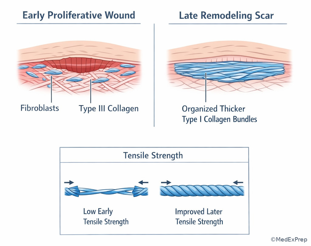
Exam takeaway: Distinguish vascular cellular granulation tissue from dense relatively hypocellular mature scar.
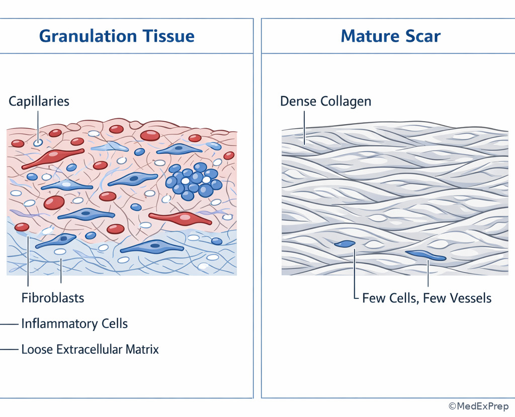
B Worked example
A 72-year-old woman undergoes closure of a pretibial wound after Mohs defect preparation. She has chronic steroids for COPD, rheumatoid arthritis, and peripheral edema. On postoperative day 8, the epidermis appears approximated, but after minor trauma the wound partially separates. ESR is mildly elevated from chronic disease, and a junior trainee blames “late infection.” Which biologic explanation best accounts for this event?
Reasoning chain: Identify task: explain dehiscence risk, not diagnose infection. Extract key facts: day 8, pretibial location, chronic steroids, edema, apparently closed surface, partial separation after stress. Apply rule: early wounds still have limited tensile strength because collagen is immature and remodeling is incomplete. Eliminate distractors: final mature type I scar is too late; platelet failure is too early; isolated keratinocyte problems do not explain load failure. Verify: weak early collagen matrix under tension is the best explanation.
C Exam trap
D Checkpoint
Question: Which statement best explains why wounds remain vulnerable to separation after the skin surface has sealed?
- A. Re-epithelialization restores full tensile strength immediately — Tempting because the surface looks closed, but it is wrong because epidermal coverage is not equivalent to deep matrix maturity.
- B. Early matrix is collagen-poor and disorganized relative to mature scar — Tempting because it matches proliferative biology, and it is the correct answer.
- C. Neutrophils provide most long-term wound strength — Tempting because they are early abundant cells, but they are wrong because they do not create durable tensile support.
- D. Platelets remain the dominant structural cells for weeks — Tempting because they initiate repair, but they are wrong because later strength depends on fibroblast-driven collagen remodeling.
1.1.1.6 Why wounds fail to progress: host factors, local factors, and chronic inflammation
A Explanation
The decision conflict is whether delayed healing is due mainly to normal slow biology or a correctable barrier. The single resolving rule: a wound that remains trapped in inflammation or lacks a viable bed will not progress until the limiting factor is corrected. Local factors include ischemia, necrotic tissue, edema, contamination, infection, foreign body, radiation injury, pressure, and desiccation. Systemic factors include diabetes, smoking, malnutrition, anemia, immunosuppression, steroids, and uremia. Chronic wounds often show prolonged inflammatory signaling, excessive MMP activity, senescent cells, and degraded growth-factor environment.
WHY THIS IS TESTED: exam stems often ask for the most important reason a wound is not granulating or why a graft/closure would fail if attempted now. The tested pattern is not “name every risk factor,” but rather “choose the dominant barrier that must be corrected first.” If the bed is necrotic or ischemic, do not choose closure. If edema and bioburden dominate, optimize those before grafting. If pressure persists, any reconstruction will be threatened. Choose the factor that prevents phase transition; reject minor abnormalities that are not the primary rate-limiting step.
For plastic surgery planning, a wound is not “ready” just because it is smaller. It must be debrided, perfused, relatively controlled from contamination, and able to support epithelialization or graft take. This biology directly anticipates the next topic on primary, secondary, and tertiary closure. See Fig 4.
Exam takeaway: Identify and correct the dominant barrier before choosing closure, graft, or flap reconstruction.
flowchart TD
A[Delayed healing] --> B{Main barrier}
B -->|Necrosis or slough| C[Debride nonviable tissue]
B -->|Ischemia| D[Assess perfusion and revascularization needs]
B -->|Edema pressure shear| E[Off-load and control edema]
B -->|Bioburden infection| F[Source control and targeted treatment]
B -->|Poor host factors| G[Optimize glucose nutrition smoking steroid issues]
C --> H[Reassess wound bed]
D --> H
E --> H
F --> H
G --> H
H --> I{Ready for closure?}
I -->|Yes| J[Choose closure graft or flap]
I -->|No| K[Continue wound-bed preparation] Exam takeaway: A stalled wound image should trigger search for dominant local barriers rather than immediate closure.
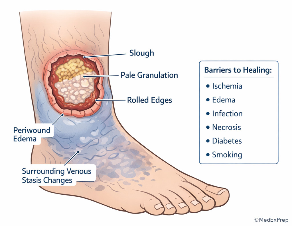
Exam takeaway: Compare a healthy granular bed with a stalled wound to decide whether reconstruction timing is appropriate.
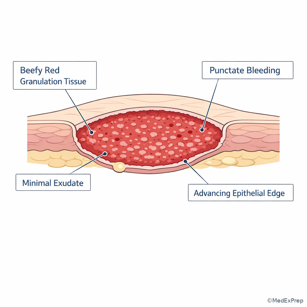
B Worked example
A 67-year-old man with diabetes, CKD, smoking history, and prior tibial radiation presents three weeks after debridement of a distal leg wound. The wound is covered with fibrinous debris and minimal granulation; cultures grow mixed colonizers without systemic illness. ABI is 0.52. The resident wants split-thickness skin grafting because the wound is “clean enough.” What is the most important reason this reconstruction choice is premature?
Reasoning chain: Identify task: choose the limiting factor before reconstruction. Extract key facts: distal leg, radiation history, diabetes, smoking, poor granulation, fibrinous bed, ABI 0.52. Apply rule: severe perfusion deficiency prevents a wound from supporting healing and graft take. Eliminate distractors: surface colonization alone is not the dominant clue; edema may matter but is secondary to the marked ischemia; time since debridement alone is not decisive. Verify: inadequate perfusion is the major barrier that must be corrected or addressed before grafting.
C Exam trap
D Checkpoint
Question: Which factor most directly traps many chronic wounds in a nonhealing inflammatory state?
- A. Excess MMP activity relative to matrix formation — Tempting because it fits chronic wound biology, and it is the correct answer.
- B. Immediate platelet aggregation — Tempting because platelets begin repair, but it is wrong because this is not the mechanism of chronic stalling.
- C. Early scar maturation with dense type I collagen — Tempting because mature collagen changes healing dynamics, but it is wrong because chronic wounds are not stalled by overmature scar in the usual board framing.
- D. Complete keratinocyte overgrowth — Tempting because epithelial edge abnormalities exist, but it is wrong because this is not the central inflammatory trap being tested.
- The most common cause of a stalled reconstructive wound bed is failure to correct the dominant local barrier.
- True or False: A wound can be colonized yet still be reconstruction-ready if it is well vascularized, debrided, and clinically uninfected. True.
- Name 3 local factors that delay healing: ischemia, necrosis, edema.
1.1.1.7 Applying biology to reconstructive timing and board-style decisions
A Explanation
The final foundational integration question is not “what phase exists?” but “what should the plastic surgeon do with that information?” The decision conflict is whether to close now, wait, debride again, graft, flap, or continue wound-bed preparation. The single rule that resolves the choice is this: reconstruction must match recipient-bed biology. A clean, perfused, granulating wound may be ready for delayed closure or grafting. A contaminated, edematous, or ischemic wound is not. A wound with exposed tendon without paratenon, exposed hardware, irradiated tissue, or compromised perfusion may need vascularized coverage rather than a graft, regardless of surface appearance.
WHY THIS IS TESTED: ABPS-style stems frequently disguise a reconstructive planning question as a biology question. The tested pattern is to infer readiness for the next step from what the wound is biologically doing. Choose continued preparation when the bed is not ready. Choose closure/graft only when tissue viability and phase progression support success. Reject closure strategies that ignore ischemia, gross contamination, or persistent necrosis.
This lesson also sets up the next two topics. From here, closure method is a consequence of wound biology, and scar behavior is a consequence of later remodeling plus mechanical and inflammatory environment. See Fig 5 for a practical bridge from phase recognition to action.
Exam takeaway: Reconstruction succeeds when the wound bed is perfused, debrided, and phase-appropriate for the chosen method.
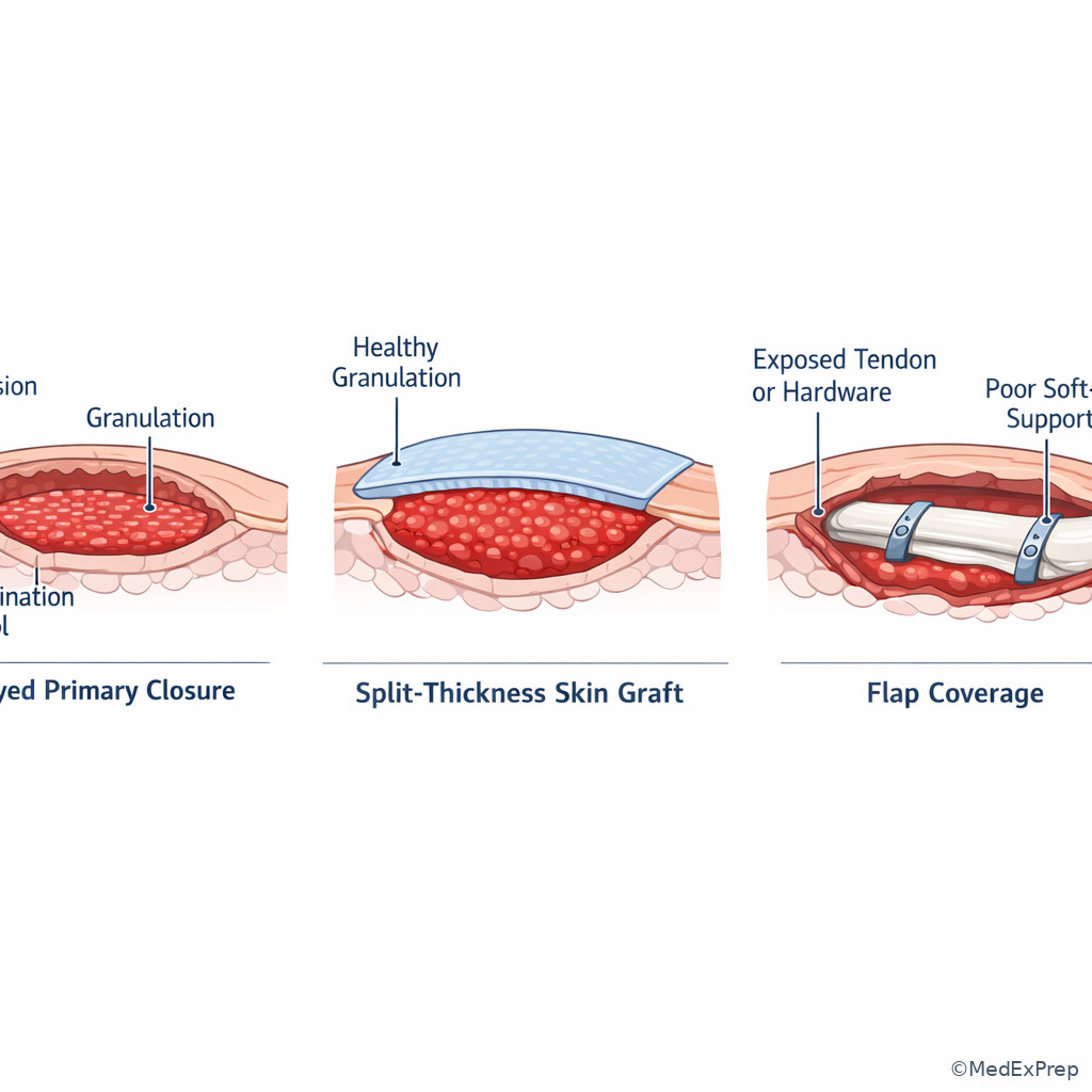
Exam takeaway: Distinguish expected early postoperative erythema from purulence, progressive pain, and systemic signs that suggest infection.
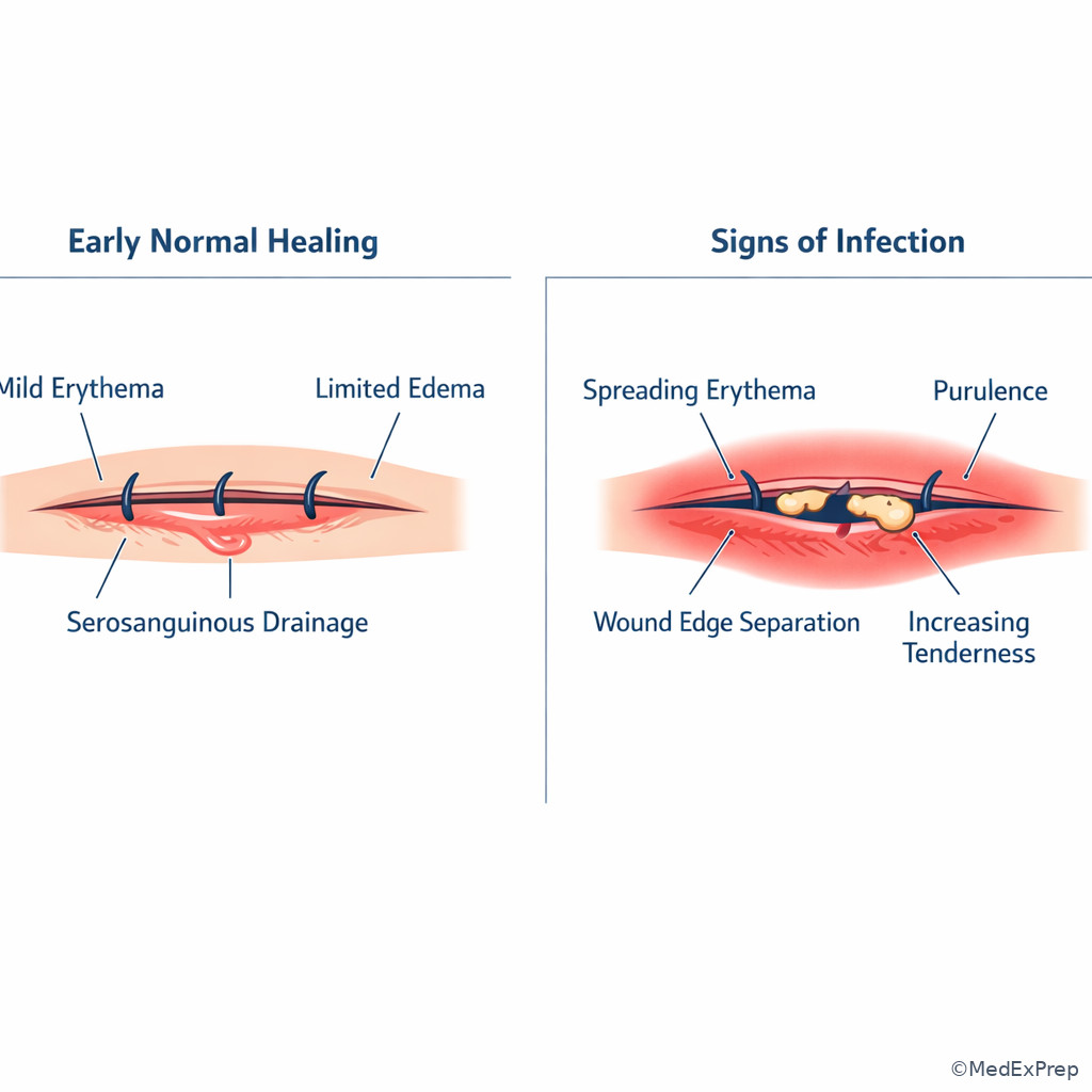
B Worked example
A 55-year-old man with obesity and type 2 diabetes has a traumatic calf wound after debridement. There is no fever. The wound shows beefy red granulation tissue with punctate bleeding, minimal fibrin, and no exposed tendon or bone. A small amount of surrounding erythema is present. A colleague wants more serial debridements because “redness means inflammation.” What is the best interpretation?
Reasoning chain: Identify task: decide if the wound is progressing normally and what that means for planning. Extract key facts: healthy granulation, punctate bleeding, minimal fibrin, no deep exposure, no systemic infection. Apply rule: this is a biologically favorable proliferative wound bed, not a stalled or infected one. Eliminate distractors: mild erythema alone is not infection; further debridement of viable bed may be counterproductive; flap coverage is not mandatory without exposed critical structure or poor bed quality. Verify: the wound appears ready for the next reconstructive step, depending on size and tension requirements.
C Exam trap
D Checkpoint
Question: Which wound finding most strongly supports readiness for grafting or delayed closure rather than continued wound-bed preparation?
- A. Thick black eschar adherent to the base — Tempting because it can cover a wound, but it is wrong because nonviable tissue blocks healthy healing.
- B. Pale base with no punctate bleeding and severe edema — Tempting because the wound is open and seemingly clean, but it is wrong because perfusion and bed quality are poor.
- C. Beefy red granulation tissue with controlled bioburden and viable edges — Tempting because it matches a healthy bed, and it is the correct answer.
- D. Persistent purulence with exposed tendon — Tempting because exposed structures often need reconstruction, but it is wrong because the wound is not yet ready and likely needs further source control and possibly flap planning.
Exam Traps & Differentiators
- Most common wrong answer and why: choosing neutrophils instead of macrophages. Tempting because both are inflammatory cells; eliminated by any clue about transition to proliferation or growth-factor coordination.
- Looks similar but isn’t: normal postoperative erythema versus infection. Normal erythema is mild, nonprogressive, and time-appropriate; infection adds purulence, progressive pain, separation, systemic findings, or concerning local deterioration. See Fig 6.
- If the stem says “day 0 to 1, clot, vasoconstriction, platelet granules,” think hemostasis.
- If the stem says “day 2 to 3, phase transition, growth-factor orchestration,” think macrophages.
- If the stem says “beefy red bed, punctate bleeding, wound fill,” think proliferative phase and granulation tissue.
- If the stem says “closed surface but poor load tolerance,” think immature collagen and inadequate tensile strength.
- If the stem says “chronic wound with protease-heavy inflammatory milieu,” think excess MMP activity and persistent inflammatory stalling.
- If the stem says “ischemic, pale, radiated distal wound,” think recipient-bed failure rather than immediate grafting.
| Look-alike pair | Why they confuse people | Single discriminator | Exam action |
|---|---|---|---|
| Normal inflammation vs infection | Both can have erythema and warmth | Purulence, progressive pain, systemic toxicity, worsening separation | Do not overcall infection from mild early erythema alone |
| Neutrophil vs macrophage answer choice | Both appear in early wound healing | Transition to repair points to macrophages | Choose macrophage when the stem asks what drives proliferation onset |
| Granulation vs mature scar | Both are “healing tissue” | Granulation is vascular and friable; mature scar is dense and relatively avascular | Granulation supports grafting; mature scar explains later scar behavior questions |
| Open wound with mild erythema vs wound needing debridement | Redness may be misread as pathologic | Viability and punctate bleeding indicate healthy tissue | Do not debride healthy granulation unnecessarily |
| Poor epithelialization vs poor vascularity | Both present as delayed closure | Pale bed with poor bleeding implicates perfusion | Correct the bed first; do not blame keratinocytes prematurely |
Tables
| Phase | Typical timing | Dominant cells | Key mediators/processes | What you see clinically |
|---|---|---|---|---|
| Hemostasis | Immediate to hours | Platelets | Clot formation, fibrin matrix, early PDGF/TGF-β signaling | Bleeding control, fresh clot |
| Inflammation | Hours to days | Neutrophils then macrophages | Debridement of bacteria/debris, cytokine signaling, transition setup | Mild erythema, edema, exudate, cellular cleanup |
| Proliferation | Days to weeks | Fibroblasts, endothelial cells, keratinocytes | Granulation, angiogenesis, collagen deposition, epithelial migration | Beefy red bed, punctate bleeding, wound fill |
| Remodeling | Weeks to months | Fibroblasts, myofibroblasts, lower cellularity overall | Collagen reorganization, scar maturation, contraction | Scar flattening, strength gains, color change over time |
| Mediator / cell | Main role | Tempting wrong association | Discriminating clue |
|---|---|---|---|
| Platelets | Immediate clot and early signaling | Any early wound event | Best answer only when the stem centers on hemostasis or immediate growth-factor release |
| Neutrophils | Early antimicrobial and debris clearance | All inflammation-phase questions | Wrong if the stem asks about transition to proliferation |
| Macrophages | Bridge inflammation to repair; growth-factor coordination | Late inflammatory cleanup only | Correct when the stem emphasizes switching into granulation/repair |
| VEGF | Angiogenesis | Any open wound process | Correct when poor capillary ingrowth or granulation is the issue |
| Fibroblasts | Matrix and collagen deposition | All long-term scar questions | Best when the stem asks about early matrix formation or wound strength development |
| Keratinocytes | Re-epithelialization | Granulation tissue formation | Choose only when surface coverage is the primary biologic task |
| MMP excess | Matrix degradation in chronic wounds | Normal remodeling | Correct when the wound is stalled in chronic inflammation |
| Delayed-healing cause | Typical clue | Biologic consequence | Reconstructive implication |
|---|---|---|---|
| Ischemia | Pale bed, poor bleeding, distal location, low ABI | Failed angiogenesis and matrix support | Do not graft/close until perfusion is addressed |
| Necrotic tissue | Eschar or adherent slough | Persistent inflammation and bacterial burden | Debride before reconstruction |
| Edema / pressure / shear | Dependent limb, compression issues, motion | Poor oxygen diffusion and mechanical disruption | Off-load and control edema first |
| Infection / uncontrolled bioburden | Purulence, worsening pain, systemic signs | Tissue destruction and stalled repair | Source control before definitive coverage |
| Diabetes / malnutrition / steroids | High glucose, low protein markers, chronic steroid use | Impaired cellular response and collagen synthesis | Optimize host factors; protect closure under low tension |
| Radiation injury | Fibrotic, poorly vascular tissue | Reduced regenerative capacity | Consider vascularized tissue when needed |
| Finding in stem | Think phase/problem | Best board move |
|---|---|---|
| Day 1 wound, serosanguinous drainage, mild edge erythema | Expected early inflammation | Do not reflexively diagnose infection |
| Day 3 question asking which cell triggers fibroblast recruitment | Macrophage-mediated transition | Choose macrophage or macrophage-associated growth factors |
| Day 7–10 wound under tension that separates | Low early tensile strength | Think immature collagen, not mature scar failure |
| Pale chronic wound with poor punctate bleeding | Ischemic/stalled bed | Correct perfusion issue before grafting |
| Beefy granular bed with minimal fibrin and viable edges | Healthy proliferation | Consider next reconstructive step |
Algorithm / Approach
Use this algorithm whenever a wound-healing question seems vague. On the ABPS exam, the answer is often unlocked by one time clue, one cell clue, and one reconstructive implication.
Rapid Review
- Hemostasis → immediate clot and platelet signaling, not day-5 granulation
- Neutrophils → early cleanup, not the main repair-transition cell
- Macrophages → inflammation-to-proliferation switch
- VEGF → angiogenesis and capillary ingrowth
- Fibroblasts → matrix deposition and early wound strength development
- Keratinocytes → surface resurfacing, not deep bed vascularity
- Healthy granulation → beefy red, moist, punctate bleeding
- Chronic wound stalling → persistent inflammation with excess protease activity
- Early closed wound → not equal to mature tensile strength
- Type III collagen → early wound matrix
- Type I collagen → later scar predominance
- Pale nonbleeding wound bed → think ischemia before graft
- Mild early erythema → can be normal healing, not automatic infection
- Recipient-bed readiness → perfusion plus viable tissue plus controlled contamination
Self-check quiz
1. A 43-year-old man is seen 12 hours after forearm laceration repair. He has mild wound-edge oozing controlled in the OR and no infection signs. Which process is most appropriate to identify as the dominant phase?
- A. Hemostasis
- B. Proliferation
- C. Remodeling
- D. Chronic inflammatory stalling
2. A 58-year-old woman on postoperative day 3 after scalp closure has mild edema and no purulence. The stem asks which cell most strongly coordinates progression into granulation tissue formation. Which is the best answer?
- A. Platelet
- B. Neutrophil
- C. Macrophage
- D. Keratinocyte
3. A 65-year-old man with diabetes and smoking history presents 9 days after lower-leg debridement. The wound is beefy red with punctate bleeding, minimal fibrin, and no exposed tendon or bone. He has mild surrounding erythema but no fever. What is the most appropriate next step?
- A. Diagnose infection and begin serial urgent debridements
- B. Consider definitive closure strategy because the wound bed appears biologically favorable
- C. Delay any reconstruction because all erythema indicates persistent inflammation
- D. Assume the wound has already reached mature scar strength
4. A 70-year-old woman with peripheral arterial disease, CKD, and prior radiation has a distal leg wound three weeks after debridement. The wound is pale, minimally bleeding, and covered by thin fibrin; ABI is 0.50. Which factor most requires immediate clarification before graft planning?
- A. Whether keratinocyte migration is delayed
- B. Whether perfusion is inadequate for wound-bed support
- C. Whether remodeling has begun too early
- D. Whether myofibroblast contraction is excessive
5. A 49-year-old man with obesity and poorly controlled diabetes is 5 days after debridement of an abdominal wound. The bed is clean but pale with little granulation tissue. Which mediator is most closely linked to the biologic process that should now increase to improve granulation?
- A. VEGF
- B. Histamine alone
- C. Type I collagen cross-linking
- D. Platelet plug stabilization
6. A 76-year-old woman with chronic steroid use, venous edema, and rheumatoid arthritis has partial wound separation on postoperative day 8 after pretibial closure. The epidermal surface had appeared approximated the day before. Which explanation best accounts for this finding?
- A. Re-epithelialization should have restored full tensile strength by one week
- B. Early wound matrix remains mechanically vulnerable despite surface closure
- C. Mature type I collagen predominance develops by day 8 in most wounds
- D. Platelets provide the principal structural support at this stage
7. A 62-year-old man with diabetes, tobacco use, and a plantar ulcer has a chronic wound that remains inflamed despite off-loading attempts. The wound has recurrent slough, high exudate, and poor durable granulation after repeated superficial improvement. Which biologic disturbance best explains the persistent nonhealing state?
- A. Excess matrix degradation relative to stable matrix deposition
- B. Premature scar maturation with low vascularity
- C. Persistent platelet dominance in the wound bed
- D. Complete absence of inflammatory signaling
8. A 59-year-old man with obesity, type 2 diabetes, and traumatic calf injury is 1 week after serial debridement. The wound is red, moist, and punctate-bleeding, with viable edges and no exposed critical structures. A trainee argues for flap coverage because the wound is still open. Which is the most appropriate interpretation?
- A. The open wound proves the proliferative phase has failed
- B. The bed appears suitable for less complex reconstruction if size and tension permit
- C. The wound must continue secondary healing until remodeling begins
- D. The mild erythema excludes any reconstructive planning
Answer key
1. Correct answer: A. Hemostasis.
- A. Hemostasis — Tempting because the stem is very early after injury, and it is correct because platelet-driven clot formation and provisional matrix creation dominate immediately after repair. Single clue: 12 hours after laceration repair. Source: Neligan, Plastic Surgery; Sabiston, Textbook of Surgery.
- B. Proliferation — Tempting because all wounds will proliferate, but incorrect because fibroplasia and angiogenesis are not the dominant earliest event in the first several hours. Single clue: immediate post-repair timing.
- C. Remodeling — Tempting because the incision has already been closed, but incorrect because scar maturation occurs much later over weeks to months. Single clue: no mature scar timeline.
- D. Chronic inflammatory stalling — Tempting because some wounds do stall, but incorrect because nothing in the case suggests prolonged nonhealing. Single clue: acute fresh repair with expected course.
2. Correct answer: C. Macrophage.
- A. Platelet — Tempting because platelets release early growth factors, but incorrect because the question asks about progression into granulation on postoperative day 3. Single clue: transition timing rather than immediate clotting. Source: Gurtner et al.; Neligan, Plastic Surgery.
- B. Neutrophil — Tempting because mild edema suggests inflammation, but incorrect because neutrophils are early responders rather than the principal coordinators of proliferative transition. Single clue: “coordinates progression into granulation tissue.”
- C. Macrophage — Tempting because it fits both cleanup and signaling, and it is correct because macrophages release mediators that drive fibroblasts and angiogenesis. Single clue: day 3 transition biology.
- D. Keratinocyte — Tempting because wounds need epithelial resurfacing, but incorrect because keratinocytes are not the main bridge out of inflammation. Single clue: granulation, not just resurfacing.
3. Correct answer: B. Consider definitive closure strategy because the wound bed appears biologically favorable.
- A. Diagnose infection and begin serial urgent debridements — Tempting because the patient has diabetes and erythema, but incorrect because the wound bed is healthy, granular, and not clinically infected. Single clue: beefy red tissue with punctate bleeding. Source: Janis, Essentials of Plastic Surgery; Wound Healing Society guidance.
- B. Consider definitive closure strategy because the wound bed appears biologically favorable — Tempting because the wound shows classic readiness features, and it is correct because healthy granulation supports reconstruction planning. Single clue: minimal fibrin, viable bed, no exposed critical structures.
- C. Delay any reconstruction because all erythema indicates persistent inflammation — Tempting because early learners overcall redness, but incorrect because mild surrounding erythema can be normal. Single clue: no fever or purulence.
- D. Assume the wound has already reached mature scar strength — Tempting because the wound looks improved, but incorrect because strength maturation lags behind appearance. Single clue: only day 9.
4. Correct answer: B. Whether perfusion is inadequate for wound-bed support.
- A. Whether keratinocyte migration is delayed — Tempting because the wound remains open, but incorrect because epithelial migration is not the primary problem in a pale, poorly bleeding ischemic bed. Single clue: ABI 0.50. Source: Neligan, Plastic Surgery; chronic wound literature.
- B. Whether perfusion is inadequate for wound-bed support — Tempting because distal leg wounds commonly fail from poor blood flow, and it is correct because ischemia prevents granulation and graft take. Single clue: pale bed with minimal bleeding and severe PAD.
- C. Whether remodeling has begun too early — Tempting because the wound is several weeks old, but incorrect because the wound is still open and not behaving like a remodeling scar problem. Single clue: persistent poor wound bed, not scar maturation.
- D. Whether myofibroblast contraction is excessive — Tempting because contraction affects wound size, but incorrect because contraction is not the major barrier in an ischemic radiated distal wound. Single clue: perfusion findings dominate.
5. Correct answer: A. VEGF.
- A. VEGF — Tempting because the stem asks about increasing granulation in a pale bed, and it is correct because angiogenesis is central to granulation tissue development. Single clue: little granulation with a pale wound base. Source: Gurtner et al., “Wound Repair and Regeneration”; Neligan.
- B. Histamine alone — Tempting because histamine participates in early vascular changes, but incorrect because it does not best explain the needed proliferative angiogenic response at day 5. Single clue: this is not an immediate permeability question.
- C. Type I collagen cross-linking — Tempting because collagen matters in healing, but incorrect because mature remodeling is not the main process needed to form new granulation. Single clue: open wound lacking proliferative tissue.
- D. Platelet plug stabilization — Tempting because the wound began with clot formation, but incorrect because hemostatic events are too early for the problem being tested now. Single clue: day 5 clean wound needing granulation.
6. Correct answer: B. Early wound matrix remains mechanically vulnerable despite surface closure.
- A. Re-epithelialization should have restored full tensile strength by one week — Tempting because the wound looked closed, but incorrect because epidermal closure does not equal structural maturity. Single clue: postoperative day 8 with mechanical separation. Source: Sabiston; Janis; wound-healing texts.
- B. Early wound matrix remains mechanically vulnerable despite surface closure — Tempting because it directly explains the case, and it is correct because collagen organization and strength are still immature at one week. Single clue: stress-induced separation in a steroid-treated edematous pretibial wound.
- C. Mature type I collagen predominance develops by day 8 in most wounds — Tempting because collagen is indeed present early, but incorrect because mature remodeling is not established this soon. Single clue: very early time course.
- D. Platelets provide the principal structural support at this stage — Tempting because platelets start the repair cascade, but incorrect because they do not provide lasting tensile support. Single clue: late first-week mechanics depend on matrix, not clotting cells.
7. Correct answer: A. Excess matrix degradation relative to stable matrix deposition.
- A. Excess matrix degradation relative to stable matrix deposition — Tempting because chronic wounds often have protease-heavy inflammatory environments, and it is correct because high MMP activity impairs durable extracellular matrix formation. Single clue: recurrent slough and failure to sustain granulation. Source: Chronic wound biology literature; Wound Healing Society.
- B. Premature scar maturation with low vascularity — Tempting because chronic wounds can look fibrotic, but incorrect because the wound is not failing from an overmature scar; it is repeatedly inflamed and unstable. Single clue: exudate and slough, not mature scar behavior.
- C. Persistent platelet dominance in the wound bed — Tempting because platelets initiate healing, but incorrect because chronic wounds are not explained by prolonged platelet dominance. Single clue: long-standing inflammatory pattern.
- D. Complete absence of inflammatory signaling — Tempting because healing is impaired, but incorrect because chronic wounds usually have too much maladaptive inflammation, not none. Single clue: persistent inflamed exudative bed.
8. Correct answer: B. The bed appears suitable for less complex reconstruction if size and tension permit.
- A. The open wound proves the proliferative phase has failed — Tempting because the wound is not yet closed, but incorrect because a healthy granulating open wound is exactly what successful proliferation looks like. Single clue: red, moist, punctate-bleeding bed. Source: Neligan; Janis; reconstructive principles texts.
- B. The bed appears suitable for less complex reconstruction if size and tension permit — Tempting because it respects the wound’s biology, and it is correct because healthy recipient-bed features may support grafting or delayed closure rather than automatic flap coverage. Single clue: viable edges and no exposed critical structures.
- C. The wound must continue secondary healing until remodeling begins — Tempting because continued healing is possible, but incorrect because waiting for remodeling is not required before reconstruction once the bed is ready. Single clue: current favorable proliferative phase.
- D. The mild erythema excludes any reconstructive planning — Tempting because erythema raises concern for infection, but incorrect because mild erythema alone in an otherwise healthy wound bed does not preclude planning. Single clue: no purulence or systemic illness.
Keep going with the full ABPS Plastic Surgery Prep Course (Plastic)
You just studied the way members do, in the same lesson interface. Full access unlocks every lesson, the question bank and flashcards for a full year.
One payment · 1 year of access · No auto-renewal