1.1.1 - Motor Pathway Localization
Why it matters
- Motor localization is the first branching decision in many neurology board stems: central lesion, peripheral lesion, neuromuscular junction disorder, or muscle disease.
- The correct diagnosis often depends less on naming a disease and more on recognizing the lesion level from tone, reflexes, pattern of weakness, and associated findings.
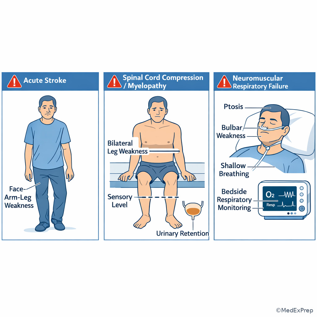
- High-stakes emergencies begin as localization questions: acute hemiparesis suggests stroke, bilateral leg weakness with a sensory level suggests spinal cord disease, and fluctuating bulbar weakness raises neuromuscular respiratory risk.
- Many distractors share weakness as a surface feature; the exam rewards the single clue that separates corticospinal tract disease from peripheral nerve, root, NMJ, or myopathy.
Key Terms & Must-Know Facts
1.1.1.1 Key terms
- Upper motor neuron (UMN): lesion above the anterior horn cell, typically involving corticospinal pathways.
- Lower motor neuron (LMN): lesion at the anterior horn cell, root, plexus, peripheral nerve, NMJ, or muscle output pathway.
- Spasticity: velocity-dependent increased tone, usually UMN.
- Hyperreflexia: increased deep tendon reflexes, usually UMN.
- Babinski sign: extensor plantar response, supports corticospinal tract dysfunction.
- Flaccidity: decreased tone, often acute UMN stage or LMN disease.
- Atrophy: loss of muscle bulk, more prominent in chronic LMN or myopathic states.
- Fasciculations: spontaneous motor unit discharges, suggest anterior horn cell or LMN disease.
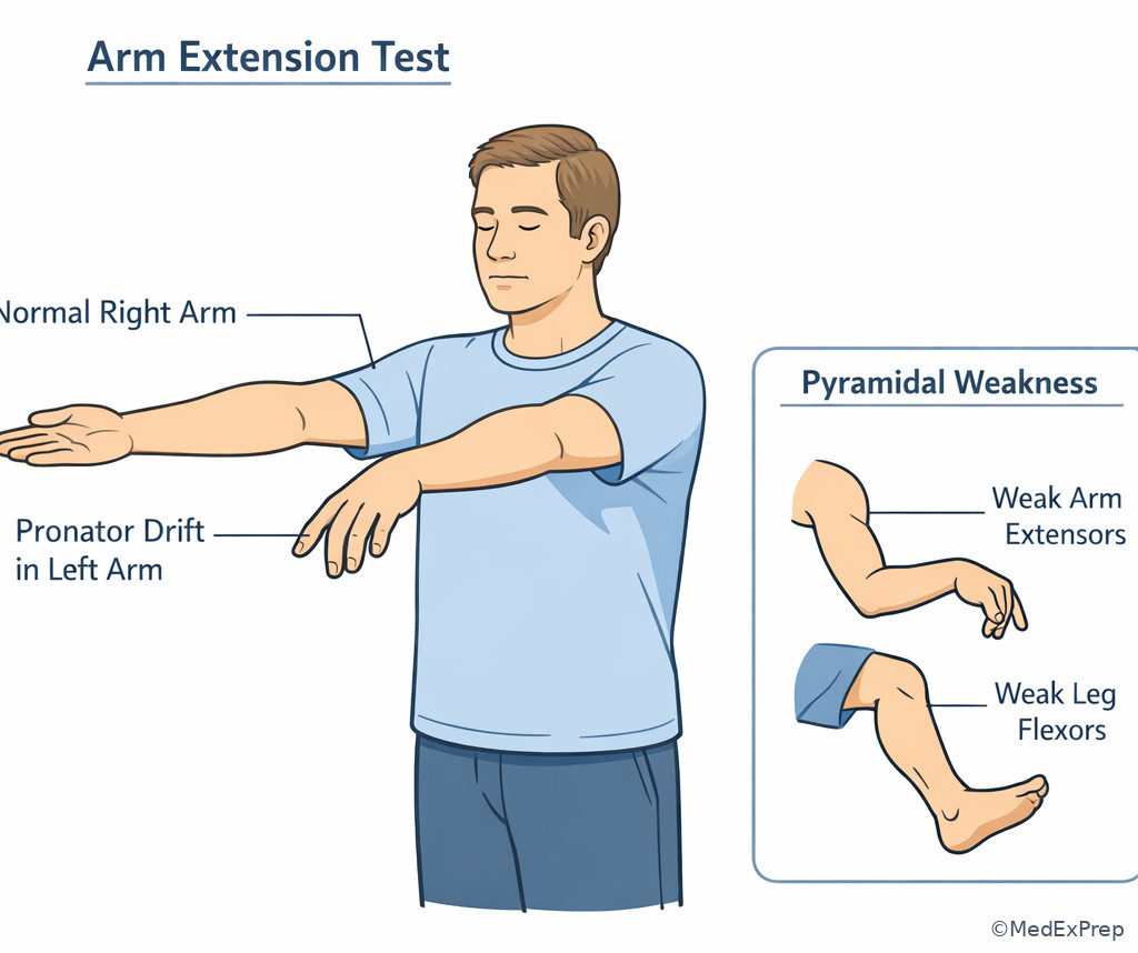
- Pronator drift: sensitive sign of mild corticospinal weakness.
- Pyramidal pattern: arm extensors/abductors and leg flexors relatively weaker in UMN lesions.
- Hemiparesis: weakness of face/arm/leg on one side, often cerebral or brainstem.
- Paraparesis: bilateral leg weakness, often spinal cord, cauda equina, polyneuropathy, or myopathy depending associated signs.
- Quadriparesis: weakness of all limbs, localizes to high cervical cord, brainstem, diffuse peripheral process, NMJ, or generalized muscle disease.
- Sensory level: abrupt horizontal sensory boundary, strongly suggests spinal cord localization.
- Crossed findings: cranial nerve deficit with contralateral body weakness, suggests brainstem.
1.1.1.2 Must-know facts
- Hyperreflexia plus Babinski beats most distractors: think corticospinal tract.
- Acute cerebral lesions can be initially hypotonic; do not reject a central lesion only because tone is not yet increased.
- Face-arm-leg weakness together points above the foramen magnum unless a peripheral mimic is obvious.
- Pure motor hemiparesis without cortical signs is classic for internal capsule or basis pontis lacunar localization.
- Weakness with aphasia, neglect, gaze deviation, or visual field loss points to cortex, not isolated internal capsule.
- Bilateral leg weakness with a sensory level or bladder dysfunction is spinal cord disease until proven otherwise.
- Reflexes are usually preserved in myasthenia gravis and often reduced in neuropathy.
- Proximal greater than distal weakness with normal sensation suggests myopathy.
- Ptosis and diplopia with fatigability suggest NMJ, especially when sensory exam is normal.
- Fasciculations plus mixed UMN/LMN signs in multiple regions suggest motor neuron disease.
Exam takeaway: Start with distribution plus reflexes to separate central corticospinal lesions from peripheral motor unit disorders.
flowchart TD
A[Weakness on exam] --> B{Reflexes and plantar response}
B -->|Hyperreflexia or Babinski| C[Central motor pathway]
B -->|Hyporeflexia or areflexia| D[Peripheral motor unit]
C --> E{Cortical signs or cranial nerve clues?}
E -->|Aphasia neglect field cut| F[Cortex]
E -->|Pure motor pattern| G[Internal capsule or brainstem or cord]
D --> H{Sensory loss present?}
H -->|Yes| I[Root plexus nerve cord]
H -->|No| J[NMJ muscle or anterior horn cell] Exam takeaway: Cortical signs localize outward to cortex, pure dense weakness often localizes inward to internal capsule, and crossed findings point to brainstem.
flowchart LR
A[Cortex] --> A1[Face arm leg weakness]
A --> A2[Aphasia neglect gaze deviation field cut]
B[Internal capsule] --> B1[Dense pure motor hemiparesis]
B --> B2[No cortical signs]
C[Brainstem] --> C1[Long tract weakness]
C --> C2[Ipsilateral CN deficit]
D[Spinal cord] --> D1[Bilateral weakness below level]
D --> D2[Sensory level bladder signs]
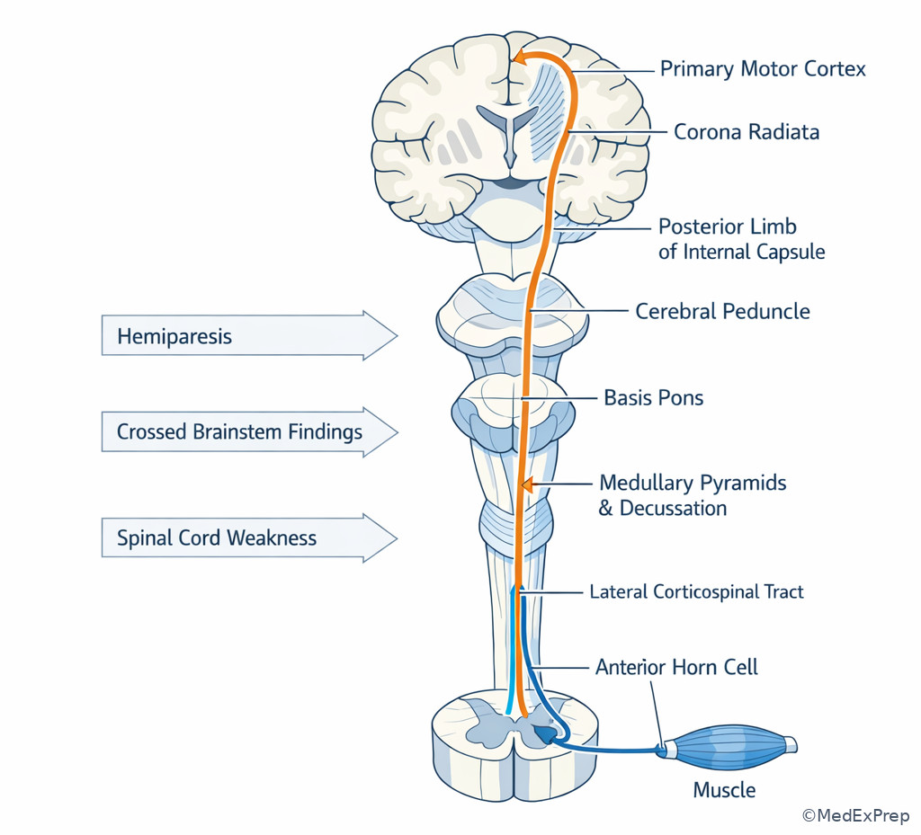
Exam takeaway: Visualize where corticospinal fibers travel so deficits can be mapped to cortex, internal capsule, brainstem, or spinal cord.
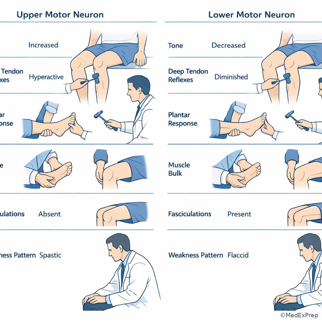
Exam takeaway: Pair tone, reflexes, plantar response, and muscle bulk to separate corticospinal from motor unit disorders.
Core content
1.1.1.3 Foundational split: upper motor neuron versus lower motor neuron weakness
A Explanation
The first decision conflict is simple but decisive: Is this weakness from the corticospinal system or from the motor unit? The single resolving rule is the examination triad of tone, reflexes, and plantar response. Choose a central/UMN localization when weakness is accompanied by hyperreflexia, spasticity, clonus, or an extensor plantar response. Reject a peripheral-first answer when those signs are convincingly present. Choose LMN or motor unit localization when weakness comes with reduced reflexes, flaccidity, early atrophy, or fasciculations.
WHY THIS IS TESTED: board stems frequently hide the diagnosis inside one or two exam details. A patient with weakness and a normal CK does not automatically have a central lesion; a patient with weakness and sensory symptoms does not automatically have neuropathy. The exam wants the examinee to identify the governing motor sign rather than follow distracting symptoms.
Important nuance: very acute corticospinal lesions can be transiently hypotonic and hyporeflexic, especially immediately after stroke or spinal shock. That is why you should use the whole pattern and time course rather than a single isolated finding. Another nuance: reflexes are often preserved in NMJ disease despite clear weakness, and sensation remains normal. Myopathy typically causes proximal weakness with normal sensation and no Babinski sign. Motor neuron disease may show both UMN and LMN signs in different regions.
Key rule: Babinski plus weakness is corticospinal until proven otherwise; fasciculations plus atrophy and hyporeflexia point away from isolated corticospinal tract disease.
Memory anchor: “\(BASH = Brain/cord Above anterior horn\)” — Babinski, Augmented reflexes, Spasticity, Hypertonia suggest UMN localization.
B Worked example
A 67-year-old man with hypertension, type 2 diabetes, and remote lumbar surgery develops sudden right arm and leg weakness while eating breakfast. He reports mild shoulder pain from yard work the day before. Examination shows right facial droop, right pronator drift, 4/5 strength in the right arm and leg, brisk right biceps and patellar reflexes, and an extensor right plantar response. Sensation is intact. CK is normal. The tempting wrong clue is the shoulder pain, which could nudge you toward plexopathy.
Reasoning chain: Identify task: localize weakness. Extract key facts: abrupt onset, face-arm-leg involvement, brisk reflexes, Babinski, intact sensation. Apply rule: hemibody weakness with UMN signs localizes centrally above the cervical cord, most likely cerebral. Eliminate distractors: brachial plexopathy would not cause facial weakness or Babinski; myopathy would not be unilateral with hyperreflexia; NMJ disease would not produce Babinski. Verify: sudden onset in a vascular-risk patient supports acute stroke.
C Exam trap
Common wrong answer: Peripheral neuropathy.
Why it is tempting: The stem highlights weakness and may add diabetes, inviting automatic association with neuropathy.
Single clue that eliminates it: An extensor plantar response or clear hyperreflexia is not explained by length-dependent peripheral neuropathy.
D Checkpoint
Question: A 58-year-old woman with hypertension, hyperlipidemia, and osteoarthritis reports sudden left-sided weakness. Examination shows increased tone in the left leg, brisk left reflexes, and an upgoing left toe. Sensation is intact. What is the most likely localization category?
- Central corticospinal pathway lesion — tempting because abrupt hemibody weakness suggests stroke; correct because Babinski and hyperreflexia identify UMN involvement.
- Peripheral neuropathy — tempting because weakness can occur in neuropathy; wrong because neuropathy should not produce unilateral spasticity and extensor plantar response.
- Neuromuscular junction disorder — tempting because weakness may fluctuate and spare sensation; wrong because NMJ disease does not cause hyperreflexia or Babinski.
- Primary myopathy — tempting because some myopathies have preserved sensation; wrong because myopathy is usually proximal and symmetric without UMN signs.
Clinician Action: Diagnose with pattern recognition from tone, reflexes, plantar response, bulk, and distribution. Order urgent brain or spine imaging when a central lesion is possible, and EMG/NCS or CK when the pattern remains peripheral. Treat the likely emergency first if onset is acute or bulbar/respiratory symptoms are present. Refer/Escalate to stroke or ICU pathways when focal deficit onset is sudden or respiratory weakness is suspected.
1.1.1.4 Foundational neuraxis map: cortex, internal capsule, brainstem, and spinal cord
A Explanation
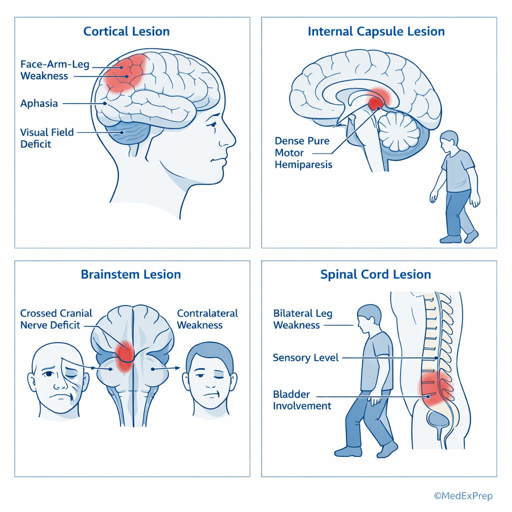
Once you have chosen a central motor pathway lesion, the next decision conflict is which level of the neuraxis is involved? The single finding that often resolves it is the presence or absence of associated localizing signs. See Fig 2. Choose cortex when weakness is accompanied by aphasia, neglect, gaze deviation, cortical sensory loss, or visual field deficit. Choose internal capsule/subcortical white matter when there is a dense pure motor hemiparesis without cortical signs. Choose brainstem when there are crossed findings: ipsilateral cranial nerve abnormality with contralateral limb weakness. Choose spinal cord when weakness is bilateral below a level, especially with a sensory level or bladder dysfunction.
WHY THIS IS TESTED: many board items give dense but incomplete neurologic data and ask for “most likely localization.” The exam expects you to prioritize pattern over exhaustive anatomy. The critical move is to ask, “What accompanying sign cannot occur at the competing level?” Aphasia excludes brainstem and spinal cord. A sensory level excludes cortex. Crossed face-body findings exclude cortex and peripheral nerve.
Recall: This is the first topic in the course, so there is no prior content to retrieve. Build the habit now: localize from distribution + associated signs before naming the disease. In the upcoming Sensory Localization Patterns lesson, the same logic will be extended using sensory levels, dissociated sensory loss, and cortical sensory deficits.
High-Yield Connection: This concept is tested alongside Anterior Circulation Stroke Syndromes and Posterior Circulation Stroke Syndromes. Know both the localization clue and the vascular syndrome because board stems often pair a motor pattern with a vessel territory.
Exam takeaway: Face-arm-leg distribution plus cortical, cranial nerve, or sensory-level clues rapidly separates cortex, internal capsule, brainstem, and spinal cord.
B Worked example
A 71-year-old woman with atrial fibrillation, chronic kidney disease, and migraine with aura presents with sudden left face, arm, and leg weakness. She also has expressive aphasia and a right gaze preference. Her daughter mentions a similar episode years ago that was “probably migraine,” which is the tempting wrong clue. Examination shows a left Babinski sign. Sensation to pin is difficult to assess because of language deficit.
Reasoning chain: Identify task: central lesion level. Extract key facts: hemiparesis plus aphasia plus gaze preference. Apply rule: cortical signs localize to cerebral cortex rather than internal capsule. Eliminate distractors: lacunar internal capsule stroke can give dense pure motor weakness but should not cause aphasia; brainstem lesions produce cranial nerve deficits, not dominant hemisphere aphasia; cervical cord lesions do not produce aphasia or gaze deviation. Verify: dominant frontal-insular or MCA cortical localization fits best.
C Exam trap
Common wrong answer: Internal capsule lacune for any pure weakness.
Why it is tempting: The examinee sees dense hemiparesis and remembers pure motor lacunar stroke as a classic board pattern.
Single clue that eliminates it: Any cortical sign—aphasia, neglect, visual field defect, cortical sensory loss, gaze deviation—pushes the lesion to cortex rather than isolated internal capsule.
D Checkpoint
Question: A 64-year-old man with hypertension and smoking history develops right face, arm, and leg weakness without aphasia, neglect, visual field deficit, or sensory loss. Examination shows brisk right reflexes. What is the most likely localization?
- Left internal capsule — tempting because sudden dense hemiparesis suggests a lacunar syndrome; correct because the pattern is pure motor without cortical signs.
- Left frontal cortex — tempting because cortical lesions can cause contralateral weakness; wrong because there are no cortical accompaniments.
- Right pons — tempting because brainstem lesions can produce weakness; wrong because there are no ipsilateral cranial nerve findings and the side is inconsistent.
- Cervical spinal cord — tempting because UMN signs occur below cord lesions; wrong because simultaneous facial weakness places the lesion above the foramen magnum.
- The classic lesion for pure motor hemiparesis is the internal capsule.
- True or False: Aphasia can localize to the brainstem. False.
- Name the 3 clue clusters that push you away from internal capsule: aphasia/neglect, visual field loss, gaze deviation.
Clinician Action: Diagnose central level using face involvement, cortical signs, crossed findings, and sensory level. Order urgent MRI brain or spinal imaging guided by the pattern. Treat sudden focal cerebral deficits as possible stroke until proven otherwise. Refer/Escalate promptly to stroke, neurosurgery, or spine services when cerebral ischemia or cord compression is suspected.
| Finding | Most helpful localization | Why it matters on boards |
|---|---|---|
| Aphasia | Dominant cerebral cortex | Rejects brainstem, spinal cord, root, nerve, NMJ, and muscle |
| Neglect | Nondominant cerebral cortex | Strong cortical clue in hemiparesis stems |
| Pure motor hemiparesis | Internal capsule or basis pons | Classic lacunar pattern when cortical signs absent |
| Crossed face-body findings | Brainstem | Separates brainstem from cortex |
| Sensory level | Spinal cord | One of the strongest spinal localizers |
| Bladder dysfunction with bilateral leg weakness | Spinal cord | Urgent cord compression/inflammatory myelopathy clue |
| Ptosis/diplopia with normal sensation | Neuromuscular junction | Rejects cortex/cord if reflexes and sensation preserved |
| Symmetric proximal weakness | Muscle | Board-favorite separation from neuropathy and UMN disease |
1.1.1.5 Application: using distribution of weakness to localize the lesion
A Explanation
After deciding central versus peripheral and roughly where on the neuraxis, the next conflict is which distribution pattern matters most? The single resolving rule is to map weakness into one of four exam shapes: hemibody, bilateral leg-predominant, distal length-dependent, or proximal symmetric. See Fig 1. Hemibody weakness involving face, arm, and leg points to the brain or upper brainstem. Bilateral leg-predominant weakness suggests spinal cord, conus/cauda equina, severe polyneuropathy, or myopathy depending sensory and reflex clues. Distal length-dependent weakness with sensory loss suggests neuropathy. Proximal symmetric weakness with preserved sensation suggests myopathy. Fluctuating ocular/bulbar weakness with preserved reflexes and sensation suggests NMJ disease.
WHY THIS IS TESTED: many distractors sound attractive because they produce weakness, but they do not produce the same shape of weakness. The board often includes a misleading comorbidity—diabetes, back pain, statin use, or fatigue—while the distribution pattern gives away the answer. Choose the localization that matches the pattern best; reject answers that require unrelated findings not present in the stem.
Pyramidal weakness is another useful concept. In UMN lesions, arm extensors/abductors and leg flexors are often more affected, producing pronator drift and circumduction. Distal hand weakness can occur in cortical “hand knob” lesions, but broader hemibody signs usually clarify the localization. Bilateral arm greater than leg weakness raises consideration of cervical cord or central cord syndromes, especially with sensory changes and bladder symptoms.
Exam takeaway: The shape of weakness often localizes the lesion before any imaging is ordered.
flowchart TD
A[Pattern of weakness] --> B{Hemibody?}
B -->|Yes| C[Brain or brainstem]
B -->|No| D{Bilateral legs?}
D -->|Yes| E[Spinal cord or cauda or neuropathy or myopathy]
D -->|No| F{Distal more than proximal with sensory loss?}
F -->|Yes| G[Peripheral neuropathy]
F -->|No| H{Proximal symmetric, sensation normal?}
H -->|Yes| I[Myopathy]
H -->|No| J[Consider NMJ or anterior horn cell] B Worked example
A 59-year-old man with diabetes, chronic alcohol use, and hypertension reports 3 months of progressive foot numbness and difficulty climbing stairs. He takes a statin. Examination shows distal weakness in ankle dorsiflexion greater than hip flexion, absent ankle reflexes, impaired vibration at the toes, and normal plantar responses. CK is mildly elevated. The tempting wrong clue is the statin, which may make myopathy seem appealing.
Reasoning chain: Identify task: localize the weakness pattern. Extract key facts: distal greater than proximal weakness, sensory loss in the feet, absent ankle jerks, normal plantar responses. Apply rule: distal symmetric weakness with sensory loss and reduced reflexes localizes to peripheral neuropathy, not primary muscle disease. Eliminate distractors: statin myopathy causes proximal more than distal weakness and usually no sensory loss; spinal cord disease would more likely produce UMN signs or sensory level; NMJ disease does not cause distal sensory loss. Verify: diabetic or alcoholic polyneuropathy fits best.
C Exam trap
Common wrong answer: Myopathy in any patient with weakness plus statin use.
Why it is tempting: Statins are a memorized cause of myopathy, so learners anchor on the medication before reading the exam pattern.
Single clue that eliminates it: Objective sensory loss or distal areflexia points away from isolated myopathy and toward neuropathy or root/cord disease.
D Checkpoint
Question: A 46-year-old woman with hypothyroidism and obesity reports progressive trouble rising from a chair and lifting objects overhead. She has no numbness, diplopia, or bowel/bladder symptoms. Examination shows symmetric proximal arm and leg weakness, normal reflexes, and normal plantar responses. What localization is most likely?
- Myopathy — tempting because proximal weakness can be caused by several processes; correct because the pattern is symmetric proximal weakness with normal sensation and no UMN signs.
- Peripheral neuropathy — tempting because weakness can accompany endocrine disease; wrong because neuropathy is usually distal and often sensory with reduced reflexes.
- Cervical spinal cord lesion — tempting because limb weakness can be bilateral; wrong because there is no sensory level, bladder issue, or UMN pattern.
- Myasthenia gravis — tempting because normal sensation and reflexes can occur; wrong because proximal fatigable weakness usually includes ocular or bulbar clues rather than isolated fixed hip/shoulder weakness.
Clinician Action: Diagnose by naming the weakness shape first. Order MRI when the pattern is central, EMG/NCS for neuropathy/NMJ/anterior horn cell, and CK/TSH/inflammatory studies for myopathy. Treat reversible contributors such as toxic, metabolic, or inflammatory causes after localization. Refer/Escalate urgently when the pattern suggests cord compression, stroke, or neuromuscular respiratory weakness.
1.1.1.6 Application: paired clues from cranial nerves, sensation, and reflexes
A Explanation
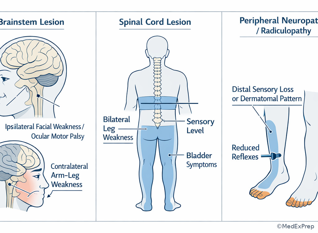
The most testable board stems add one extra clue to force a finer localization: cranial nerve findings, a sensory pattern, or a reflex pattern. The conflict becomes: Do these additional findings move me toward brainstem, spinal cord, root/nerve, NMJ, or muscle? The single resolving rule is to look for the clue that cannot be produced by the competing lesion. Cranial nerve deficits plus contralateral weakness localize to the brainstem. A horizontal sensory level suggests spinal cord disease. Dermatomal pain or sensory loss with depressed reflex in one limb suggests radiculopathy. Length-dependent sensory loss suggests neuropathy. Preserved sensation with fatigable ocular/bulbar weakness suggests NMJ. Preserved sensation with proximal fixed weakness suggests muscle.
WHY THIS IS TESTED: when two answer choices seem plausible, the exam expects you to use the one finding with the highest localization value. If the patient has diplopia and dysarthria, do not stop at “generalized weakness”; ask whether there are fluctuating ocular findings and normal reflexes. If the patient has leg weakness and urinary retention, ask whether there is a sensory level. If the patient has unilateral weakness plus facial numbness, ask whether these are crossed or same-sided.
High-Yield Connection: This concept is tested alongside the upcoming Brainstem And Cerebellar Syndromes. Know that cranial nerve findings with contralateral motor deficits localize to the brainstem, while ataxia alone may instead reflect cerebellar, sensory, or vestibular disease.
Memory anchor: “FACE CROSSES STEM” — if a cranial nerve sign is on one side and limb weakness is on the other, think brainstem.
Exam takeaway: Cranial nerve involvement, sensory level, and reflex pattern are the tie-breakers when weakness alone is not enough.
B Worked example
A 62-year-old man with hypertension, coronary disease, and prior Bell palsy presents with acute diplopia and left facial weakness followed by right arm and leg weakness. He also complains of chronic diabetic toe numbness, which is the distracting clue. Examination shows a left abducens palsy, left facial weakness, and right-sided hyperreflexia. Plantars are extensor on the right.
Reasoning chain: Identify task: localize lesion level. Extract key facts: ipsilateral cranial nerve deficits with contralateral body weakness. Apply rule: crossed findings localize to the brainstem. Eliminate distractors: diabetic neuropathy explains chronic toe numbness but cannot explain abducens palsy plus contralateral UMN signs; cortex does not usually produce isolated ipsilateral lower cranial nerve-type crossed syndromes; cervical cord does not affect abducens nerve. Verify: pontine localization is most likely.
C Exam trap
Common wrong answer: Cervical spinal cord lesion in any patient with bilateral or severe limb weakness.
Why it is tempting: Cord disease is a familiar cause of UMN signs below the lesion, so learners may overuse it.
Single clue that eliminates it: A cranial nerve finding places the lesion above the spinal cord.
D Checkpoint
Question: A 55-year-old woman with diabetes and asthma develops diplopia, dysarthria, and fluctuating ptosis that worsens late in the day. Limb strength is mildly weak proximally, sensation is normal, and reflexes are preserved. Which localization is most likely?
- Brainstem — tempting because cranial symptoms suggest a central lesion; wrong because fluctuation with preserved sensation and reflexes favors NMJ, not a fixed brainstem lesion.
- Neuromuscular junction — tempting because ocular and bulbar fatigability are classic; correct because normal sensation and preserved reflexes support NMJ localization.
- Peripheral neuropathy — tempting because diabetes raises that possibility; wrong because neuropathy causes sensory loss and usually distal patterns rather than fatigable ocular weakness.
- Myopathy — tempting because proximal weakness can occur; wrong because ptosis and diplopia are far more characteristic of NMJ disease than typical myopathy.
- The most localizing clue for brainstem weakness is crossed cranial nerve and body findings.
- True or False: A sensory level strongly suggests peripheral neuropathy. False.
- Name the 3 clues that favor NMJ over myopathy: ptosis, diplopia, fatigability.
Clinician Action: Diagnose with the highest-yield tie-breaker clue: crossed CN findings, sensory level, dermatomal loss, or fatigability. Order MRI brainstem or spine for central patterns and EMG/NCS with serology for NMJ/peripheral patterns. Treat respiratory or bulbar compromise first. Refer/Escalate urgently for possible myasthenic crisis, brainstem stroke, or spinal cord compression.
| Pattern | Best localization | Key discriminator | Common trap |
|---|---|---|---|
| Face-arm-leg weakness with aphasia | Cerebral cortex | Dominant cortical sign | Internal capsule lacune |
| Dense pure motor hemiparesis | Internal capsule | No cortical signs | Cortical MCA stroke |
| Ipsilateral CN deficit + contralateral limb weakness | Brainstem | Crossed findings | Cortical stroke |
| Bilateral leg weakness + sensory level | Spinal cord | Horizontal sensory boundary | Polyneuropathy |
| Distal weakness + stocking sensory loss + areflexia | Polyneuropathy | Length-dependent sensory pattern | Myopathy |
| Ptosis/diplopia + normal sensation | Neuromuscular junction | Fatigability | Brainstem lesion |
| Symmetric proximal weakness + normal sensation | Myopathy | Shoulder/hip pattern | Neuropathy |
1.1.1.7 Integration: choosing the best next test after bedside localization
A Explanation
Localization is not the endpoint; the next board conflict is what should be ordered first? The single rule is to let the localization decide the test. Choose MRI or urgent neuroimaging for suspected central lesions, especially acute onset or spinal cord patterns. Choose EMG/NCS for suspected peripheral nerve, root, anterior horn cell, or NMJ disorders when the patient is stable and the diagnosis is not already obvious. Choose CK and targeted laboratory testing for suspected myopathy. Choose pulmonary function monitoring and blood gas assessment when weakness threatens respiration, especially with bulbar or NMJ disease.
WHY THIS IS TESTED: board stems often ask for the “most appropriate next step,” and the wrong answers are usually reasonable tests ordered at the wrong time. MRI brain is correct for acute hemiparesis but not for obvious diabetic polyneuropathy. EMG is useful in neuromuscular disease but not the first priority in acute spinal cord compression. The exam rewards sequence: localize first, then choose the test that confirms that level or addresses the emergency.
Urgency matters. Sudden hemiparesis demands stroke evaluation. Bilateral weakness with a sensory level and urinary retention demands emergent spinal imaging. Fluctuating bulbar weakness with reduced vital capacity suggests myasthenic crisis and requires respiratory monitoring before confirmatory serologies. Progressive LMN signs in multiple regions may eventually require EMG to support motor neuron disease. Do not over-order broad testing before you have chosen the lesion level.
Exam takeaway: The correct next step depends on lesion level and urgency, not on ordering every possible neurologic test.
flowchart TD
A[Bedside localization] --> B{Acute central pattern?}
B -->|Yes| C[Urgent brain imaging or spine MRI]
B -->|No| D{Peripheral motor unit pattern?}
D -->|Yes| E[EMG NCS +/- targeted labs]
D -->|No| F{Proximal symmetric weakness?}
F -->|Yes| G[CK endocrine inflammatory myopathy workup]
F -->|No| H{Bulbar or respiratory risk?}
H -->|Yes| I[Respiratory monitoring urgent escalation]
H -->|No| J[Refine differential and targeted testing] B Worked example
A 48-year-old man with ulcerative colitis and hypertension reports 2 days of progressive bilateral leg weakness, band-like trunk numbness, and urinary retention. He also has chronic carpal tunnel symptoms, a tempting distracting detail. Examination shows a T8 sensory level, brisk knee reflexes, and extensor plantar responses bilaterally.
Reasoning chain: Identify task: best next test after localization. Extract key facts: bilateral leg weakness, sensory level, bladder involvement, UMN signs. Apply rule: this localizes to spinal cord and is urgent. Eliminate distractors: EMG/NCS would be delayed and nonurgent for a clear cord syndrome; CK does not explain sensory level; brain MRI misses the likely emergency site. Verify: emergent MRI of the spine is the best next step, with escalation for possible compressive or inflammatory myelopathy.
C Exam trap
Common wrong answer: EMG/NCS for any weakness of uncertain cause.
Why it is tempting: EMG feels broadly neurologic and confirmatory, so learners select it reflexively.
Single clue that eliminates it: A sensory level or acute focal cerebral deficit requires urgent imaging first because the problem may be time-sensitive stroke or cord compression.
D Checkpoint
Question: A 39-year-old woman with Graves disease and asthma develops fluctuating ptosis, diplopia, and nasal speech over several weeks. Reflexes and sensation are normal. What is the most appropriate next diagnostic direction after bedside localization?
- MRI thoracic spine — tempting because weakness evaluation sometimes begins with imaging; wrong because there is no sensory level, UMN sign, or bladder clue.
- EMG/NCS with NMJ-focused testing — tempting because the pattern suggests NMJ disease; correct because ocular-bulbar fatigability with normal sensation/reflexes points to NMJ localization.
- CK and muscle biopsy — tempting because weakness may reflect muscle disease; wrong because diplopia and ptosis are much more typical of NMJ than primary myopathy.
- Routine lumbar MRI — tempting because weakness often triggers spine imaging; wrong because the presentation is not radicular, myelopathic, or cauda equina-like.
Clinician Action: Diagnose the lesion level before ordering broad tests. Order emergent imaging for suspected stroke or cord lesions, EMG/NCS for peripheral/NMJ questions, and CK-targeted labs for suspected myopathy. Treat immediately when localization indicates a time-sensitive process. Refer/Escalate to stroke, ICU, or spine services if there is respiratory threat, sudden onset, or cord compression risk.
1.1.1.8 Integration: common board differentials and emergency motor patterns
A Explanation
The final integration step is choosing between close-call differentials and recognizing when localization itself mandates urgent action. The conflict is which weak patient can wait for outpatient workup and which one needs immediate escalation? The single resolving rule is to look for time-sensitive motor patterns: sudden hemiparesis, rapidly progressive bulbar weakness, acute ascending weakness, and bilateral leg weakness with sensory level or sphincter dysfunction. Sudden hemiparesis is stroke until proven otherwise. Sensory level plus bladder dysfunction is spinal cord until proven otherwise. Fluctuating bulbar weakness with dyspnea suggests myasthenic crisis. Ascending weakness with areflexia suggests Guillain-Barré syndrome and respiratory monitoring needs.
WHY THIS IS TESTED: ABPN-style items frequently convert localization into action. It is not enough to say “probably NMJ” if the patient cannot count to 20 or has weak neck flexion. It is not enough to say “spinal cord lesion” if urinary retention is present; you must choose urgent spine MRI and escalation. Close distractors are often correct diagnoses for a slightly different patient. Use the one clue that changes urgency.
Key rule: Weakness plus respiratory, bulbar, or sphincter compromise is never routine outpatient neurology.
Exam takeaway: Sudden hemiparesis, sensory level with bladder signs, and bulbar-respiratory weakness require immediate escalation rather than delayed outpatient testing.
Exam takeaway: Mild corticospinal weakness may appear first as pronator drift before obvious loss on formal strength testing.
B Worked example
A 33-year-old woman with rheumatoid arthritis and obesity presents with 1 day of worsening leg weakness and urinary hesitancy. She also reports chronic migraines and recently started a gym program, both distracting details. Examination reveals bilateral leg spasticity, brisk reflexes, extensor plantar responses, and decreased pin sensation below the umbilicus.
Reasoning chain: Identify task: determine urgent localization and next step. Extract key facts: bilateral leg UMN signs, sensory level, bladder symptom. Apply rule: spinal cord process until proven otherwise. Eliminate distractors: deconditioning does not cause a sensory level; migraine does not cause bilateral myelopathic signs; peripheral neuropathy would not create a sharp truncal level with Babinski signs. Verify: emergent spinal imaging and escalation for myelopathy are required.
C Exam trap
Common wrong answer: Outpatient workup for “possible neuropathy” when bilateral leg weakness is subacute.
Why it is tempting: Symmetric leg complaints sound like peripheral neuropathy, especially if diabetes or alcohol use appears in the stem.
Single clue that eliminates it: A sensory level, spasticity, or bladder dysfunction upgrades the problem to spinal cord localization and urgent evaluation.
D Checkpoint
Question: Which patient requires the most immediate escalation based on motor localization?
- A 61-year-old man with months of distal numbness, bilateral foot drop, and absent ankle jerks — tempting because gait failure is serious; wrong because this pattern suggests chronic neuropathy, not the most urgent threat here.
- A 42-year-old woman with proximal weakness, normal sensation, and elevated CK over several weeks — tempting because inflammatory myopathy can require treatment; wrong because the time course is less emergent than airway or cord compromise.
- A 56-year-old man with fluctuating ptosis, dysarthria, neck flexor weakness, and shortness of breath — tempting because ocular findings may seem less dramatic than hemiparesis; correct because this pattern suggests NMJ-related respiratory compromise requiring urgent monitoring.
- A 68-year-old woman with chronic radicular pain, absent ankle reflex on one side, and mild foot weakness — tempting because focal weakness needs evaluation; wrong because the pattern is outpatient radiculopathy unless red flags appear.
Clinician Action: Diagnose emergency localization from sudden onset, sensory level, ascending areflexia, or bulbar-respiratory weakness. Order immediate imaging or respiratory monitoring before slower confirmatory studies. Treat stroke, cord, or respiratory threats promptly while refining cause. Refer/Escalate to ICU, stroke, or spine teams when airway, breathing, rapid progression, or sphincter compromise is present.
Exam Traps & Differentiators
Common traps & distractors: The exam often supplies a true but irrelevant clue—diabetes, statin use, back pain, migraine, or chronic numb feet—to pull you away from the lesion level established by the motor exam.
1.1.1.9 Most common wrong answer and why
Most common wrong answer: peripheral neuropathy. It is tempting because weakness plus diabetes or distal symptoms appears in many stems. It becomes wrong when the stem includes hyperreflexia, Babinski, facial involvement, aphasia, a sensory level, or bladder dysfunction. Those clues push the lesion away from peripheral nerve.
1.1.1.10 Looks similar but isn't
| Confusable pair | Why they look similar | Single clue that separates them |
|---|---|---|
| Cortical stroke vs internal capsule lacune | Both can cause contralateral hemiparesis | Aphasia/neglect/field cut/gaze deviation means cortex |
| Brainstem lesion vs myasthenia gravis | Both can produce diplopia and dysarthria | Crossed long-tract signs suggest brainstem; fluctuation with normal sensation suggests NMJ |
| Spinal cord lesion vs polyneuropathy | Both can cause bilateral leg weakness | Sensory level or bladder dysfunction means cord |
| Myopathy vs neuropathy | Both can cause gait difficulty | Proximal symmetric weakness with intact sensation means myopathy |
| Radiculopathy vs cortical monoparesis | Both can present with focal limb weakness | Dermatomal pain/reflex loss favors root; pronator drift/UMN signs favor cortex |
| ALS vs cervical myelopathy | Both can show weakness and brisk reflexes | Sensory level/bladder symptoms favor myelopathy; widespread fasciculations with normal sensation favor ALS |
1.1.1.11 If the stem says ___, think ___
- If the stem says aphasia with contralateral weakness, think cerebral cortex.
- If the stem says pure motor hemiparesis without cortical signs, think internal capsule lacunar pattern.
- If the stem says ipsilateral cranial nerve deficit with contralateral body weakness, think brainstem.
- If the stem says bilateral leg weakness with a sensory level, think spinal cord.
- If the stem says ptosis and diplopia with normal sensation, think neuromuscular junction.
- If the stem says symmetric proximal weakness and elevated CK, think myopathy.
Forward link: in the upcoming Sensory Localization Patterns topic, these motor traps will be paired with sensory discriminators. That is especially important for separating spinal cord from thalamic, peripheral nerve, and cortical lesions. In the upcoming Brainstem And Cerebellar Syndromes topic, crossed findings and ocular motor clues will be refined further.
Algorithm / Approach
Rapid Review
- Babinski sign → corticospinal tract, not peripheral neuropathy.
- Face involved with body weakness → lesion above the cervical cord.
- Aphasia → cortex, not internal capsule alone.
- Dense pure motor hemiparesis → internal capsule until a cortical sign appears.
- Crossed CN-body deficits → brainstem.
- Sensory level → spinal cord, not length-dependent neuropathy.
- Urinary retention with leg weakness → urgent cord/conus-cauda evaluation.
- Distal weakness plus areflexia plus numb feet → neuropathy.
- Proximal symmetric weakness, sensation spared → myopathy.
- Ptosis/diplopia fluctuating through day → NMJ.
- Fasciculations in wasted muscles → LMN/anterior horn cell clue.
- Pronator drift → subtle corticospinal weakness.
- Acute flaccidity does not exclude stroke → very early UMN lesions may be hypotonic.
- EMG/NCS → peripheral/NMJ/anterior horn cell confirmation, not first-line for acute cord syndrome.
Memory anchor: “FACES” for high-yield central clues — Face involved, Aphasia, Crossed findings, Extensor plantar, Sensory level. Not every central lesion has all five, but each one should make you stop and localize centrally.
Self-check quiz
1. A 63-year-old man with hypertension and hyperlipidemia develops sudden right-sided weakness while watching television. Examination shows brisk right reflexes and an extensor right plantar response. Which localization category is most likely?
- Central corticospinal pathway
- Peripheral neuropathy
- Neuromuscular junction
- Primary muscle disease
2. A 52-year-old woman with obesity and hypothyroidism reports months of difficulty rising from chairs and lifting laundry baskets. Sensation is normal, reflexes are preserved, and weakness is symmetric in the shoulders and hips. Which localization is most likely?
- Myopathy
- Polyneuropathy
- Internal capsule lesion
- Neuromuscular junction disorder
3. A 70-year-old man with atrial fibrillation and diabetes presents with sudden left face, arm, and leg weakness. He also has expressive aphasia; chronic low back pain is noted in the chart. Which localization is most likely?
- Left cervical radiculopathy
- Right cerebral cortex
- Right internal capsule
- Left pons
4. A 45-year-old woman with Graves disease and allergic rhinitis has fluctuating ptosis, diplopia, and dysarthria that worsen by evening. Sensation is intact and reflexes are normal. What is the most appropriate next diagnostic study direction?
- MRI lumbar spine
- EMG/NCS with NMJ-focused testing
- CK and muscle biopsy as first step
- MRI thoracic spine
5. A 54-year-old man with ulcerative colitis and hypertension develops bilateral leg weakness, a band-like numb sensation around the torso, and urinary retention over 24 hours. He also mentions chronic hand tingling at night. What is the most appropriate next step?
- EMG/NCS
- Outpatient CK level
- Emergent spinal MRI
- Routine head CT only
6. A 66-year-old woman with hypertension, diabetes, and prior cataract surgery presents with dense right face, arm, and leg weakness. She has no aphasia, neglect, gaze deviation, or visual field deficit. Sensation is intact. What is the most likely localization?
- Left internal capsule
- Left frontal cortex
- Right medulla
- Cervical spinal cord
7. A 60-year-old man with coronary disease and diabetes presents with acute diplopia, left facial weakness, and right arm and leg weakness. He also reports chronic numb toes. Which localization is most likely?
- Left brainstem
- Right cerebral cortex
- Cervical spinal cord
- Peripheral neuropathy
8. A 41-year-old woman with rheumatoid arthritis and obesity presents with subacute bilateral leg weakness, extensor plantar responses, decreased pin sensation below the umbilicus, and urinary urgency. She recently increased her exercise routine, and CK is mildly elevated. What is the best localization?
- Inflammatory myopathy
- Thoracic spinal cord
- Length-dependent polyneuropathy
- Neuromuscular junction disorder
Answer key
1.1.1.12 1. Answer: A. Central corticospinal pathway
Why A is correct: Hyperreflexia and an extensor plantar response identify UMN/corticospinal dysfunction. In a patient with sudden unilateral weakness, that pattern is most consistent with a central lesion such as acute stroke. The single discriminating clue is the Babinski sign. Sources: Daroff, Jankovic, Mazziotta, and Pomeroy; Adams and Victor's Principles of Neurology. Powers et al.
Why B is tempting but wrong: Peripheral neuropathy is tempting because weakness can occur and vascular risk factors often coexist with diabetes. It is incorrect here because peripheral neuropathy does not explain unilateral brisk reflexes and an upgoing toe. The single discriminating clue is unilateral UMN pattern. Sources: Daroff et al.; Harrison's Principles of Internal Medicine.
Why C is tempting but wrong: NMJ disease can cause weakness with relatively preserved sensation. It is incorrect because NMJ disorders do not produce Babinski signs or focal hemisyndromes. The single discriminating clue is the extensor plantar response. Sources: Harrison's; Daroff et al.
Why D is tempting but wrong: Muscle disease causes weakness and may preserve sensation, making it superficially plausible. It is incorrect because myopathies are usually symmetric and proximal without hyperreflexia or Babinski. The single discriminating clue is sudden unilateral UMN weakness. Sources: Harrison's; Daroff et al.
1.1.1.13 2. Answer: A. Myopathy
Why A is correct: Symmetric proximal weakness involving shoulder and hip girdles with preserved sensation and preserved reflexes strongly favors muscle disease. The single discriminating clue is the proximal pattern without sensory loss. Sources: Harrison's Principles of Internal Medicine; Daroff et al.
Why B is tempting but wrong: Polyneuropathy is tempting because hypothyroidism can contribute to neuropathic complaints. It is incorrect because neuropathy is typically distal and sensory, often with reduced reflexes. The single discriminating clue is proximal symmetric weakness. Sources: Harrison's; Daroff et al.
Why C is tempting but wrong: Internal capsule lesions can cause weakness without sensory loss. It is incorrect because those lesions cause acute unilateral hemiparesis, not chronic symmetric shoulder/hip weakness. The single discriminating clue is symmetry. Sources: Adams and Victor's; Powers et al.
Why D is tempting but wrong: NMJ disease can preserve sensation and reflexes, making it superficially plausible. It is incorrect because isolated chronic shoulder/hip weakness without fatigable ocular or bulbar symptoms is less characteristic of NMJ disease than myopathy. The single discriminating clue is absence of ptosis/diplopia/fatigability. Sources: Harrison's; Daroff et al.
1.1.1.14 3. Answer: B. Right cerebral cortex
Why B is correct: Aphasia with contralateral face-arm-leg weakness localizes to the dominant cerebral cortex. The single discriminating clue is aphasia, which is a cortical sign. Sources: Daroff et al.; Powers et al.; Adams and Victor's.
Why A is tempting but wrong: Radiculopathy is tempting because the chart mentions back pain, a classic distractor. It is incorrect because radiculopathy does not cause aphasia or face-arm-leg hemiparesis. The single discriminating clue is aphasia. Sources: Harrison's; Daroff et al.
Why C is tempting but wrong: Internal capsule is tempting because it causes contralateral weakness and is a classic lacunar localization. It is incorrect because internal capsule lesions should not produce aphasia if the lesion is isolated. The single discriminating clue is the cortical language deficit. Sources: Adams and Victor's; Powers et al.
Why D is tempting but wrong: Pontine lesions can cause weakness and dysarthria, so brainstem may appear plausible. It is incorrect because a left pontine lesion would not explain aphasia, and brainstem lesions usually produce cranial nerve findings rather than dominant cortical language dysfunction. The single discriminating clue is aphasia. Sources: Daroff et al.; Adams and Victor's.
1.1.1.15 4. Answer: B. EMG/NCS with NMJ-focused testing
Why B is correct: Fluctuating ptosis, diplopia, and dysarthria with normal sensation and reflexes localize to the neuromuscular junction. After bedside localization, NMJ-focused electrodiagnostic testing is the appropriate next diagnostic direction. The single discriminating clue is fatigable ocular-bulbar weakness. Sources: Harrison's Principles of Internal Medicine; Daroff et al.
Why A is tempting but wrong: Lumbar spine imaging is tempting because weakness often leads to spinal imaging in practice. It is incorrect because there are no radicular symptoms, sensory level, or LMN leg-predominant findings. The single discriminating clue is ocular involvement. Sources: Daroff et al.; Harrison's.
Why C is tempting but wrong: CK and muscle biopsy are tempting because muscle disease can present with weakness. They are incorrect because diplopia and ptosis are far more typical of NMJ disease than primary myopathy. The single discriminating clue is ocular fatigability. Sources: Harrison's; Daroff et al.
Why D is tempting but wrong: Thoracic spine MRI is tempting when weakness is not yet fully explained. It is incorrect because no myelopathic sensory level, bladder symptoms, or UMN signs are present. The single discriminating clue is preserved sensation with fluctuating ocular-bulbar weakness. Sources: Daroff et al.; Harrison's.
1.1.1.16 5. Answer: C. Emergent spinal MRI
Why C is correct: Bilateral leg weakness plus band-like sensory change and urinary retention localize to the spinal cord and represent a time-sensitive syndrome. The best next step is emergent spinal MRI. The single discriminating clue is the sensory level with sphincter dysfunction. Sources: Daroff et al.; Harrison's Principles of Internal Medicine.
Why A is tempting but wrong: EMG/NCS is tempting because weakness with hand tingling may suggest a peripheral process. It is incorrect because chronic nocturnal hand tingling is distracting noise, while the acute trunk sensory level and urinary retention demand cord imaging first. The single discriminating clue is the sensory level. Sources: Daroff et al.; Harrison's.
Why B is tempting but wrong: CK is tempting because inflammatory or toxic myopathy can cause weakness. It is incorrect because myopathy does not produce a truncal sensory level or urinary retention. The single discriminating clue is bladder involvement. Sources: Harrison's; Daroff et al.
Why D is tempting but wrong: Head CT is tempting because central causes of weakness often need imaging. It is incorrect because the pattern localizes below the brain, and isolated head imaging would miss the likely lesion site. The single discriminating clue is bilateral leg weakness with band-like trunk numbness. Sources: Daroff et al.; Adams and Victor's.
1.1.1.17 6. Answer: A. Left internal capsule
Why A is correct: Dense contralateral face-arm-leg weakness without aphasia, neglect, gaze deviation, visual field cut, or sensory loss is classic for pure motor hemiparesis from an internal capsule lesion. The single discriminating clue is absence of cortical signs despite severe weakness. Sources: Powers et al.; Adams and Victor's; Daroff et al.
Why B is tempting but wrong: Frontal cortex is tempting because cortical strokes can produce contralateral weakness. It is incorrect because an isolated frontal cortical lesion causing dense hemiparesis would more often bring some cortical accompaniment such as gaze deviation or higher cortical dysfunction depending location. The single discriminating clue is pure motor pattern. Sources: Powers et al.; Daroff et al.
Why C is tempting but wrong: Medullary lesions can cause long-tract weakness and cranial findings, so brainstem may seem possible. It is incorrect because no crossed cranial nerve findings are present, and the side listed would not fit the pattern. The single discriminating clue is absence of brainstem signs. Sources: Adams and Victor's; Daroff et al.
Why D is tempting but wrong: Cervical cord lesions can produce UMN signs in the limbs. They are incorrect here because facial weakness places the lesion above the spinal cord. The single discriminating clue is face involvement. Sources: Daroff et al.; Harrison's.
1.1.1.18 7. Answer: A. Left brainstem
Why A is correct: Acute diplopia and ipsilateral facial weakness with contralateral limb weakness are crossed findings that localize to the brainstem, most likely on the left. The single discriminating clue is ipsilateral cranial nerve deficit with contralateral body weakness. Sources: Daroff et al.; Adams and Victor's Principles of Neurology.
Why B is tempting but wrong: Right cerebral cortex is tempting because cortical lesions cause contralateral weakness. It is incorrect because cortex does not produce the classic crossed cranial nerve-body pattern seen here. The single discriminating clue is facial and ocular cranial nerve involvement on the opposite side from the limb weakness. Sources: Adams and Victor's; Daroff et al.
Why C is tempting but wrong: Cervical spinal cord is tempting because it can produce limb weakness and UMN signs. It is incorrect because the cervical cord cannot explain diplopia or facial weakness. The single discriminating clue is cranial nerve involvement. Sources: Harrison's; Daroff et al.
Why D is tempting but wrong: Peripheral neuropathy is tempting because the patient has diabetes and chronic toe numbness. It is incorrect because chronic diabetic numbness is incidental noise and cannot explain acute diplopia plus crossed weakness. The single discriminating clue is the acute crossed brainstem pattern. Sources: Harrison's; Daroff et al.
1.1.1.19 8. Answer: B. Thoracic spinal cord
Why B is correct: Bilateral leg weakness with extensor plantar responses, a sensory level below the umbilicus, and urinary urgency localizes to the thoracic spinal cord. The single discriminating clue is the sensory level with UMN signs and sphincter involvement. Sources: Daroff et al.; Harrison's Principles of Internal Medicine.
Why A is tempting but wrong: Inflammatory myopathy is tempting because exercise change and mild CK elevation may suggest muscle injury or myositis. It is incorrect because myopathy does not produce a truncal sensory level, Babinski signs, or urinary symptoms. The single discriminating clue is the sensory level. Sources: Harrison's; Daroff et al.
Why C is tempting but wrong: Polyneuropathy is tempting because bilateral leg symptoms are common in neuropathy. It is incorrect because length-dependent neuropathy does not create a sharp sensory level or extensor plantar responses. The single discriminating clue is bilateral UMN pattern. Sources: Harrison's; Daroff et al.
Why D is tempting but wrong: NMJ disorder is tempting because sensation can be normal in NMJ disease. It is incorrect because NMJ disorders do not cause sensory loss below a level or UMN plantar responses. The single discriminating clue is the sensory level with Babinski signs. Sources: Harrison's; Daroff et al.
Keep going with the full ABPN Neurology Prep Course (Neurology)
You just studied the way members do, in the same lesson interface. Full access unlocks every lesson, the question bank and flashcards for a full year.
One payment · 1 year of access · No auto-renewal