1.1.1 - Surgical Anatomy Landmarks
Why it matters
- Board stems often hide the answer inside location language: posterior to the medial epicondyle, surgical neck of the humerus, fibular neck, or snuffbox tenderness.
- Landmarks are not memorization trivia; they drive reduction safety, incision choice, splint placement, compartment checks, and neurovascular reassessment.
- A candidate who localizes anatomy quickly can separate stable from dangerous patterns before advanced imaging is available.
- Many “management” questions are actually anatomy questions in disguise: the right treatment depends on knowing what structure is threatened.
Key Terms & Must-Know Facts
1.1.1.1 Key terms
- Acromion
- Coracoid process
- Surgical neck of humerus
- Medial and lateral epicondyles
- Anatomic snuffbox
- Lister tubercle
- ASIS and PSIS
- Greater trochanter
- Femoral triangle
- Fibular head/neck
- Tibial tubercle
- Medial malleolus
- Lateral malleolus
- Calcaneal tuberosity
- Tarsometatarsal/Lisfranc region
1.1.1.2 Must-know facts
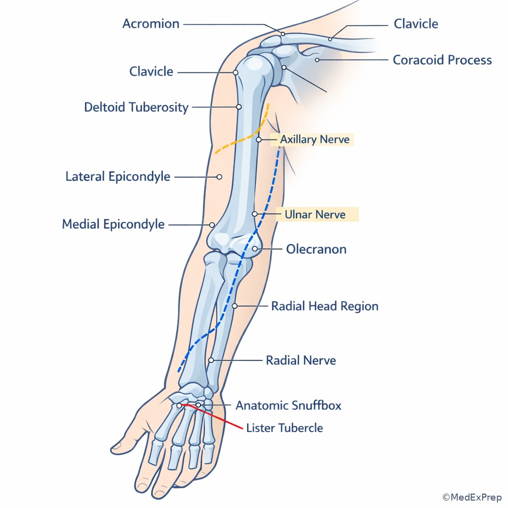
- Axillary nerve risk is classically tied to the surgical neck of the humerus and inferior glenohumeral dislocation patterns.
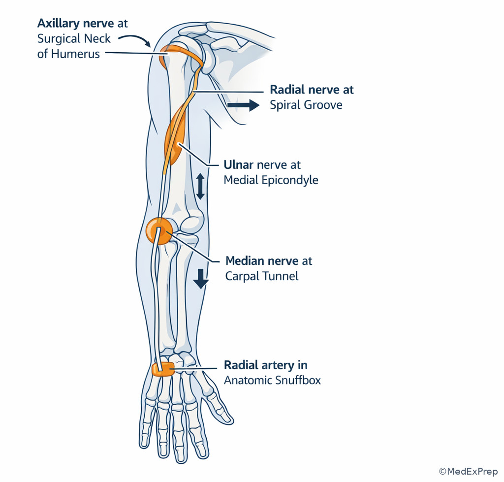
- Ulnar nerve runs posterior to the medial epicondyle; the clue is sensory change in the small finger or intrinsic weakness.
- Radial nerve is tightly linked to the humeral shaft, especially spiral groove injuries.
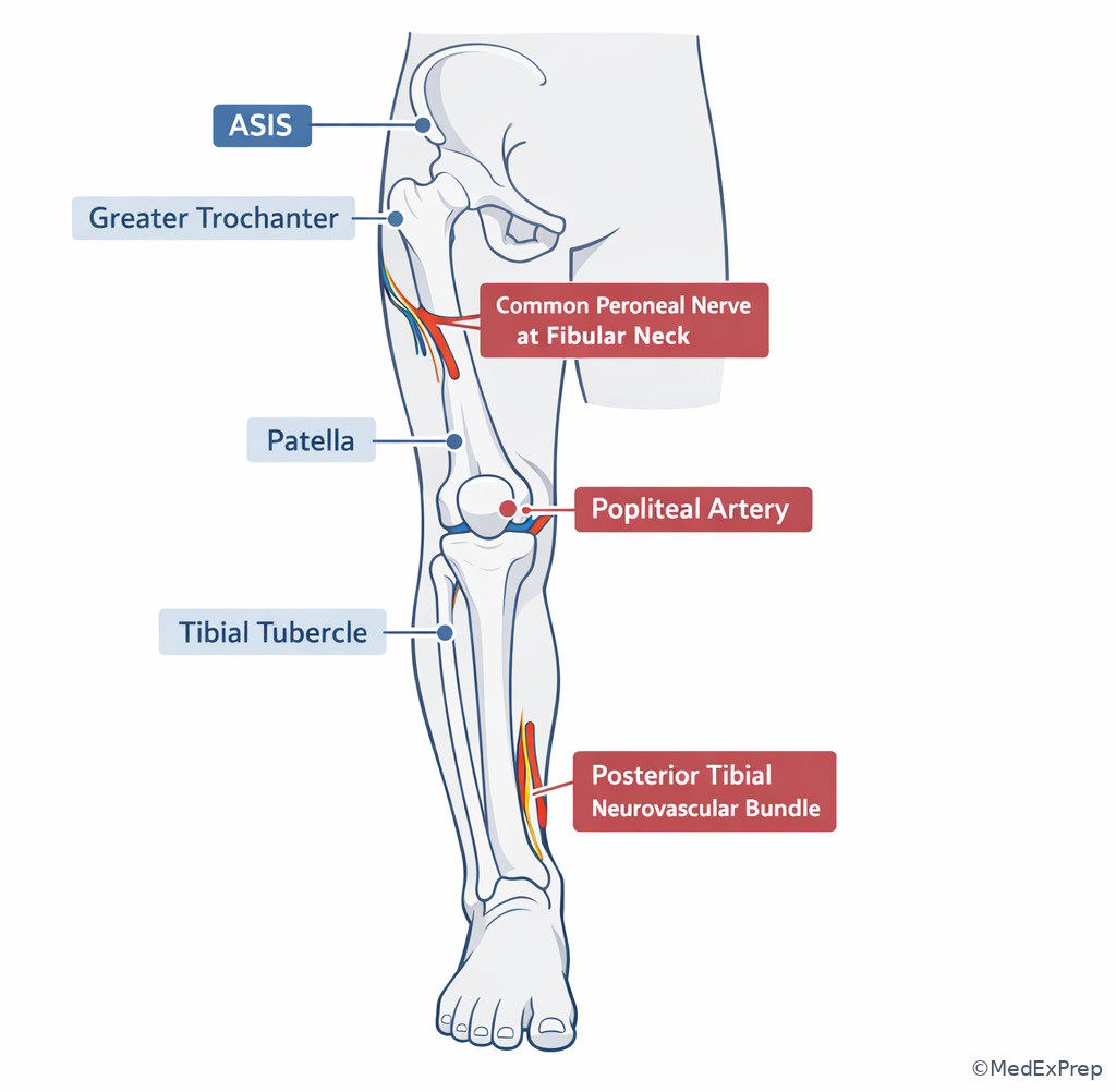
- Common peroneal nerve is vulnerable at the fibular neck; foot drop after knee trauma localizes here until proven otherwise.
- Posterior tibial artery and tibial nerve run behind the medial malleolus.
- Anatomic snuffbox tenderness after a FOOSH should make you think occult scaphoid fracture even if initial radiographs are negative.
- Pulse presence does not exclude vascular injury; landmark-based mechanism still matters.
- Hip dislocation, knee dislocation, supracondylar humerus injury, and elbow dislocation are classic landmark-linked neurovascular danger patterns.
| Landmark | What to palpate | Board use | Nearby structure at risk |
|---|---|---|---|
| Acromion | Lateral shoulder roof | Shoulder offset, dislocation contour | Axillary nerve deeper/inferior |
| Coracoid | Anterior shoulder prominence | Anterior shoulder approach, instability localization | Brachial plexus/axillary vessels medially |
| Medial epicondyle | Medial distal humerus | Elbow trauma localization | Ulnar nerve posterior |
| Lateral epicondyle | Lateral distal humerus | Extensor origin, elbow alignment | Radial tunnel nearby distal/anterior |
| Anatomic snuffbox | Dorsoradial wrist hollow | Occult scaphoid screening | Radial artery floor |
| Fibular neck | Lateral proximal leg | Knee trauma neuro exam | Common peroneal nerve |
| Medial malleolus | Medial ankle | Tarsal tunnel, ankle fracture mapping | Posterior tibial neurovascular bundle posterior |
| Greater trochanter | Lateral proximal femur | Hip pain localization | Abductor insertion |
Exam takeaway: Use landmark location to predict the single most threatened nerve or vessel before choosing management.
flowchart TD
A[Shoulder landmark] --> B[Acromion contour loss]
A --> C[Coracoid prominence]
B --> D[Think glenohumeral dislocation]
D --> E[Check axillary nerve sensation over lateral deltoid]
C --> F[Think anterior shoulder relationship]
G[Distal humerus landmark] --> H[Medial epicondyle]
G --> I[Lateral column or shaft]
H --> J[Check ulnar nerve]
I --> K[Check radial nerve pattern if shaft injury]
L[Wrist landmark] --> M[Anatomic snuffbox tenderness]
M --> N[Immobilize for possible scaphoid injury]
Exam takeaway: Recognize the palpable relationship of shoulder, elbow, and wrist landmarks to localize injury on exam stems.
Core content
1.1.1.3 Foundational upper-extremity landmarks: shoulder and humerus
A Explanation
The decision conflict in shoulder and proximal humerus questions is usually this: Is this simply pain around the shoulder, or is the landmark telling you which structure must be checked immediately? The resolving rule is simple: when the stem names the acromion, coracoid, surgical neck, or deltoid patch, you should localize to the glenohumeral/proximal humeral region and deliberately screen for axillary nerve function. If the stem moves down the humeral shaft, especially spiral groove language, reject an axillary-only answer and think radial nerve.
WHY THIS IS TESTED: exam writers package nerve localization inside fracture wording. A candidate who identifies the landmark can choose the correct neurovascular exam, reduction urgency, and counseling point. See Fig 1 and Fig 2.
The acromion forms the palpable roof of the shoulder. A squared-off shoulder contour suggests dislocation. The coracoid is the anterior bony landmark often more prominent in anterior dislocation. The surgical neck is the classic exam association for the axillary nerve and posterior circumflex humeral artery. The practical board question is not to recite anatomy, but to know what to document: sensation over the lateral deltoid, deltoid firing if possible, and distal perfusion.
The humeral shaft introduces a different nerve-risk corridor. Midshaft fractures, especially spiral or Holstein-Lewis type distal third patterns, point toward radial nerve risk. The discriminating clue is wrist/finger extension weakness rather than isolated deltoid sensory change. On ABOS-style stems, the wrong answer becomes tempting because “humerus fracture” sounds like one anatomy bucket. Do not pool proximal humerus and shaft injuries together.
B Worked example
A 67-year-old woman with osteoporosis and type 2 diabetes falls on her outstretched hand while gardening. She reports severe shoulder pain and cannot abduct the arm. Examination shows a visibly flattened lateral shoulder contour. She also has chronic cervical spondylosis and mentions intermittent hand numbness at baseline. Radiographs describe an anterior glenohumeral dislocation with an associated minimally displaced greater tuberosity fracture. Which neurologic finding is most important to document before and after reduction?
Reasoning chain: Identify task: the question is asking for the most relevant structure at risk based on landmark/anatomic zone, not the patient’s chronic neuropathy. Extract key facts: flattened shoulder contour, anterior dislocation, proximal shoulder location, distracting baseline hand numbness. Apply rule: shoulder dislocation/proximal humerus region threatens the axillary nerve. Eliminate distractors: median neuropathy would fit hand numbness but does not match the injury zone; radial nerve fits humeral shaft injuries; ulnar nerve fits medial elbow. Verify: the best focused exam is sensation over the lateral deltoid and deltoid activation. The wrong clue is the baseline hand numbness, which is noise.
C Exam trap
D Checkpoint
Question: A 29-year-old man is brought after a motorcycle crash with shoulder pain, a prominent coracoid region, and numbness over the lateral deltoid. Which structure is most likely injured?
- Axillary nerve
- Radial nerve
- Ulnar nerve
- Musculocutaneous nerve
Answer: A. Axillary nerve.
- A: Tempting because the symptoms match classic shoulder-dislocation anatomy; right because the lateral deltoid sensory patch is the discriminator.
- B: Tempting because high-energy upper-extremity trauma can injure the radial nerve; wrong because radial injury gives wrist/finger extension deficits and aligns with shaft-level injury.
- C: Tempting because sensory symptoms can suggest peripheral nerve injury broadly; wrong because ulnar symptoms localize to medial elbow or hand distribution, not the regimental badge area.
- D: Tempting because it is an upper-arm nerve; wrong because musculocutaneous injury does not explain the classic shoulder-dislocation sensory finding.
1.1.1.4 Elbow, forearm, and wrist landmarks that change management
A Explanation
The key decision conflict here is: Which elbow or wrist landmark represents a simple tenderness point, and which one demands a specific unstable injury workup? The single resolving rule is to link each landmark to a named high-yield injury. Posterior to the medial epicondyle means ulnar nerve territory. Radial head tenderness with painful forearm rotation means think radial head injury. Anatomic snuffbox tenderness means treat as occult scaphoid fracture until proven otherwise. Lister tubercle is a dorsal distal radius landmark relevant to EPL tendon mechanics and dorsal wrist anatomy.
WHY THIS IS TESTED: board stems love “normal x-ray, but focal tenderness at one landmark” questions. The answer is often immobilization and follow-up, not reassurance. See Fig 2.
At the elbow, the three-point relationship of medial epicondyle, lateral epicondyle, and olecranon changes with flexion and helps orient injury. A medial-sided injury plus paresthesias in the ring and small fingers should not be dismissed as swelling alone. At the wrist, the scaphoid is the classic trap because blood supply concerns and initial radiograph occultness make delayed diagnosis costly. The snuffbox is not just an anatomy term; it is an action cue.
A practical exam distinction: lateral wrist pain after FOOSH can suggest distal radius sprain, de Quervain-type symptoms, or occult scaphoid injury. The landmark breaks the tie. Snuffbox or scaphoid tubercle tenderness should push you toward thumb-spica immobilization and repeat imaging or advanced imaging when indicated. Reject the “negative x-ray means discharge without immobilization” option in a classic stem.
B Worked example
A 21-year-old collegiate midfielder with ADHD and mild asthma falls during a match onto an extended wrist. He has mild dorsal wrist swelling, full finger motion, and normal sensation. Standard wrist radiographs are read as negative in the emergency department. He is eager to return to play because the pain seems “just like a sprain.” On exam, he has point tenderness in the anatomic snuffbox and pain with axial thumb loading. What is the most appropriate next step?
Reasoning chain: Identify task: management after a classic occult injury pattern. Extract key facts: FOOSH, negative initial x-rays, snuffbox tenderness, distracting athlete-return pressure. Apply rule: snuffbox tenderness after FOOSH should be treated as scaphoid fracture until excluded. Eliminate distractors: immediate unrestricted return ignores occult fracture risk; simple soft dressing is insufficient; carpal tunnel or de Quervain diagnoses do not fit the mechanism and landmark. Verify: thumb-spica immobilization with repeat imaging or advanced imaging is the appropriate orthopaedic action.
C Exam trap
D Checkpoint
Question: A 35-year-old woman falls on an outstretched hand and has negative wrist radiographs but marked snuffbox tenderness. Which is the best initial orthopaedic management?
- Reassurance and activity as tolerated
- Thumb-spica immobilization and interval reassessment
- Counterforce brace for lateral epicondylitis
- Immediate carpal tunnel release referral
Answer: B. Thumb-spica immobilization and interval reassessment.
- A: Tempting because the radiographs are negative; wrong because occult scaphoid injury is specifically screened by snuffbox tenderness.
- B: Tempting because it may seem overly cautious; right because the landmark and mechanism strongly support a possible scaphoid fracture.
- C: Tempting because lateral elbow and wrist pain are both common overuse complaints; wrong because the mechanism is acute trauma and the tenderness is in the wrist.
- D: Tempting because hand symptoms can lead learners toward median nerve pathology; wrong because there is no median nerve pattern and the trauma-localized landmark dominates.
- The landmark most associated with occult scaphoid fracture is the anatomic snuffbox.
- True or False: A negative initial wrist radiograph excludes scaphoid fracture. False.
- Name the nerve posterior to the medial epicondyle: ulnar nerve.
Exam takeaway: Local wrist tenderness pattern can determine immobilization even when radiographs do not yet prove the injury.
flowchart TD
A[FOOSH wrist pain] --> B{Where is focal tenderness?}
B --> C[Snuffbox or scaphoid tubercle]
B --> D[Dorsal distal radius]
B --> E[Ulnar fovea]
C --> F[Possible scaphoid fracture]
F --> G[Thumb-spica and follow-up imaging]
D --> H[Consider distal radius injury pattern]
E --> I[Consider TFCC/ulnar-sided pathology]
G --> J[Do not dismiss because initial x-rays can be negative] Exam takeaway: Pair each palpable landmark with the highest-yield nerve at risk to avoid choosing the wrong localization on exam day.
1.1.1.5 Pelvic and hip landmarks: localization, safe zones, and hidden urgency
A Explanation
Pelvic and hip stems test a different decision conflict: Which pain location represents a local musculoskeletal structure, and which one implies deeper instability or neurovascular risk? The resolving rule is to separate ASIS/PSIS/iliac crest landmarks from groin/greater trochanter/femoral triangle landmarks. Lateral pain over the greater trochanter suggests abductor insertion or peritrochanteric pathology; groin pain suggests intra-articular hip pathology. The femoral triangle matters because puncture wounds, expanding hematoma, or fracture-dislocation patterns near this region raise concern for major vascular structures.
WHY THIS IS TESTED: board stems often ask for the most likely pain generator or structure at risk from a wound or approach. A precise landmark keeps you from confusing lumbar, pelvic, and hip sources.
The ASIS is important for pelvic orientation, graft harvest discussions, and approach planning. The PSIS helps identify posterior pelvic and sacroiliac relationships. The greater trochanter is the palpable lateral proximal femur marker and an anchor for lateral hip localization. In hip trauma, however, the single landmark that changes urgency is often not a bony prominence but the relationship of the injury to the hip joint and groin. Hip dislocation with groin deformity requires rapid reduction to reduce the risk of femoral head complications and demands a documented sciatic nerve exam, especially peroneal division function.
The exam trap is to overcall every lateral hip pain complaint as “hip joint” pathology. If the tenderness is directly over the greater trochanter and the story is atraumatic or overuse, that is different from deep groin pain with limited internal rotation. In trauma, deformity and limb position matter more: posterior dislocation classically presents with a flexed, adducted, internally rotated limb; the anatomy cue should trigger urgency, not prolonged workup.
B Worked example
A 34-year-old man with obesity and well-controlled hypertension is involved in a dashboard injury during a motor vehicle collision. His right lower limb is shortened, flexed, adducted, and internally rotated. He also has a superficial abrasion over the iliac crest and complains of low back soreness. Distal pulses are present. Which focused neurologic structure is most important to document before and after reduction?
Reasoning chain: Identify task: urgent dislocation anatomy, not the abrasion. Extract key facts: dashboard mechanism, flexed/adducted/internally rotated limb = posterior hip dislocation, distracting iliac crest abrasion, pulses present. Apply rule: posterior hip dislocation places the sciatic nerve, especially the peroneal division, at risk. Eliminate distractors: femoral nerve fits anterior hip or groin surgery patterns more than this position; obturator nerve is not the classic acute dislocation concern; pulse presence does not resolve nerve risk. Verify: dorsiflexion and sensation in the peroneal distribution should be documented.
C Exam trap
D Checkpoint
Question: Direct tenderness over the greater trochanter in an otherwise stable patient most strongly localizes to which region?
- Peritrochanteric lateral hip structures
- Sacroiliac joint only
- Central lumbar canal
- Pubic symphysis
Answer: A. Peritrochanteric lateral hip structures.
- A: Tempting because the landmark is directly lateral; right because the greater trochanter is the key lateral hip palpation point.
- B: Tempting because pelvic pain can radiate; wrong because the SI joint is posterior and not defined by focal trochanteric tenderness.
- C: Tempting because spine pathology can refer to the hip; wrong because this question is about direct landmark localization.
- D: Tempting because it is another pelvic landmark; wrong because pubic symphysis pain localizes anteriorly, not laterally.
1.1.1.6 Knee and leg landmarks: where nerves, vessels, and compartments become testable
A Explanation
The major decision conflict in the knee and proximal leg is: Which landmark implies a predictable nerve injury, and which pattern means vascular threat even if pulses are present? The rule is to split your reasoning between fibular neck and knee dislocation/popliteal region. The fibular neck is a common peroneal nerve landmark. Knee dislocation or gross instability raises popliteal artery concern, and pulse presence cannot fully reassure you.
WHY THIS IS TESTED: these are classic orthopaedic emergency stems disguised as anatomy localization. If you miss the landmark, you choose observation when the question is really testing escalation.
The tibial tubercle is another useful landmark, especially for extensor mechanism localization in adolescents and adults. The joint line helps localize meniscal or collateral tenderness. But the highest-yield exam landmark on boards is the fibular head/neck. A fracture here or high-energy posterolateral knee trauma plus foot drop should make you think common peroneal nerve injury first. The distinction from L5 radiculopathy will come later in the course; at this stage, the landmark is enough to localize.
The popliteal fossa is anatomically tethered and vulnerable in knee dislocations. Even after spontaneous reduction, the landmark pattern matters: significant swelling, ecchymosis, instability, or mechanism can imply vascular injury. The board-level move is not to become a vascular surgeon; it is to recognize the danger, document the exam, stabilize, and coordinate urgent evaluation. See Fig 4.
B Worked example
A 46-year-old man with tobacco use disorder and prior ACL reconstruction twists his knee during a fall from a loading dock. The knee appears grossly deformed at the scene but looks more aligned on arrival after splinting. He has severe pain, lateral proximal leg tenderness near the fibular neck, weakness with ankle dorsiflexion, and intact dorsalis pedis pulse. A small abrasion overlies the patella. What is the best anatomic localization for the neurologic deficit?
Reasoning chain: Identify task: localize nerve deficit by landmark. Extract key facts: fibular neck tenderness, foot dorsiflexion weakness, distracting intact pulse and patellar abrasion. Apply rule: common peroneal nerve wraps around fibular neck and is vulnerable in knee trauma. Eliminate distractors: tibial nerve would affect plantarflexion and sole sensation; femoral nerve is proximal/anterior thigh; isolated L5 radiculopathy is less supported than a direct local traumatic landmark. Verify: common peroneal nerve is the best localization.
C Exam trap
D Checkpoint
Question: A patient with a proximal fibular fracture develops foot drop. Which nerve is most likely involved?
- Tibial nerve
- Common peroneal nerve
- Femoral nerve
- Obturator nerve
Answer: B. Common peroneal nerve.
- A: Tempting because it is also a major leg nerve; wrong because tibial nerve injury produces plantarflexion and plantar sensory deficits, not classic foot drop.
- B: Tempting because the fibular neck is the classic board association; right because dorsiflexion weakness localizes here.
- C: Tempting because proximal lower-extremity trauma can involve multiple nerves; wrong because femoral nerve affects quadriceps and anterior thigh sensation.
- D: Tempting because it is a lower-extremity nerve around the pelvis; wrong because obturator nerve affects adduction, not ankle dorsiflexion.
- The nerve most classically injured at the fibular neck is the common peroneal nerve.
- True or False: A normal distal pulse excludes popliteal artery injury after knee dislocation. False.
- Name the landmark for extensor mechanism insertion below the knee: tibial tubercle.
Exam takeaway: Use fibular neck, popliteal fossa, malleolar, and hindfoot landmarks to identify the next exam step and the threatened structure.
1.1.1.7 Ankle, foot, and hindfoot landmarks: the decisive clues in trauma stems
A Explanation
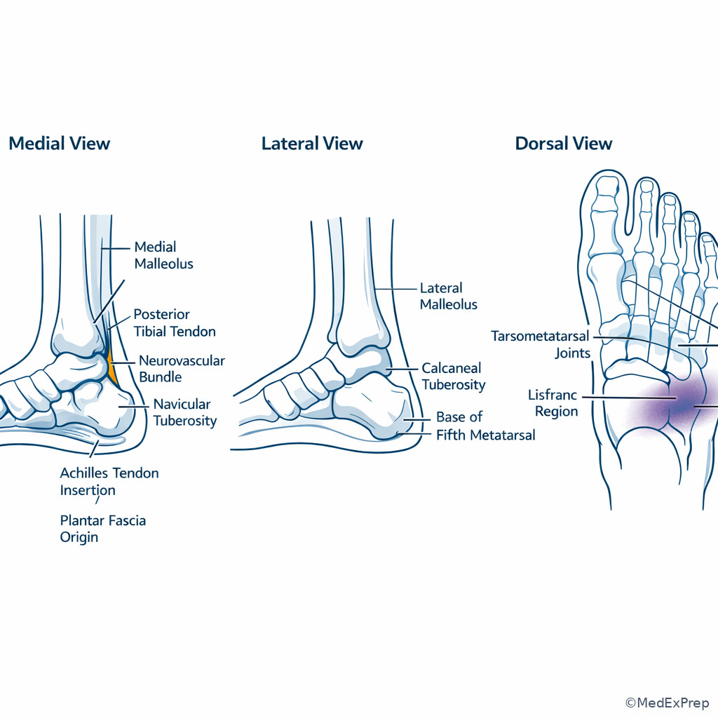
The decision conflict in foot and ankle anatomy is usually: Which exact tenderness point or wound location changes the injury category? The rule is to distinguish malleolar, base of fifth metatarsal, Lisfranc/tarsometatarsal, Achilles insertion, and posterior to the medial malleolus patterns. Each directs a different line of reasoning.
WHY THIS IS TESTED: foot and ankle stems often appear crowded with swelling and bruising; the true answer depends on one landmark, not the whole paragraph. Midfoot plantar ecchymosis and tarsometatarsal tenderness should make you think Lisfranc injury. Base-of-fifth tenderness after inversion can indicate an avulsion or Jones-region issue depending on exact location. Tenderness posterior to the medial malleolus localizes the tarsal tunnel region where the posterior tibial tendon, artery, nerve, and flexor tendons pass in sequence.
For hindfoot anatomy, the calcaneal tuberosity is the Achilles insertion region posteriorly, while the plantar fascia originates from the medial calcaneal tubercle. Board writers may give heel pain and ask for the structure involved; the landmark sorts insertional Achilles pathology from plantar fascial complaints. In trauma, however, the higher-yield concern is missed midfoot instability. Lisfranc injuries are notorious because standard ankle-style thinking can miss them. The clue is midfoot tenderness, swelling, plantar ecchymosis, or pain with forefoot stress rather than isolated malleolar pain.
The practical orthopaedic move is to reject simplistic “ankle sprain” answers when the tenderness is not centered at the lateral ligaments. See Fig 4 and Fig 5.
B Worked example
A 39-year-old warehouse worker with obesity and gout steps off a curb, twists his foot, and presents with “ankle sprain” pain. He has swelling but minimal tenderness over the lateral malleolus. The strongest tenderness is over the tarsometatarsal region with plantar ecchymosis. He can weakly dorsiflex and has normal capillary refill. An urgent care radiograph report says “no obvious ankle fracture.” What is the most important injury to suspect?
Reasoning chain: Identify task: detect a commonly missed injury based on landmark. Extract key facts: midfoot tenderness, plantar ecchymosis, distracting ankle label, irrelevant gout history, normal perfusion. Apply rule: plantar ecchymosis plus TMT tenderness suggests Lisfranc injury. Eliminate distractors: routine lateral ankle sprain would center over ATFL region; Achilles rupture causes posterior heel/weak plantarflexion; isolated malleolar fracture does not fit the main tenderness zone. Verify: the key next step is immobilization and instability-focused evaluation.
C Exam trap
D Checkpoint
Question: A patient has posterior tenderness just behind the medial malleolus. Which structure group lies in that region?
- Posterior tibial tendon and posterior tibial neurovascular bundle
- Common peroneal nerve and fibularis brevis insertion
- Achilles tendon only
- Anterior tibial artery and deep peroneal nerve
Answer: A. Posterior tibial tendon and posterior tibial neurovascular bundle.
- A: Tempting because it matches the tarsal tunnel zone; right because this is the classic posterior-medial ankle landmark relationship.
- B: Tempting because it includes lateral ankle structures; wrong because the fibular neck and lateral foot are different regions.
- C: Tempting because Achilles is posterior; wrong because it is central posterior heel, not specifically behind the medial malleolus.
- D: Tempting because those structures cross the ankle; wrong because they are anterior, not in the posteromedial retromalleolar tunnel.
Exam takeaway: The exact tenderness point determines whether to treat as ankle sprain, occult fracture, tendon injury, or midfoot instability.
flowchart TD
A[Foot or ankle trauma] --> B{Main tenderness site}
B --> C[Lateral malleolus region]
B --> D[Base of fifth metatarsal]
B --> E[Tarsometatarsal midfoot]
B --> F[Posterior heel]
E --> G[Think Lisfranc injury]
G --> H[Immobilize and assess stability]
D --> I[Differentiate avulsion versus Jones-region injury]
F --> J[Think Achilles insertion or rupture pattern]
C --> K[Assess standard ankle injury pathway]
H --> L[Do not call this a routine sprain] Exam takeaway: Midfoot, malleolar, retromalleolar, and posterior heel landmarks sort common board-tested injuries quickly.
1.1.1.8 Integration: using landmarks to choose the next orthopaedic action
A Explanation
The final board-level skill is integration: Given a landmark, what is the next action—observe, immobilize, reduce, document, or escalate? The resolving rule is to sort landmarks into four operational buckets: document a nerve exam, suspect occult fracture and immobilize, recognize urgent reduction need, or escalate for vascular/compartment threat. Landmarks are not endpoints. They are action triggers.
WHY THIS IS TESTED: ABOS-style reasoning rewards the candidate who converts anatomy into management, not the one who merely names structures. The same fact pattern can ask for diagnosis, imaging, reduction urgency, or documentation. See Fig 1, Fig 3, Fig 4, and Fig 5.
Examples: shoulder dislocation landmarks trigger axillary nerve documentation; snuffbox tenderness triggers thumb-spica immobilization; classic hip dislocation posture triggers urgent reduction and sciatic/peroneal documentation; fibular neck trauma with foot drop triggers common peroneal localization; knee dislocation patterns trigger vascular reassessment even if pulses remain palpable; plantar ecchymosis at the midfoot triggers Lisfranc evaluation and protected weight bearing.
The biggest integration error is to stop once you localize the structure. On boards, localization is only half-credit thinking. The full answer usually adds urgency and a specific next step. Another common error is being distracted by comorbidities, chronic baseline numbness, or a minor abrasion. Landmark-based board stems intentionally include noise. The correct move is to ask: Which named anatomic zone has the highest consequence if missed?
B Worked example
A 58-year-old man with coronary artery disease and chronic lumbar stenosis falls from a ladder. He has a shoulder deformity, snuffbox tenderness on the same side, and lateral knee bruising near the fibular head. He also reports chronic bilateral toe numbness from prior spine disease. Which abnormality requires the most immediate focused documentation because it most directly changes urgent orthopaedic management?
Reasoning chain: Identify task: choose the finding that most immediately alters action. Extract key facts: multiple injuries, chronic sensory noise, shoulder deformity, snuffbox tenderness, fibular head bruising. Apply rule: shoulder deformity suggesting dislocation requires pre- and post-reduction axillary nerve documentation because urgent reduction is likely. Eliminate distractors: snuffbox tenderness is important but usually enters an immobilization pathway rather than immediate reduction; fibular head bruising matters, but if no foot drop or gross knee instability is given, the shoulder dislocation drives the urgent action. Verify: focused axillary nerve and perfusion documentation around reduction is highest yield.
C Exam trap
D Checkpoint
Question: Which landmark pattern most strongly requires an occult-fracture pathway rather than immediate reduction or vascular escalation?
- Snuffbox tenderness after FOOSH with negative initial radiographs
- Flexed, adducted, internally rotated hip after dashboard injury
- Grossly unstable knee after dislocation mechanism
- Shoulder flattening with loss of contour after trauma
Answer: A. Snuffbox tenderness after FOOSH with negative initial radiographs.
- A: Tempting because it may appear less dramatic than the other choices; right because it maps to suspected occult scaphoid fracture and immobilization/follow-up.
- B: Tempting because hip trauma can include associated fractures; wrong because the landmark-position pattern points first to urgent reduction.
- C: Tempting because fractures can accompany knee trauma; wrong because the dominant issue is vascular threat in a dislocation pattern.
- D: Tempting because proximal humerus fractures can coexist; wrong because the contour change chiefly signals shoulder dislocation and reduction planning.
Exam Traps & Differentiators
- “Humerus fracture = radial nerve” always — tempting because the association is famous; eliminate it when the stem specifically localizes to the surgical neck or shoulder dislocation, which points to axillary nerve.
- Negative wrist radiographs exclude scaphoid injury — tempting because imaging feels definitive; eliminate it with snuffbox tenderness after FOOSH.
- Normal pulses rule out limb threat after knee trauma — tempting because pulse checks are memorable; eliminate it when the mechanism suggests knee dislocation or the exam shows major instability.
- Any twisting ankle injury is a sprain — tempting because sprains are common; eliminate it with plantar ecchymosis and TMT tenderness suggesting Lisfranc injury.
- Lateral hip pain means hip joint pathology — tempting because patients say “my hip hurts”; eliminate it when tenderness is directly over the greater trochanter rather than deep groin pain.
| Looks similar but isn't | Tempting overlap | Single discriminator | Best orthopaedic implication |
|---|---|---|---|
| Shoulder dislocation vs humeral shaft fracture nerve injury | Both can follow high-energy arm trauma | Lateral deltoid sensory loss vs wrist drop | Check axillary vs radial nerve specifically |
| Scaphoid injury vs wrist sprain | Both follow FOOSH and may have subtle x-rays | Snuffbox/scaphoid tubercle tenderness | Thumb-spica rather than simple discharge |
| Lateral hip pain vs intra-articular hip pathology | Both described as “hip pain” by patients | Greater trochanter tenderness vs deep groin pain | Localize before ordering and counseling |
| Fibular neck nerve injury vs lumbar radicular complaint | Both can cause dorsiflexion weakness | Direct local trauma at fibular neck | Local traumatic nerve pattern takes priority |
| Lisfranc injury vs ankle sprain | Both can follow twist and swelling | Plantar ecchymosis/midfoot tenderness | Protect midfoot and evaluate stability |
| If the stem says... | Think... |
|---|---|
| “Regimental badge numbness” | Axillary nerve |
| “Posterior to medial epicondyle” | Ulnar nerve |
| “Snuffbox tenderness with negative x-ray” | Occult scaphoid fracture pathway |
| “Flexed, adducted, internally rotated hip” | Posterior hip dislocation |
| “Fibular neck pain with foot drop” | Common peroneal nerve injury |
| “Plantar ecchymosis in twisted foot” | Lisfranc injury |
Related upcoming traps matter for continuity. In Peripheral Nerve Injury Patterns, the exam will no longer be satisfied with naming the danger zone; you will need the exact motor and sensory deficit. In Compartment And Vascular Threats, the landmark becomes the opening clue, but the answer turns on urgency recognition. Use this lesson to build the first step: accurate localization before higher-order decision making.
Algorithm / Approach
Exam takeaway: Convert a named landmark into the correct action bucket before you get distracted by noise in the stem.
flowchart TD
A[Read the stem] --> B[Find the named landmark or tenderness point]
B --> C{What does the landmark imply?}
C --> D[Nerve-risk zone]
C --> E[Occult fracture zone]
C --> F[Urgent reduction pattern]
C --> G[Vascular-threat pattern]
D --> H[Document focused motor and sensory exam]
E --> I[Immobilize and arrange follow-up imaging]
F --> J[Reduce promptly and recheck neurovascular status]
G --> K[Escalate urgently and perform serial vascular checks]
H --> L[Do not mix landmarks from adjacent regions]
I --> L
J --> L
K --> L Rapid Review
- Acromion contour loss → think shoulder dislocation, then check axillary nerve
- Coracoid prominence after trauma → anterior shoulder relationship clue
- Surgical neck of humerus → axillary nerve risk, not radial by default
- Humeral shaft spiral groove region → radial nerve pattern with wrist/finger extension deficit
- Posterior to medial epicondyle → ulnar nerve localization
- Snuffbox tenderness → occult scaphoid pathway despite negative initial films
- Greater trochanter tenderness → lateral hip/peritrochanteric localization
- Groin pain with hip motion → intra-articular hip source over lateral bursal source
- Flexed adducted internally rotated limb → posterior hip dislocation until proven otherwise
- Fibular neck trauma → common peroneal nerve risk with foot drop
- Knee dislocation mechanism → vascular concern persists even with pulses
- Posterior to medial malleolus → tarsal tunnel region and posterior tibial bundle
- Plantar ecchymosis plus TMT tenderness → Lisfranc until excluded
- Posterior heel insertion pain → Achilles region, not midfoot or malleolar injury
Self-check quiz
1. A 24-year-old man falls during basketball and has a flattened shoulder contour with numbness over the lateral deltoid. Which structure is most likely involved?
- Axillary nerve
- Radial nerve
- Ulnar nerve
- Median nerve
2. A 19-year-old woman falls from a skateboard onto an outstretched hand. Wrist radiographs are negative, but she has focal tenderness in the anatomic snuffbox. What is the most appropriate initial management?
- Soft wrap and return to activity as tolerated
- Thumb-spica immobilization with repeat assessment
- Corticosteroid injection for de Quervain tenosynovitis
- Immediate wrist arthroscopy
3. A 41-year-old man with obesity and prediabetes is evaluated after a dashboard injury. His right leg is shortened, flexed, adducted, and internally rotated. He also has a minor lateral thigh abrasion and normal pedal pulses. Which neurologic exam finding most requires immediate documentation before reduction?
- Ankle dorsiflexion strength and dorsal foot sensation
- Quadriceps strength and anterior thigh sensation
- Hip abduction strength only
- Great toe plantar callus sensation only
4. A 52-year-old woman with rheumatoid arthritis and chronic cervical spondylosis falls at home and sustains a midshaft humeral fracture. She reports long-standing intermittent fingertip paresthesias. Which focused motor exam is most appropriate to prioritize based on the injury landmark?
- Deltoid contraction during shoulder abduction
- Wrist and finger extension
- Finger abduction only
- Thumb opposition only
5. A 37-year-old warehouse employee twists his foot stepping off a pallet. He has swelling, plantar ecchymosis, and maximal tenderness over the tarsometatarsal region; urgent care ankle radiographs are reported as negative. What is the most appropriate orthopaedic next step?
- Treat as a routine lateral ankle sprain with immediate weight bearing
- Immobilize and evaluate for Lisfranc instability
- Begin Achilles tendon rupture protocol
- Provide shoe inserts for plantar fasciitis
6. A 47-year-old man with prior lumbar radiculopathy sustains a knee injury in a motorcycle crash. The knee was grossly deformed at the scene but appears reduced on arrival. He has tenderness at the fibular neck, weak ankle dorsiflexion, intact pulses, and mild chronic toe numbness bilaterally. Which diagnosis best explains the new focal deficit?
- Common peroneal nerve injury at the fibular neck
- Tibial nerve injury in the popliteal fossa
- Isolated recurrence of L5 radiculopathy
- Femoral neuropathy from pelvic trauma
7. A 63-year-old woman with osteoporosis and hypothyroidism falls from standing and has shoulder pain, visible loss of shoulder contour, and mild tenderness over the distal humerus from a previous bruise. Which finding most strongly favors axillary nerve risk rather than radial nerve risk?
- Numbness over the lateral deltoid patch
- Inability to extend the wrist
- Numbness in the small finger
- Loss of thumb opposition
8. A 28-year-old soccer player presents after a twist injury with ankle swelling, mild tenderness at the anterior talofibular region, and marked pain at the base of the fifth metatarsal. He wants to return for the second half. Which detail most strongly requires you to reject a simple isolated ankle sprain answer?
- Base-of-fifth metatarsal focal tenderness
- Immediate swelling after inversion
- Ability to plantarflex the ankle
- Mild lateral malleolar tenderness
Answer key
1.1.1.9 1. Question 1 answer: A. Axillary nerve
Correct-answer logic: Shoulder contour loss after trauma localizes to glenohumeral dislocation, and lateral deltoid sensory loss is the classic axillary nerve clue. Mechanistically, the axillary nerve courses around the surgical neck/inferior shoulder region, making it vulnerable in dislocation and proximal humerus injury patterns. This is why pre- and post-reduction documentation matters. Sources: Rockwood and Green's Fractures in Adults; Campbell's Operative Orthopaedics.
- A. Axillary nerve: Tempting because the landmark and sensory zone fit perfectly; correct because the single discriminator is numbness over the lateral deltoid patch after shoulder deformity.
- B. Radial nerve: Tempting because upper-arm trauma often brings radial nerve to mind; incorrect because the stem lacks humeral shaft localization and wrist/finger extension weakness.
- C. Ulnar nerve: Tempting because trauma can cause paresthesias; incorrect because the small-finger/medial hand pattern is absent and the injury zone is wrong.
- D. Median nerve: Tempting because hand numbness is common in trauma differentials; incorrect because there is no carpal tunnel/thenar pattern and the acute landmark is the shoulder.
1.1.1.10 2. Question 2 answer: B. Thumb-spica immobilization with repeat assessment
Correct-answer logic: Snuffbox tenderness after FOOSH is an occult scaphoid-fracture pattern until excluded. Initial radiographs may be negative; the orthopaedic action is protective immobilization with interval reassessment or advanced imaging when appropriate. Missing this injury risks nonunion and avascular complications. Sources: AAOS educational standards; Green's Operative Hand Surgery; Campbell's Operative Orthopaedics.
- A. Soft wrap and return to activity: Tempting because the x-rays are negative; incorrect because the discriminator is focal snuffbox tenderness, which overrides false reassurance from initial films.
- B. Thumb-spica immobilization with repeat assessment: Tempting because it is the classic cautious pathway; correct because this is the standard response to suspected occult scaphoid injury.
- C. Corticosteroid injection for de Quervain tenosynovitis: Tempting because both can cause radial wrist pain; incorrect because the mechanism is acute trauma and the landmark points to the scaphoid, not the first dorsal compartment.
- D. Immediate wrist arthroscopy: Tempting because occult carpal pathology exists; incorrect because this is too invasive and not the first orthopaedic step for a classic suspected scaphoid fracture.
1.1.1.11 3. Question 3 answer: A. Ankle dorsiflexion strength and dorsal foot sensation
Correct-answer logic: The limb position indicates posterior hip dislocation, which threatens the sciatic nerve, especially its peroneal division. The most discriminating bedside documentation is dorsiflexion strength and dorsal foot sensation. The mechanism-to-treatment link is urgent reduction plus focused pre- and post-reduction neuro exam. Sources: Rockwood and Green's Fractures in Adults; AAOS trauma principles.
- A. Ankle dorsiflexion strength and dorsal foot sensation: Tempting because it may look distal for a hip problem; correct because the peroneal division of the sciatic nerve is the classic neurologic concern in posterior hip dislocation.
- B. Quadriceps strength and anterior thigh sensation: Tempting because femoral nerve injuries can occur around the hip; incorrect because the stem’s limb position favors posterior, not anterior, dislocation.
- C. Hip abduction strength only: Tempting because abductor function relates to hip anatomy; incorrect because it does not capture the highest-yield threatened nerve in this injury pattern.
- D. Great toe plantar callus sensation only: Tempting because it samples distal sensation; incorrect because it is too narrow and does not target the expected sciatic/peroneal deficit.
1.1.1.12 4. Question 4 answer: B. Wrist and finger extension
Correct-answer logic: Midshaft humeral fractures localize to the radial nerve danger corridor. The most useful focused motor exam is wrist and finger extension. Chronic fingertip paresthesias are distracting noise; the landmark controls the priority exam. Sources: Campbell's Operative Orthopaedics; Rockwood and Green's Fractures in Adults.
- A. Deltoid contraction during shoulder abduction: Tempting because proximal humeral injuries can injure the axillary nerve; incorrect because the stem specifically says midshaft humeral fracture.
- B. Wrist and finger extension: Tempting because it aligns with the famous nerve-fracture association; correct because radial nerve function is the best discriminator here.
- C. Finger abduction only: Tempting because hand weakness may suggest nerve injury; incorrect because this tests ulnar nerve, not the shaft-level association.
- D. Thumb opposition only: Tempting because it tests a named motor function; incorrect because it localizes to the median nerve, which is not the main risk from a midshaft humeral fracture.
1.1.1.13 5. Question 5 answer: B. Immobilize and evaluate for Lisfranc instability
Correct-answer logic: Tarsometatarsal tenderness with plantar ecchymosis is a classic Lisfranc clue. The mechanism-to-treatment logic is that unstable midfoot injuries are easily missed if labeled as ankle sprains, so the next orthopaedic step is protection and instability-focused imaging/evaluation. Sources: Campbell's Operative Orthopaedics; Mann's Surgery of the Foot and Ankle.
- A. Treat as routine lateral ankle sprain: Tempting because twist injuries are commonly sprains; incorrect because the tenderness is centered in the midfoot and plantar ecchymosis is the key discriminator.
- B. Immobilize and evaluate for Lisfranc instability: Tempting because it may feel aggressive for a “negative ankle x-ray”; correct because the stem is designed to test a missed midfoot injury, not an ankle sprain.
- C. Begin Achilles tendon rupture protocol: Tempting because ankle/hindfoot injuries can alter gait; incorrect because there is no posterior heel pain or plantarflexion deficit.
- D. Provide shoe inserts for plantar fasciitis: Tempting because foot pain often leads to nontraumatic plantar diagnoses; incorrect because the mechanism and location are acute traumatic midfoot findings.
1.1.1.14 6. Question 6 answer: A. Common peroneal nerve injury at the fibular neck
Correct-answer logic: The new focal deficit follows direct trauma at the fibular neck and presents with weak ankle dorsiflexion. Although the patient has prior lumbar symptoms, the anatomic landmark and new trauma-localized finding outweigh the chronic history. This is common peroneal nerve injury until proven otherwise. Sources: Campbell's Operative Orthopaedics; Hoppenfeld, Physical Examination of the Spine and Extremities.
- A. Common peroneal nerve injury at the fibular neck: Tempting because it matches the classic landmark; correct because the single discriminator is focal fibular neck tenderness with new foot-drop pattern.
- B. Tibial nerve injury in the popliteal fossa: Tempting because knee trauma can affect multiple structures; incorrect because tibial injury would emphasize plantarflexion and plantar sensory loss, not dorsiflexion weakness.
- C. Isolated recurrence of L5 radiculopathy: Tempting because he has prior lumbar disease and toe numbness; incorrect because the acute local traumatic landmark better explains the new deficit.
- D. Femoral neuropathy from pelvic trauma: Tempting because polytrauma can produce proximal nerve injury; incorrect because there is no pelvic-localized clue and the motor pattern is wrong.
1.1.1.15 7. Question 7 answer: A. Numbness over the lateral deltoid patch
Correct-answer logic: The stem is separating proximal shoulder anatomy from distal arm distractors. Visible loss of shoulder contour points to glenohumeral dislocation; the sensory patch over the lateral deltoid is the classic discriminator for axillary nerve injury. Sources: Rockwood and Green's Fractures in Adults; Campbell's Operative Orthopaedics.
- A. Numbness over the lateral deltoid patch: Tempting because it is the classic board phrase; correct because this is the defining sensory territory associated with axillary injury.
- B. Inability to extend the wrist: Tempting because radial nerve injury is common after humerus trauma; incorrect because the landmark pattern is shoulder dislocation, not shaft fracture.
- C. Numbness in the small finger: Tempting because trauma can cause neuropathy broadly; incorrect because that localizes to the ulnar nerve, usually around the elbow/hand distribution.
- D. Loss of thumb opposition: Tempting because it is a discrete motor sign; incorrect because it indicates median nerve dysfunction, not the expected shoulder-dislocation nerve.
1.1.1.16 8. Question 8 answer: A. Base-of-fifth metatarsal focal tenderness
Correct-answer logic: The question asks which detail should stop you from accepting a simple isolated ankle sprain. Focal tenderness at the base of the fifth metatarsal identifies a different injury zone that may represent a fracture pattern requiring a separate management pathway. The stem includes true ankle-sprain features as distractors, but the precise landmark is the discriminator. Sources: Mann's Surgery of the Foot and Ankle; Campbell's Operative Orthopaedics.
- A. Base-of-fifth metatarsal focal tenderness: Tempting because it may seem like minor adjacent pain; correct because this landmark moves the diagnosis away from isolated ligament sprain toward a possible fifth metatarsal injury.
- B. Immediate swelling after inversion: Tempting because it sounds dramatic; incorrect because it is common in routine ankle sprains and does not discriminate.
- C. Ability to plantarflex the ankle: Tempting because preserved motion can reassure; incorrect because it neither excludes ankle sprain nor addresses the metatarsal landmark.
- D. Mild lateral malleolar tenderness: Tempting because it is a classic ankle sprain location; incorrect because this actually supports, rather than rejects, a sprain pattern.
Keep going with the full ABOS Orthopaedic Surgery Prep Course (Ortho)
You just studied the way members do, in the same lesson interface. Full access unlocks every lesson, the question bank and flashcards for a full year.
One payment · 1 year of access · No auto-renewal