1.1.1 - Gram Stain And Morphology
Why it matters
- A Gram stain is often the earliest microbiology result that changes management in sepsis, meningitis, pneumonia, device infection, and abscesses.
- Board stems frequently give only partial data: “gram-positive cocci in clusters” or “curved gram-negative rods.” Your job is to choose the most appropriate next step before speciation returns.
- Misreading morphology causes predictable errors: overcalling contaminants, missing anaerobes, or choosing coverage that ignores the most likely cell wall class.
- Gram stain reasoning is not memorization alone; it is a triage tool that integrates specimen quality, host setting, and syndrome likelihood.
Key Terms & Must-Know Facts
1.1.1.1 Key terms
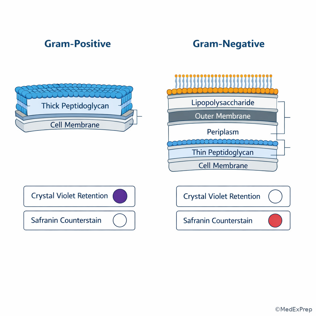
- Gram-positive: thick peptidoglycan retains crystal violet.
- Gram-negative: thin peptidoglycan with outer membrane, appears pink/red.
- Coccus: spherical bacterium.
- Bacillus/rod: cylindrical bacterium.
- Coccobacillus: short plump rod, intermediate appearance.
- Diplococci: paired cocci.
- Chains: linear streptococcal arrangement.
- Clusters: grape-like staphylococcal arrangement.
- Palisading rods: angular “Chinese letter” arrangement classically seen with Corynebacterium.
- Curved rods: comma-shaped morphology seen with organisms such as Vibrio or Campylobacter.
- Fusiform rods: spindle-shaped rods, often anaerobic.
- Pleomorphic: variable shape and size.
- Intracellular organisms: organisms visible within neutrophils or host cells on smear.
- Decolorization artifact: technical issue causing false gram-variable appearance.
- Polymicrobial smear: multiple morphologies suggesting mixed infection, contamination, or poor-quality specimen depending on source.
1.1.1.2 Must-know facts
- Gram stain narrows the field; it rarely gives definitive species identification by itself.
- Arrangement matters as much as color: clusters, chains, pairs, and branching patterns are exam-defining clues.
- Good respiratory specimens have many neutrophils and few squamous epithelial cells; poor specimens can mislead.
- Gram-positive cocci in clusters from blood culture should immediately raise concern for Staphylococcus aureus until proven otherwise.
- Gram-positive cocci in chains or pairs suggest streptococci or enterococci, but the clinical syndrome decides which is more likely.
- Gram-negative diplococci in CSF or urethral exudate strongly narrow the differential.
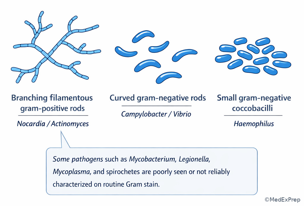
- Branching filamentous gram-positive rods raise Nocardia or Actinomyces; oxygen status and acid-fast staining help separate them.
- Not all important bacteria stain well with Gram stain: think mycobacteria, spirochetes, mycoplasma, chlamydia, and legionella.
- Prior antibiotics can reduce yield or distort morphology without excluding true infection.
- Polymicrobial Gram stain is common in abscesses and intra-abdominal infection but less typical in meningitis or hematogenous bacteremia.
Core content
1.1.1.3 Foundational Gram stain logic: what the stain is really telling you
A Explanation
The central decision conflict is whether the stain result should immediately narrow therapy around a gram-positive or gram-negative cell envelope pattern, or whether the result is too nonspecific to drive a major treatment change. The single rule that resolves this is: use Gram stain as an early directional tool, not as final organism proof. Thick peptidoglycan retains crystal violet, so gram-positive organisms appear purple. Gram-negative organisms lose crystal violet during decolorization and take up the counterstain, appearing pink. This matters because the cell envelope predicts not only appearance but also broad treatment logic, toxin/endotoxin associations, and which resistance mechanisms are likely to matter in the next topic.
WHY THIS IS TESTED: board items reward candidates who can act on incomplete microbiology data without overcommitting. A stem may ask for the best empiric adjustment after a report of “gram-negative rods in blood cultures” or “gram-positive diplococci in CSF.”
Choose morphology-guided action when the specimen is from a normally sterile site, the stain matches the syndrome, and the arrangement is classic. Reject over-interpretation when the specimen is poor quality, mixed, or from a site prone to colonization. See Fig 1 for the basic decision map and Fig 2 for how arrangement adds discriminatory value.
Exam takeaway: Start with specimen reliability, then use Gram reaction plus shape plus arrangement to narrow the board-style differential.
flowchart TD
A[Clinical specimen] --> B{Sterile site or colonized site?}
B -->|Sterile site| C[Give Gram stain high decision value]
B -->|Colonized site| D[Check specimen quality before acting]
C --> E{Purple or pink?}
D --> E
E -->|Purple| F[Gram-positive]
E -->|Pink| G[Gram-negative]
F --> H{Cocci or rods?}
G --> I{Cocci or rods?}
H -->|Cocci| J[Clusters vs chains/pairs]
H -->|Rods| K[Spore-forming? branching? pleomorphic?]
I -->|Cocci| L[Diplococci strongly narrow differential]
I -->|Rods| M[Enteric vs nonfermenter vs curved rod patterns]
J --> N[Adjust empiric therapy and verify with culture]
K --> N
L --> N
M --> N Exam takeaway: The thick peptidoglycan of gram-positive bacteria retains crystal violet, whereas the outer membrane and thin peptidoglycan of gram-negative bacteria lead to pink counterstaining.
B Worked example
A 68-year-old man with diabetes mellitus, stage 3 chronic kidney disease, and a tunneled hemodialysis catheter is admitted with fever and rigors. He has mild bibasilar crackles on exam and a chest radiograph with chronic interstitial changes that tempt the team toward pneumonia. Two blood culture bottles drawn from separate sites flag positive at 11 hours and show gram-positive cocci in clusters. The task is to decide the most appropriate empiric interpretation.
Identify task: Is this likely contamination, pneumonia-associated bacteremia, or true catheter-associated bloodstream infection due to a high-risk organism category? Extract key facts: multiple positive blood cultures, rapid time to positivity, sterile-site specimen, and clustered gram-positive cocci. Apply rule: gram-positive cocci in clusters from blood are S. aureus until proven otherwise, especially with intravascular hardware. Eliminate distractors: coagulase-negative staphylococcal contamination is tempting because of the catheter and skin flora, but multiple early positives with sepsis physiology argue against dismissal. Streptococci would more often appear in chains or pairs. Verify: the safest next step is anti-staphylococcal bacteremia management while confirming source and awaiting speciation.
C Exam trap
D Checkpoint
Question: A 59-year-old woman with cirrhosis and a new femoral central line develops fever and hypotension. Blood cultures show gram-positive cocci in clusters. Which interpretation is most appropriate?
- A. Most likely streptococcal bacteremia because cocci in chains can fragment and mimic clusters.
Tempting because both are gram-positive cocci; wrong because the arrangement clue specifically favors staphylococci, not streptococci. - B. Most likely staphylococcal bacteremia and should be managed as clinically significant pending speciation.
Tempting because it acts before full data are back; right because clusters in blood from an unstable patient are high-yield for staphylococci, especially S. aureus. - C. Most likely enterococcal bacteremia because enterococci commonly arise from healthcare exposure.
Tempting because of hospitalization and line placement; wrong because enterococci more often appear in pairs and short chains, not clusters. - D. Most likely contaminant because skin flora commonly enter the bottle during phlebotomy.
Tempting because skin organisms can contaminate cultures; wrong because the patient is unstable and the morphology is a dangerous pattern that should not be dismissed.
1.1.1.4 Morphology and arrangement: the fastest way to narrow the organism class
A Explanation
The decision conflict here is whether morphology truly distinguishes likely pathogen groups or whether you are forcing memorized associations onto ambiguous data. The resolving rule is: shape plus arrangement plus clinical source is more powerful than color alone. Gram-positive cocci in clusters point toward staphylococci. Gram-positive cocci in chains or pairs point toward streptococci or enterococci. Gram-negative diplococci suggest Neisseria in the right setting. Gram-negative rods are broad but can still be narrowed by context: enteric, nonfermenting, curved, or coccobacillary patterns. Gram-positive rods force you to ask spore-forming, branching, club-shaped, or contaminant-like. See Fig 2.
WHY THIS IS TESTED: the exam often gives a morphology clue before asking for the syndrome-level next step. If you misread the arrangement, you start the entire reasoning chain in the wrong lane.
Choose the narrowest morphology-supported bucket first, then widen only if the syndrome requires it. Reject species-level certainty unless the pattern is classic and the source is highly informative. Example: “gram-negative diplococci in CSF” is much tighter than “gram-negative rods in sputum.”
Exam takeaway: Arrangement is the key discriminator that converts a broad Gram category into the most likely organism family.
flowchart LR
A[Gram-positive cocci] --> B[Clusters]
A --> C[Chains or pairs]
B --> D[Staphylococci]
C --> E[Streptococci]
C --> F[Enterococci]
G[Gram-negative cocci] --> H[Diplococci]
H --> I[Neisseria species]
J[Gram-positive rods] --> K[Spore-forming]
J --> L[Branching filamentous]
J --> M[Palisading pleomorphic]
K --> N[Bacillus or Clostridium context]
L --> O[Nocardia or Actinomyces]
M --> P[Corynebacterium species]
Q[Gram-negative rods] --> R[Enteric rods]
Q --> S[Nonfermenters]
Q --> T[Curved rods]
Q --> U[Coccobacilli]
Exam takeaway: Recognizing clusters, chains, diplococci, rods, coccobacilli, and branching forms allows faster syndrome-based narrowing before culture finalization.

B Worked example
A 33-year-old man with HIV well controlled on antiretroviral therapy, hypertension, and recent urethral discharge presents with dysuria and purulent urethral exudate. He took one leftover dose of doxycycline at home, and the intern is tempted to attribute the partial treatment to a “gram-variable” smear. Gram stain of urethral discharge shows intracellular gram-negative diplococci within neutrophils. The question is the most likely organism class and best immediate treatment direction.
Identify task: classify the pathogen from morphology in a syndrome-specific specimen. Extract key facts: urethritis, purulent exudate, intracellular gram-negative diplococci, neutrophilic inflammation. Apply rule: in symptomatic urethritis, intracellular gram-negative diplococci strongly support Neisseria gonorrhoeae. Eliminate distractors: enteric gram-negative rods can cause UTI but would not appear as diplococci within neutrophils in urethral exudate. Moraxella is a gram-negative diplococcus but does not fit this syndrome. Verify: treat gonorrhea per guideline-concordant therapy and assess for coinfections.
C Exam trap
D Checkpoint
Question: A 71-year-old man with a prosthetic aortic valve, benign prostatic hyperplasia, and recent urinary instrumentation has bacteremia. Gram stain shows gram-positive cocci in chains. Which organism group is most strongly suggested?
- A. Staphylococci
Tempting because prosthetic material raises concern for staphylococcal infection; wrong because clusters, not chains, are the usual morphology. - B. Streptococci or enterococci
Tempting because the wording is broad; right because chains support this bucket, and recent GU instrumentation raises enterococcal probability. - C. Neisseria species
Tempting because some cocci appear in pairs; wrong because these are gram-negative diplococci, not gram-positive chains. - D. Corynebacterium species
Tempting because prosthetic valves can seed unusual organisms; wrong because Corynebacterium are pleomorphic gram-positive rods, not cocci in chains.
- The classic arrangement for staphylococci is clusters.
- True or False: Gram-negative diplococci in the right syndrome can strongly narrow the differential. True.
- Name the 2 major groups suggested by gram-positive cocci in chains or pairs: streptococci and enterococci.
1.1.1.5 Gram stain interpretation in real specimens: quality, contamination, and clinical context
A Explanation
The decision conflict is whether the organisms seen on Gram stain represent the true pathogen, background flora, or technical noise. The single resolving rule is: the value of the Gram stain depends on specimen quality and whether the site is normally sterile. A blood culture smear is high-stakes. A poor sputum Gram stain full of squamous epithelial cells is low-value. Polymicrobial morphology in a drained abscess may be meaningful, but polymicrobial sputum often reflects colonization. See Fig 3 for a quality-centered respiratory specimen algorithm.
WHY THIS IS TESTED: ABIM-style items frequently ask whether to trust a microbiology result. Infectious diseases practice is full of distinguishing contamination from infection and specimen quality from false reassurance.
For respiratory specimens, many neutrophils suggest inflammation; many squamous epithelial cells suggest oral contamination. For wound cultures, superficial swabs are weaker than deep aspirates or operative specimens. For blood cultures, the pattern across bottles and sets matters more than any single bottle. Prior antibiotics may make organisms sparse or distorted, but that does not convert a strong clinical syndrome into “no infection.”
Exam takeaway: In sputum, specimen quality determines whether morphology should influence diagnosis or empiric therapy.
flowchart TD
A[Sputum Gram stain] --> B{Many squamous epithelial cells?}
B -->|Yes| C[Poor specimen quality]
B -->|No| D[Proceed]
D --> E{Many neutrophils?}
E -->|Yes| F[Inflammatory specimen]
E -->|No| G[Lower confidence]
F --> H{Predominant single morphology?}
H -->|Yes| I[Use morphology to support likely pathogen class]
H -->|No| J[Consider mixed flora or aspiration pattern]
C --> K[Repeat or obtain better sample]
G --> K
J --> K Exam takeaway: Neutrophil-rich sputum with few squamous epithelial cells is more trustworthy than a saliva-contaminated sample.

B Worked example
A 74-year-old woman with COPD, heart failure, and chronic prednisone use is admitted with fever, productive cough, and a right lower lobe infiltrate. She also has long-standing poor dentition and a proton pump inhibitor, which tempts the team toward aspiration alone. Sputum Gram stain shows many squamous epithelial cells, few neutrophils, and mixed gram-positive cocci with gram-negative rods. The question is whether this result should steer pathogen-specific therapy.
Identify task: determine the reliability of a respiratory Gram stain. Extract key facts: colonized site, many squamous cells, low inflammatory signal, mixed morphologies. Apply rule: poor-quality sputum should not drive narrow diagnostic claims or de-escalation. Eliminate distractors: aspiration is tempting because of dentition and lower lobe disease, but the Gram stain does not confirm oral anaerobe-dominant infection; it is simply low quality. Verify: the best next step is to interpret the pneumonia clinically, repeat or improve sampling if needed, and avoid over-reading the smear.
C Exam trap
D Checkpoint
Question: Which sputum Gram stain finding most strongly supports that the specimen is interpretable?
- A. Numerous squamous epithelial cells and mixed bacterial morphologies
Tempting because there are many visible organisms; wrong because epithelial cells indicate oropharyngeal contamination. - B. Few squamous epithelial cells, many neutrophils, and a predominant organism morphology
Tempting because it sounds idealized; right because this is the classic interpretable pattern. - C. No visible bacteria and no inflammatory cells
Tempting because prior antibiotics can suppress organisms; wrong because it does not support a useful microbiologic interpretation. - D. Mixed gram-positive cocci and gram-negative rods in a saliva-rich sample
Tempting because aspiration may be polymicrobial; wrong because the sample quality prevents confident inference.
1.1.1.6 High-yield special morphologies: branching rods, coccobacilli, curved rods, and organisms that evade Gram stain
A Explanation
The decision conflict is whether an unusual morphology should trigger a specific narrow differential or whether the organism may not be reliably visible on standard Gram stain at all. The resolving rule is: when the morphology is atypical, ask what special structural clue or alternative stain explains it. Branching filamentous gram-positive rods raise Nocardia or Actinomyces; partial acid-fastness and aerobic pulmonary/CNS disease favor Nocardia, while cervicofacial, thoracic, or pelvic disease with sulfur granules and anaerobic tissue invasion favor Actinomyces. Coccobacilli suggest organisms like Haemophilus, Acinetobacter, or zoonotic pathogens depending on syndrome. Curved gram-negative rods point toward Campylobacter or Vibrio depending on exposure and syndrome.
Equally important is knowing what not to expect on Gram stain. Mycobacteria require acid-fast methods; spirochetes are too thin for routine Gram stain; mycoplasma lacks a cell wall; chlamydia is intracellular and not diagnosed by routine smear; legionella stains poorly and often requires urinary antigen, culture on special media, or molecular methods. See Fig 4.
WHY THIS IS TESTED: these are classic board pivots. The candidate who knows when Gram stain fails can choose the correct next diagnostic test instead of chasing the wrong culture pathway.
Exam takeaway: Unusual morphology or absent organisms on routine stain should trigger a short list of special pathogens and the correct alternative test.
flowchart TD
A[Unusual or absent Gram stain pattern] --> B{Branching filamentous gram-positive rods?}
B -->|Yes| C[Nocardia vs Actinomyces]
B -->|No| D{Curved gram-negative rods?}
D -->|Yes| E[Campylobacter or Vibrio context]
D -->|No| F{Coccobacilli?}
F -->|Yes| G[Haemophilus, Acinetobacter, zoonotic pathogens]
F -->|No| H{Strong syndrome but poor routine stain?}
H -->|Yes| I[Consider Mycobacteria, Legionella, Chlamydia, Mycoplasma, spirochetes]
I --> J[Use acid-fast stain, special media, antigen, or NAAT]
C --> J
E --> J
G --> J Exam takeaway: Atypical bacterial shapes are board-level shortcuts to the next best diagnostic step, especially when routine Gram stain performance is limited.
B Worked example
A 56-year-old kidney transplant recipient on tacrolimus and prednisone presents with subacute cough, weight loss, a ring-enhancing brain lesion, and a cavitary right upper lobe infiltrate. He also reports recent gardening, a clue that tempts a fungal-only differential. Sputum Gram stain shows branching filamentous gram-positive rods. The task is the most likely organism category and the next diagnostic refinement.
Identify task: determine the significance of branching rods in an immunocompromised host with pulmonary and CNS disease. Extract key facts: transplant, cavitary lung lesion, brain lesion, branching gram-positive rods. Apply rule: this pattern strongly raises Nocardia; modified acid-fast staining and culture support the diagnosis. Eliminate distractors: Actinomyces can branch but more often causes chronic cervicofacial or thoracic disease associated with aspiration and does not classically present with disseminated CNS disease in this way. Fungal infection remains possible, but the smear has already narrowed the list. Verify: pursue testing and therapy consistent with nocardiosis while culture and speciation proceed.
C Exam trap
D Checkpoint
Question: A 47-year-old man with alcoholism, poor dentition, and chronic aspiration risk has indolent mandibular swelling with draining sinus tracts. Gram stain shows branching filamentous gram-positive rods. Which feature most strongly favors Actinomyces over Nocardia?
- A. Disseminated pulmonary and CNS lesions
Tempting because both are branching organisms; wrong because this pattern favors nocardiosis. - B. Weak acid-fast positivity on modified stain
Tempting because it is a specialized clue; wrong because weak acid-fastness supports Nocardia, not Actinomyces. - C. Chronic cervicofacial disease with sinus tracts after mucosal disruption
Tempting because the syndrome is very characteristic; right because this is the classic actinomycosis presentation. - D. Requirement for airborne isolation
Tempting because cavitary disease can raise concern for transmissible infection; wrong because this is not the distinguishing clue between these two organisms.
- The organism class suggested by intracellular gram-negative diplococci in urethral exudate is Neisseria gonorrhoeae.
- True or False: Mycoplasma is reliably identified on routine Gram stain. False.
- Name one clue that favors Nocardia over Actinomyces: weak acid-fastness or pulmonary/CNS dissemination in an immunocompromised host.
1.1.1.7 Integrating morphology with syndrome and empiric therapy: what Gram stain should change today
A Explanation
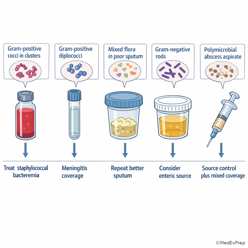
The final decision conflict is practical: when should a morphology result actually change empiric therapy, isolation thinking, or further testing? The resolving rule is: act decisively when the stain comes from a meaningful specimen and points to a dangerous narrow category; stay broad or get better data when the stain is low-quality or broad. Gram-positive cocci in clusters from blood should push anti-staphylococcal coverage and prompt evaluation for endovascular source. Gram-positive diplococci in CSF argue for pneumococcal meningitis coverage. Gram-negative diplococci in CSF raise meningococcal disease and infection prevention implications. Gram-negative rods in urine or bacteremia support enteric coverage, but species and resistance risk remain crucial. Polymicrobial abscess fluid supports source control and anaerobic/mixed coverage rather than fixation on a single organism.
WHY THIS IS TESTED: the exam is less interested in naming microscopic shapes than in whether you translate them into the safest next clinical step.
Choose to escalate confidence when morphology and syndrome align. Reject premature narrowing when morphology is broad, the host has multidrug resistance risk, or the site is colonized. Forward-looking note: this same reasoning will connect directly to the next topics on virulence factors and resistance mechanisms.
Exam takeaway: Gram stain should change therapy only when specimen quality, syndrome, and morphology all point in the same direction.
flowchart TD
A[Gram stain result available] --> B{Meaningful specimen?}
B -->|No| C[Do not over-narrow; improve sampling]
B -->|Yes| D{Classic narrow morphology?}
D -->|Yes| E[Link to syndrome]
D -->|No| F[Keep broader empiric framework]
E --> G{Blood with clusters}
E --> H{CSF with diplococci}
E --> I{Abscess with mixed rods/cocci}
G --> J[Think staphylococcal bacteremia and endovascular source]
H --> K[Think meningococcal or pneumococcal pattern based on Gram reaction]
I --> L[Think polymicrobial infection and source control]
J --> M[Refine therapy and pursue source evaluation]
K --> M
L --> M
F --> M Exam takeaway: The same morphology has different meaning in blood, CSF, sputum, urine, and abscess fluid because pretest probability and contamination risk differ.
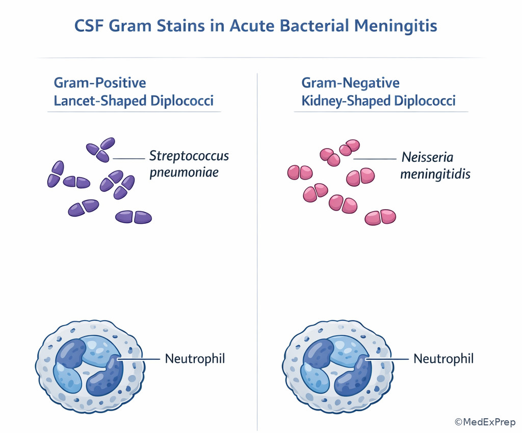
Exam takeaway: In acute meningitis, Gram stain patterns such as gram-positive diplococci or gram-negative diplococci can immediately narrow the highest-yield pathogen class before culture finalization.
B Worked example
A 62-year-old woman with rheumatoid arthritis on chronic prednisone and methotrexate presents with fever, headache, neck stiffness, and confusion. She also has a chronic diabetic foot ulcer, a distracting clue that could pull attention toward polymicrobial sepsis. CSF shows neutrophilic pleocytosis, low glucose, high protein, and Gram stain with gram-positive lancet-shaped diplococci. The question is the best organism-level interpretation for immediate management.
Identify task: turn a CSF Gram stain into empiric meningitis reasoning. Extract key facts: acute bacterial meningitis pattern in CSF, gram-positive diplococci, adult immunosuppressed host. Apply rule: this morphology strongly suggests Streptococcus pneumoniae. Eliminate distractors: the foot ulcer is noise and does not explain classic CSF morphology. Staphylococcus aureus would more likely appear in clusters. Neisseria meningitidis is gram-negative diplococci. Verify: maintain appropriate meningitis coverage and adjunctive measures while culture confirmation proceeds.
C Exam trap
D Checkpoint
Question: Which Gram stain result is most likely to justify immediate narrowing toward anti-staphylococcal bacteremia management?
- A. Mixed gram-positive cocci and gram-negative rods in poor-quality sputum
Tempting because pneumonia can seed blood; wrong because the specimen is low quality and nonspecific. - B. Gram-positive cocci in clusters from two blood culture sets in a febrile patient with a vascular catheter
Tempting because it may still be skin flora; right because the source is sterile and the morphology-syndrome pair strongly supports significant staphylococcal bacteremia. - C. Rare gram-negative rods in urine from an asymptomatic catheterized patient
Tempting because the urinary tract is a common source of gram-negative bacteremia; wrong because asymptomatic bacteriuria and colonization are common and this does not justify that specific inference. - D. Polymicrobial morphology in superficial wound swab from a chronic ulcer
Tempting because chronic wounds are often infected; wrong because superficial swabs poorly distinguish colonization from invasive infection.
Exam Traps & Differentiators
- Most common wrong answer: dismissing a dangerous sterile-site morphology as “nonspecific” because Gram stain is not definitive. Why: candidates know culture is the gold standard and underuse preliminary data.
- Looks similar but isn’t: gram-positive cocci in clusters versus chains. Both are “gram-positive cocci,” but only arrangement separates likely staphylococci from streptococci/enterococci.
- Looks similar but isn’t: polymicrobial poor sputum versus polymicrobial abscess aspirate. The former may be contamination; the latter may represent true mixed infection.
- Looks similar but isn’t: branching bacteria versus fungal hyphae. On boards, branching gram-positive rods should make you think bacterial branching organisms first.
- Related upcoming trap: in Virulence Factors And Toxins, a morphology clue may be paired with capsule or toxin effects; do not confuse structure-based identification with toxin-mediated syndrome selection.
- Related upcoming trap: in Resistance Mechanisms And Selection, the Gram category predicts some resistance logic, but you still must not assume a specific susceptibility pattern without local or final microbiology data.
If the stem says “blood cultures with gram-positive cocci in clusters,” think clinically significant staphylococcal bacteremia until proven otherwise.
If the stem says “many squamous epithelial cells in sputum,” think poor specimen quality before choosing a pathogen.
If the stem says “intracellular gram-negative diplococci in urethral exudate,” think gonococcal infection.
If the stem says “branching filamentous gram-positive rods with pulmonary plus CNS lesions,” think nocardiosis and order the right confirmatory testing.
If the stem says “acute meningitis with gram-positive diplococci in CSF,” think pneumococcal pattern.
If the stem says “strong bacterial syndrome but routine Gram stain is unrevealing,” think whether the pathogen class is poorly visualized by Gram stain.
| Stem clue | Tempting wrong interpretation | Correct board move |
|---|---|---|
| Gram-positive cocci in clusters in blood | Likely contaminant; wait | Treat as significant staphylococcal bacteremia pending speciation |
| Mixed flora in sputum with many squamous cells | Confirmed aspiration pneumonia microbiology | Downgrade the specimen; improve sampling |
| Gram-negative diplococci in urethral smear | Meningococcal disease | Interpret in source context as gonococcal pattern |
| Branching gram-positive rods | Fungal hyphae | Consider Nocardia or Actinomyces and order targeted tests |
| No organisms on routine Gram stain despite compatible syndrome | No infection | Consider pathogens poorly seen on Gram stain and choose alternative testing |
Tables
| Pattern | Most likely organism group | High-yield syndrome settings | Immediate board implication |
|---|---|---|---|
| Gram-positive cocci in clusters | Staphylococci | Bacteremia, line infection, endocarditis, skin/soft tissue infection | Do not dismiss sterile-site results |
| Gram-positive cocci in chains/pairs | Streptococci or enterococci | Endocarditis, cellulitis, meningitis, GU-source bacteremia | Use syndrome to separate likely family |
| Gram-positive diplococci in CSF | Streptococcus pneumoniae pattern | Acute bacterial meningitis | Supports pneumococcal coverage |
| Gram-negative diplococci | Neisseria species pattern | Meningitis, gonorrhea | Specimen source is critical |
| Gram-negative rods | Enterics, nonfermenters, others | UTI, sepsis, pneumonia, intra-abdominal infection | Often still broad; avoid over-narrowing |
| Branching filamentous gram-positive rods | Nocardia or Actinomyces | Chronic cervicofacial disease, pulmonary/CNS disease in immunocompromise | Consider modified acid-fast testing |
| Curved gram-negative rods | Campylobacter or Vibrio pattern | Gastroenteritis, seafood exposure, wound exposure to water | Use exposure history to narrow |
| Comparison point | Staphylococci | Streptococci/Enterococci | Neisseria |
|---|---|---|---|
| Typical Gram stain appearance | Gram-positive cocci in clusters | Gram-positive cocci in chains or pairs | Gram-negative diplococci |
| High-yield specimen clue | Blood cultures, abscesses, device infection | Blood, CSF, valve infection, cellulitis source | CSF or urethral/cervical exudate |
| Common board error | Dismissing as contaminant | Confusing chains with clusters | Ignoring specimen source |
| Immediate action style | Think bacteremia/endovascular source if sterile site | Use syndrome to separate pneumococcus, viridans group, beta-hemolytic strep, enterococcus | Treat based on meningitis or STI context |
| Need for further narrowing | Species and susceptibility essential | Species/group and susceptibility essential | Species often heavily suggested by body site |
| Differential/classification problem | Key morphology | Discriminating clue | Best next diagnostic thought |
|---|---|---|---|
| Nocardia vs Actinomyces | Branching gram-positive rods | Weak acid-fastness and pulmonary/CNS spread favor Nocardia; sinus tracts after mucosal disruption favor Actinomyces | Add modified acid-fast stain and culture context |
| Pneumococcal vs meningococcal meningitis | Diplococci in CSF | Gram-positive favors pneumococcus; gram-negative favors meningococcus | Keep urgent meningitis therapy while confirming |
| True lower respiratory pathogen vs oral contamination | Mixed sputum flora | Squamous epithelial cells indicate poor quality | Repeat or improve specimen |
| Significant staphylococcal bacteremia vs skin contaminant | Clusters in blood | Multiple sets, rapid positivity, clinical sepsis, hardware | Treat seriously and investigate source |
| Routine Gram stain-negative bacterial syndrome | No helpful morphology | Consider pathogen classes poorly visualized by Gram stain | Select acid-fast, antigen, or molecular testing |
| Specimen type | How much to trust Gram stain | Most useful added clue | Board pearl |
|---|---|---|---|
| Blood culture | High | Number of positive sets and time to positivity | Never casually dismiss dangerous morphology in unstable patients |
| CSF | High | Cell count, glucose, protein, clinical meningitis pattern | Rapid morphology can shape immediate therapy and precautions |
| Sputum | Variable | Squamous epithelial cells and neutrophils | Quality first, organism second |
| Urine | Moderate | Symptoms and pyuria | Colonization is common, especially with catheters |
| Abscess aspirate | High | Deep aspirate versus superficial swab | Polymicrobial findings may be real and support source control |
| Superficial wound swab | Low | Depth and tissue invasion evidence | Colonization often overwhelms interpretation |
Algorithm / Approach
Exam takeaway: The safest exam approach is specimen first, quality second, morphology third, and treatment refinement only after syndrome matching.

Use this exact sequence under time pressure. See Fig 6. Most board misses come from skipping step 1 or step 2 and jumping straight to memorized organism names.
Rapid Review
- Clusters → staphylococcal bucket, not streptococcal bucket
- Chains/pairs → streptococci or enterococci, then use syndrome to split
- Gram-negative diplococci → source determines gonococcal versus meningococcal thinking
- Sputum with many squamous epithelial cells → contamination signal, not pathogen signal
- Polymicrobial abscess aspirate → true mixed infection more likely than with sputum
- Branching gram-positive rods → think Nocardia/Actinomyces before fungus
- Weak acid-fast branching rods → nocardiosis clue
- Cervicofacial sinus tracts after mucosal disruption → actinomycosis clue
- Gram-positive diplococci in CSF → pneumococcal pattern
- Gram-negative diplococci in CSF → meningococcal pattern
- No useful Gram stain despite strong syndrome → consider organisms poorly seen on routine stain
- Blood culture morphology from multiple sets → more actionable than morphology from colonized surfaces
- Generic gram-negative rods → often too broad for aggressive narrowing without resistance-risk context
- Superficial wound swab → colonization-heavy, low specificity for invasive pathogen
Self-check quiz
1. A 64-year-old man with diabetes and chronic hemodialysis presents with fever after dialysis. Two blood culture sets show gram-positive cocci in clusters. Which organism group is most likely?
- A. Staphylococci
- B. Streptococci
- C. Enterococci
- D. Neisseria species
2. A 29-year-old man with HIV on ART, recent new sexual partner, and purulent urethral discharge has Gram stain showing intracellular gram-negative diplococci. Which organism is most likely?
- A. Neisseria gonorrhoeae
- B. Escherichia coli
- C. Moraxella catarrhalis
- D. Enterococcus faecalis
3. A 76-year-old woman with COPD, GERD, and recent influenza has pneumonia. Sputum Gram stain shows many squamous epithelial cells, few neutrophils, and mixed gram-positive cocci plus gram-negative rods. What is the most appropriate next step?
- A. Narrow therapy to oral anaerobes based on mixed flora
- B. Interpret the Gram stain as diagnostic of aspiration pneumonia
- C. Downgrade the specimen quality and obtain a better respiratory sample if microbiologic narrowing is needed
- D. Stop antibiotics because the stain is nonlocalizing
4. A 58-year-old kidney transplant recipient on tacrolimus and prednisone presents with cough, weight loss, and a brain abscess. Sputum Gram stain shows branching filamentous gram-positive rods. Which additional test is most appropriate to clarify the leading diagnosis?
- A. Modified acid-fast stain
- B. Heterophile antibody test
- C. Serum cryptococcal antigen only
- D. Rapid influenza PCR
5. A 61-year-old woman with confusion, nuchal rigidity, hypertension, and rheumatoid arthritis on prednisone has CSF with neutrophilic pleocytosis and gram-positive diplococci. Which organism is most likely driving the immediate empiric focus?
- A. Streptococcus pneumoniae
- B. Neisseria meningitidis
- C. Staphylococcus aureus
- D. Listeria monocytogenes
6. A 67-year-old man with cirrhosis, a new central line, and septic shock has one peripheral blood culture bottle positive for gram-positive cocci in clusters at 9 hours and a second set that turns positive shortly after. He also has bibasilar opacities on chest radiograph. Which is the most appropriate interpretation?
- A. Likely contamination because skin flora commonly grow in blood cultures
- B. Likely true staphylococcal bacteremia requiring immediate management as clinically significant infection
- C. Likely streptococcal pneumonia with incidental bloodstream contamination
- D. Insufficient data to act until species identification returns
7. A 45-year-old man with alcohol use disorder, poor dentition, and recurrent aspiration has chronic jaw swelling with draining sinus tracts after dental work. Gram stain shows branching filamentous gram-positive rods. Which diagnosis is most likely?
- A. Nocardiosis
- B. Actinomycosis
- C. Invasive aspergillosis
- D. Tuberculosis
8. A 72-year-old woman with neurogenic bladder, an indwelling catheter, and chronic kidney disease has cloudy urine but no dysuria, fever, flank pain, or leukocytosis. Urine Gram stain shows gram-negative rods. Which is the most appropriate next step?
- A. Start broad gram-negative therapy because gram-negative rods in urine prove active infection
- B. Remove the catheter immediately and begin antifungal therapy
- C. Interpret the finding in clinical context because colonization/asymptomatic bacteriuria is common in catheterized patients
- D. Diagnose pyelonephritis because gram-negative rods are present
Answer key
1. Correct answer: A. Staphylococci.
- A. Staphylococci — Tempting because clusters are the classic morphology and the patient has vascular access risk; correct because gram-positive cocci in clusters in blood strongly indicate staphylococci, especially in dialysis patients. Single discriminating clue: clusters. Sources: Harrison’s Principles of Internal Medicine; Mandell, Douglas, and Bennett’s Principles and Practice of Infectious Diseases.
- B. Streptococci — Tempting because they are also gram-positive cocci; incorrect because streptococci more typically appear in chains or pairs. Single discriminating clue: arrangement is not chains. Sources: Harrison’s; Mandell.
- C. Enterococci — Tempting because healthcare-associated bacteremia is common; incorrect because enterococci usually appear in pairs and short chains, not clusters. Single discriminating clue: cluster morphology. Sources: Harrison’s; Mandell.
- D. Neisseria species — Tempting only if one focuses on coccus shape alone; incorrect because Neisseria are gram-negative diplococci. Single discriminating clue: gram-positive stain. Sources: Harrison’s; Mandell.
2. Correct answer: A. Neisseria gonorrhoeae.
- A. Neisseria gonorrhoeae — Tempting because it is the classic STI organism; correct because intracellular gram-negative diplococci in purulent urethral exudate are highly suggestive of gonorrhea. Single discriminating clue: intracellular gram-negative diplococci in urethral discharge. Sources: CDC STI Treatment Guidelines; Harrison’s.
- B. Escherichia coli — Tempting because it is a common urogenital pathogen; incorrect because it is a gram-negative rod, not a diplococcus, and fits UTI more than urethral exudate microscopy. Single discriminating clue: rod versus diplococcus. Sources: CDC STI Guidelines; Harrison’s.
- C. Moraxella catarrhalis — Tempting because it is also a gram-negative diplococcus; incorrect because the syndrome is purulent urethritis, not respiratory infection. Single discriminating clue: specimen source. Sources: Harrison’s; Mandell.
- D. Enterococcus faecalis — Tempting because of genitourinary association; incorrect because enterococci are gram-positive cocci in pairs/chains. Single discriminating clue: gram-negative diplococcal morphology. Sources: Harrison’s; Mandell.
3. Correct answer: C. Downgrade the specimen quality and obtain a better respiratory sample if microbiologic narrowing is needed.
- A. Narrow therapy to oral anaerobes based on mixed flora — Tempting because aspiration and oral flora are linked; incorrect because poor specimen quality prevents reliable interpretation. Single discriminating clue: many squamous epithelial cells. Sources: IDSA/ATS community-acquired pneumonia guidance; Harrison’s.
- B. Interpret the Gram stain as diagnostic of aspiration pneumonia — Tempting because influenza and GERD increase aspiration risk; incorrect because the stain is contaminated and nondiagnostic. Single discriminating clue: few neutrophils plus many squamous cells. Sources: IDSA/ATS guidance; Harrison’s.
- C. Downgrade the specimen quality and obtain a better respiratory sample if microbiologic narrowing is needed — Tempting because it avoids overcalling the result; correct because sputum quality governs interpretability. Single discriminating clue: poor-quality sputum markers. Sources: Harrison’s; Mandell.
- D. Stop antibiotics because the stain is nonlocalizing — Tempting because the microbiology is unhelpful; incorrect because a bad sputum stain does not exclude pneumonia. Single discriminating clue: clinical pneumonia still exists despite poor sample quality. Sources: IDSA/ATS guidance; Harrison’s.
4. Correct answer: A. Modified acid-fast stain.
- A. Modified acid-fast stain — Tempting because the host and morphology suggest nocardiosis; correct because branching filamentous gram-positive rods with pulmonary and CNS disease in a transplant recipient should prompt evaluation for weakly acid-fast Nocardia. Single discriminating clue: transplant host plus pulmonary/CNS spread. Sources: Harrison’s; Mandell.
- B. Heterophile antibody test — Tempting only as a broad infectious workup idea; incorrect because it does not address the smear or syndrome. Single discriminating clue: branching rods are unrelated to EBV mononucleosis testing. Sources: Harrison’s.
- C. Serum cryptococcal antigen only — Tempting because of brain involvement in an immunocompromised host; incorrect because the smear already points toward a branching bacterial pathogen rather than encapsulated yeast as the leading diagnosis. Single discriminating clue: branching gram-positive rods. Sources: Harrison’s; Mandell.
- D. Rapid influenza PCR — Tempting because respiratory symptoms are present; incorrect because it does not clarify the leading morphology-based diagnosis. Single discriminating clue: CNS lesion plus branching rods. Sources: Harrison’s.
5. Correct answer: A. Streptococcus pneumoniae.
- A. Streptococcus pneumoniae — Tempting because it is the classic adult bacterial meningitis organism; correct because gram-positive diplococci in CSF strongly support pneumococcal meningitis. Single discriminating clue: gram-positive diplococci in CSF. Sources: IDSA bacterial meningitis guidance; Harrison’s.
- B. Neisseria meningitidis — Tempting because meningococcal disease also presents with acute meningitis; incorrect because meningococci are gram-negative diplococci. Single discriminating clue: gram-positive result. Sources: IDSA guidance; Harrison’s.
- C. Staphylococcus aureus — Tempting because chronic illness can predispose to invasive staphylococcal disease; incorrect because staphylococci appear in clusters, not diplococci. Single discriminating clue: arrangement. Sources: Harrison’s; Mandell.
- D. Listeria monocytogenes — Tempting because steroids and older age raise listeriosis risk; incorrect because Listeria is a gram-positive rod/coccobacillus, not gram-positive diplococcus. Single discriminating clue: morphology class. Sources: IDSA guidance; Harrison’s.
6. Correct answer: B. Likely true staphylococcal bacteremia requiring immediate management as clinically significant infection.
- A. Likely contamination because skin flora commonly grow in blood cultures — Tempting because coagulase-negative staphylococci often contaminate blood cultures; incorrect because septic shock, a central line, and more than one rapidly positive set make true bacteremia far more likely. Single discriminating clue: multiple early positive cultures in a septic patient. Sources: IDSA intravascular catheter-related infection guidance; Harrison’s.
- B. Likely true staphylococcal bacteremia requiring immediate management as clinically significant infection — Tempting because it acts before full speciation; correct because gram-positive cocci in clusters from blood in this context demand immediate attention as staphylococcal bacteremia. Single discriminating clue: clusters in multiple blood cultures from an unstable patient with a line. Sources: IDSA catheter-related infection guidance; Harrison’s.
- C. Likely streptococcal pneumonia with incidental bloodstream contamination — Tempting because bibasilar opacities can anchor the reader on pneumonia; incorrect because the blood culture morphology is clusters, not chains/pairs, and the opacities are distracting noise. Single discriminating clue: cluster morphology. Sources: Harrison’s; Mandell.
- D. Insufficient data to act until species identification returns — Tempting because Gram stain is preliminary; incorrect because preliminary blood culture morphology can and should influence immediate management. Single discriminating clue: sterile-site dangerous morphology in shock. Sources: IDSA guidance; Harrison’s.
7. Correct answer: B. Actinomycosis.
- A. Nocardiosis — Tempting because Nocardia also forms branching filamentous gram-positive rods; incorrect because nocardiosis more often presents with pulmonary disease, CNS dissemination, and weak acid-fastness in immunocompromised hosts. Single discriminating clue: chronic cervicofacial sinus tracts after dental work. Sources: Harrison’s; Mandell.
- B. Actinomycosis — Tempting because the syndrome is textbook; correct because poor dentition, dental manipulation, chronic indurated jaw infection, and draining sinus tracts strongly favor actinomycosis. Single discriminating clue: cervicofacial disease after mucosal disruption. Sources: Harrison’s; Mandell.
- C. Invasive aspergillosis — Tempting because filamentous organisms evoke hyphae; incorrect because the smear specifies branching gram-positive rods, which indicates bacteria, not mold hyphae. Single discriminating clue: gram-positive bacterial morphology. Sources: Harrison’s; Mandell.
- D. Tuberculosis — Tempting because chronic draining lesions occur in TB; incorrect because TB is an acid-fast bacillus and does not appear as branching gram-positive rods on routine Gram stain. Single discriminating clue: morphology is not acid-fast bacillary pattern. Sources: Harrison’s; Mandell.
8. Correct answer: C. Interpret the finding in clinical context because colonization/asymptomatic bacteriuria is common in catheterized patients.
- A. Start broad gram-negative therapy because gram-negative rods in urine prove active infection — Tempting because gram-negative rods commonly cause UTI; incorrect because bacteriuria in catheterized patients may represent colonization or asymptomatic bacteriuria, not a treatment-requiring infection. Single discriminating clue: no symptoms or systemic signs. Sources: IDSA asymptomatic bacteriuria guideline; Harrison’s.
- B. Remove the catheter immediately and begin antifungal therapy — Tempting because device issues matter in urinary infection; incorrect because the microscopy shows gram-negative rods, not fungi, and the question is interpretation of significance. Single discriminating clue: bacterial morphology plus lack of symptoms. Sources: IDSA asymptomatic bacteriuria guideline; Harrison’s.
- C. Interpret the finding in clinical context because colonization/asymptomatic bacteriuria is common in catheterized patients — Tempting because it respects stewardship; correct because urine morphology alone does not define infection in an asymptomatic catheterized patient. Single discriminating clue: absence of dysuria, fever, flank pain, or leukocytosis. Sources: IDSA asymptomatic bacteriuria guideline; Harrison’s.
- D. Diagnose pyelonephritis because gram-negative rods are present — Tempting because pyelonephritis is often gram-negative; incorrect because pyelonephritis requires compatible symptoms or systemic findings. Single discriminating clue: no clinical syndrome of upper tract infection. Sources: IDSA guidance; Harrison’s.
Keep going with the full ABIM Infectious Disease Prep Course (ID)
You just studied the way members do, in the same lesson interface. Full access unlocks every lesson, the question bank and flashcards for a full year.
One payment · 1 year of access · No auto-renewal