1.1.1 - CBC Indices And Patterns
Why it matters
- Board stems often hide the diagnosis in the CBC pattern before they reveal smear, iron studies, marrow data, or molecular testing.
- A single discriminator such as MCV, RDW, RBC count, or multilineage cytopenia can eliminate several attractive but wrong answers.
- Urgent conditions in hematology-oncology commonly begin as “just an abnormal CBC,” including acute leukemia, hemolysis, DIC-associated consumption, drug toxicity, and marrow infiltration.
- Correct CBC interpretation prevents wasted testing: you should know when to order iron studies, reticulocyte count, hemolysis labs, smear review, marrow evaluation, or simply repeat the specimen.
Key Terms & Must-Know Facts
1.1.1.1 Key terms
- Hemoglobin (Hgb): Oxygen-carrying concentration; primary anemia severity marker.
- Hematocrit (Hct): Fraction of blood volume occupied by RBCs.
- MCV: Mean corpuscular volume; microcytic, normocytic, macrocytic classification anchor.
- MCHC: Mean corpuscular hemoglobin concentration; helps flag spherocytosis or artifact when elevated.
- RDW: Red cell distribution width; size variability marker.
- Reticulocyte response: Marrow output clue; distinguishes underproduction from destruction/loss.
- Leukopenia/neutropenia: Decreased WBC/ANC; infection-risk relevance depends on degree and context.
- Leukocytosis: Elevated WBC; can be reactive or clonal.
- Thrombocytopenia: Low platelet count; can be destructive, consumptive, sequestration-related, dilutional, or production-related.
- Bicytopenia/pancytopenia: Two or three cell lines low; often more concerning than isolated anemia.
- Reticulocytosis-associated macrocytosis: Larger young RBCs can raise MCV without megaloblastic disease.
- Spurious CBC abnormality: Artifact from agglutination, clotting, EDTA effect, lipemia, or sample handling.
1.1.1.2 Must-know facts
- Low MCV does not automatically mean iron deficiency; the RBC count and RDW help separate iron deficiency from thalassemia trait.
- Macrocytosis without anemia may still reflect alcohol use, liver disease, hypothyroidism, medications, or early marrow disease.
- A high reticulocyte count pushes the differential toward blood loss or hemolysis, not marrow failure.
- Multilineage cytopenia raises concern for marrow infiltration, aplasia, MDS, severe nutritional deficiency, hypersplenism, or treatment toxicity.
- Marked leukocytosis with anemia or thrombocytopenia is more worrisome for leukemia/myeloproliferative disease than isolated reactive leukocytosis.
- Thrombocytosis can be reactive; iron deficiency is a classic board-tested cause.
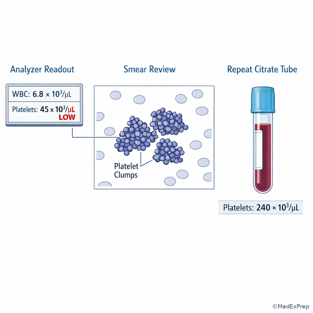
- Pseudothrombocytopenia from platelet clumping should be excluded before diagnosing immune thrombocytopenia.
- High RDW suggests mixed populations or evolving deficiency; normal RDW with microcytosis points toward thalassemia trait more than iron deficiency.
Core content
1.1.1.3 Foundational RBC index logic: classify first, then choose the next test
A Explanation
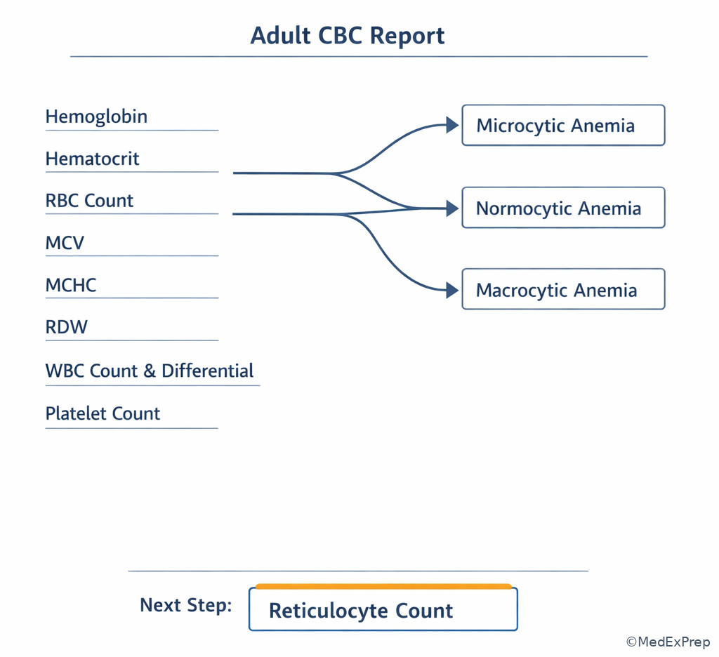
The first decision conflict is simple but highly tested: Do you classify the anemia by MCV first, or jump straight to etiology? On boards, choose classification first. MCV groups the problem into microcytic, normocytic, or macrocytic patterns; RDW, RBC count, and the reticulocyte response then resolve which pathway to pursue. If the CBC shows microcytosis with an elevated or preserved RBC count and relatively normal RDW, reject iron deficiency as the reflex answer and think thalassemia trait. If the MCV is normal but the reticulocyte count is high, reject “underproduction anemia” and think blood loss or hemolysis. If the MCV is high, distinguish true megaloblastic or marrow causes from reticulocytosis, alcohol, liver disease, hypothyroidism, and medication effect.
WHY THIS IS TESTED: Exam stems frequently give only Hgb, MCV, RBC count, and RDW, expecting you to choose the next step without overcalling a diagnosis.
For anemia pattern recognition, the most powerful pair is MCV + reticulocyte response. MCV tells you the bucket; reticulocytes tell you whether the marrow is compensating. A low reticulocyte response means underproduction until proven otherwise. A brisk response means loss or destruction unless recent treatment changed the trajectory. See Fig 1.
Exam takeaway: Use MCV first, then RBC count, RDW, and reticulocyte response to choose the next diagnostic branch.
flowchart TD
A[Low hemoglobin] --> B{MCV}
B -->|Low| C[Microcytic]
B -->|Normal| D[Normocytic]
B -->|High| E[Macrocytic]
C --> F{RBC count and RDW}
F -->|High RBC count, normal RDW| G[Think thalassemia trait]
F -->|Low RBC count, high RDW| H[Think iron deficiency]
D --> I{Reticulocyte count}
I -->|High| J[Blood loss or hemolysis]
I -->|Low| K[Underproduction, chronic disease, renal, marrow]
E --> L{Reticulocytes or classic causes}
L -->|High retics| M[Recovery, hemolysis, blood loss]
L -->|Low retics| N[B12 or folate deficiency, alcohol, liver, MDS, drugs] Exam takeaway: Read the CBC as linked cell lines and indices rather than isolated numbers.
B Worked example
A 38-year-old woman with rheumatoid arthritis and menorrhagia presents for fatigue. She takes ibuprofen and a proton pump inhibitor and has a history of Hashimoto thyroiditis. CBC shows Hgb 9.8 g/dL, MCV 69 fL, RBC count 5.4 million/mcL, RDW 12.8%, platelets 470,000/mcL. Ferritin from six months earlier was borderline low, and the stem mentions pica in high school. The task is the most appropriate next diagnostic interpretation. Identify key facts: marked microcytosis, relatively high RBC count, normal RDW, thrombocytosis. Apply the rule: microcytosis with high-normal RBC count argues for thalassemia trait more than classic iron deficiency, even though menorrhagia and thrombocytosis are tempting. Eliminate distractors: anemia of inflammation usually is not this microcytic with a high RBC count; aplastic or marrow failure states do not cause thrombocytosis. Verify: the best next step is targeted iron studies and hemoglobin electrophoresis rather than assuming iron deficiency alone.
C Exam trap
Common wrong answer: “Iron deficiency anemia” whenever the MCV is low.
Why it is tempting: Microcytosis, fatigue, and reactive thrombocytosis are classic iron-deficiency clues.
Single clue that eliminates it: A relatively high RBC count with normal RDW points away from iron deficiency and toward thalassemia trait or another microcytic production pattern.
D Checkpoint
Question: A 29-year-old man with no bleeding history is found to have Hgb 11.2 g/dL, MCV 65 fL, RBC count 6.1 million/mcL, RDW 12.4%, and normal platelets. Which interpretation is most appropriate?
- Iron deficiency anemia — tempting because profound microcytosis is common in iron deficiency, but the preserved-high RBC count and normal RDW argue against it.
- Thalassemia trait — correct because severe microcytosis out of proportion to anemia with high RBC count is the classic discriminator.
- Anemia of chronic inflammation — tempting because some inflammatory states produce mild microcytosis, but the RBC count pattern does not fit.
- Acute blood loss — tempting because anemia can be normocytic early, but isolated marked microcytosis makes this wrong.
1.1.1.4 Microcytic, normocytic, and macrocytic patterns: the board-relevant differential
A Explanation
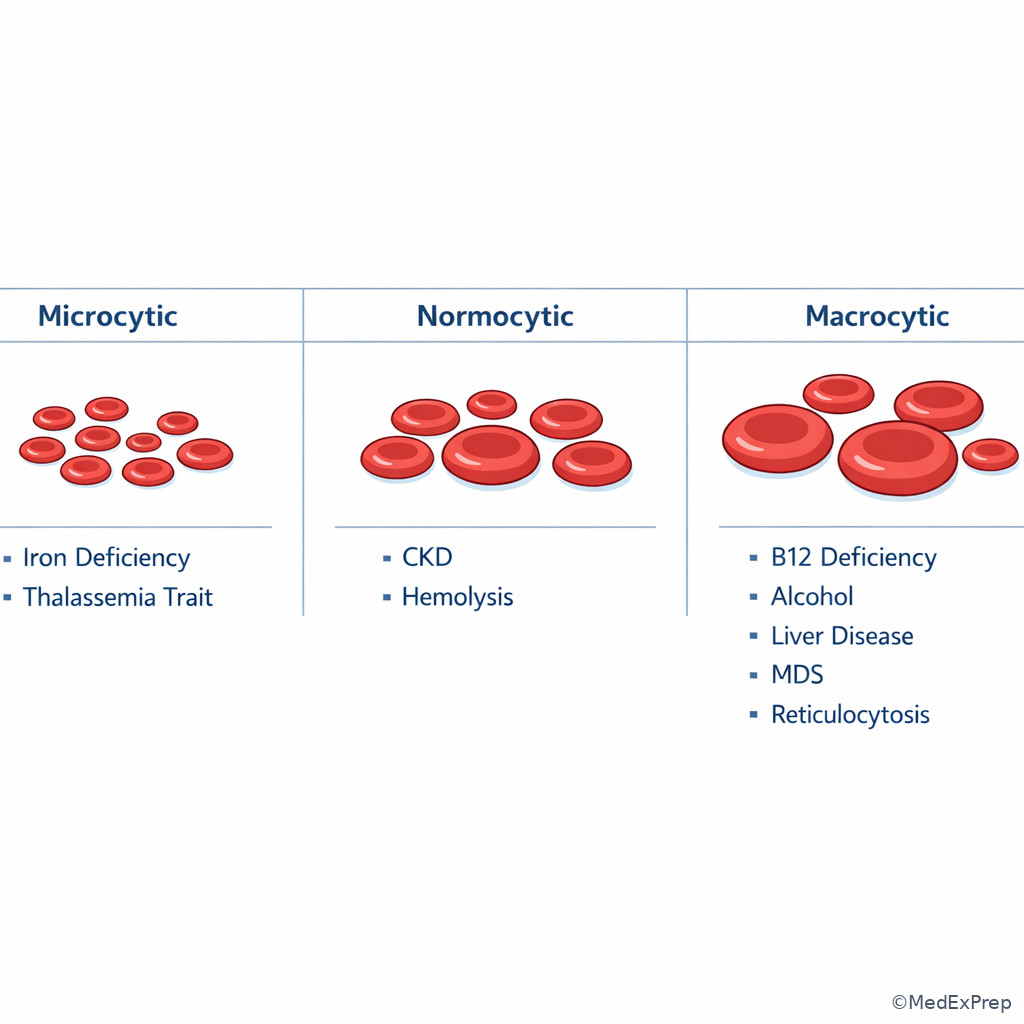
The next decision conflict is which causes belong in each MCV bucket, and which single clue separates the closest distractors? For microcytosis, the high-yield differential is iron deficiency, thalassemia trait, anemia of inflammation, sideroblastic patterns, and less commonly lead-related disease. For normocytic anemia, think early deficiency, CKD/low EPO states, inflammation, marrow suppression, mixed disorders, acute blood loss, and hemolysis. For macrocytosis, decide whether the marrow is making large cells because of reticulocytosis or because of a maturation problem such as B12/folate deficiency, alcohol use, liver disease, hypothyroidism, medications, or MDS.
The exam rule is to avoid overusing MCV as a diagnosis. Microcytosis narrows the field but does not finish the work. Macrocytosis also misleads: many stems tempt you to pick megaloblastic anemia when the actual discriminator is alcohol, hydroxyurea, methotrexate, zidovudine, liver disease, or a rising reticulocyte count after treatment or hemolysis. See Fig 2.
WHY THIS IS TESTED: ABIM-style questions often provide one extra clue—RBC count, RDW, reticulocyte count, renal function, or medication history—to force a precise pattern call rather than a broad category.
Exam takeaway: The best answer usually comes from one tie-breaker added to the MCV bucket, not from MCV alone.
flowchart LR
A[MCV low] --> A1[Iron deficiency]
A --> A2[Thalassemia trait]
A --> A3[Inflammation]
A --> A4[Sideroblastic process]
B[MCV normal] --> B1[CKD or low EPO]
B --> B2[Inflammation]
B --> B3[Acute blood loss]
B --> B4[Hemolysis]
B --> B5[Marrow failure or infiltration]
C[MCV high] --> C1[B12 or folate deficiency]
C --> C2[Alcohol use]
C --> C3[Liver disease]
C --> C4[Hypothyroidism]
C --> C5[Medications]
C --> C6[MDS]
C --> C7[Reticulocytosis]
Memory anchor: “TAILS” for common microcytic causes: Thalassemia, Anemia of inflammation, Iron deficiency, Lead, Sideroblastic.
Exam takeaway: Visually linking microcytosis, normocytosis, and macrocytosis improves rapid differential selection on stems.
B Worked example
A 67-year-old man with metastatic prostate cancer on androgen deprivation therapy, chronic kidney disease stage 3, and daily alcohol use presents with fatigue. CBC shows Hgb 10.4 g/dL, MCV 104 fL, platelets 230,000/mcL, WBC 6,200/mcL. Creatinine is 1.9 mg/dL. He recently completed palliative radiation and started folic acid supplementation on his own. The tempting wrong answer is “folate deficiency” because the MCV is high and he drinks alcohol. The task is to choose the best next diagnostic direction. Extract key facts: isolated macrocytosis, no leukopenia or thrombocytopenia, multiple potential causes, CKD present but does not explain macrocytosis. Apply the rule: macrocytosis requires distinction between megaloblastic and nonmegaloblastic causes; medication/alcohol history and reticulocytes matter. Eliminate distractors: CKD usually causes normocytic anemia; myelodysplasia is possible in older adults but isolated stable macrocytosis without other cytopenias is not enough. Verify: measure B12, reticulocyte count, liver-associated clues, and review medications/alcohol before concluding MDS.
C Exam trap
Common wrong answer: “Megaloblastic anemia” for every macrocytic CBC.
Why it is tempting: Macrocytosis strongly cues B12 or folate deficiency, and exam writers know this reflex.
Single clue that eliminates it: A medication history, alcohol use, liver disease, or reticulocytosis can explain the macrocytosis without a maturation-defect anemia.
D Checkpoint
Question: A 72-year-old woman with hypothyroidism and remote breast cancer treatment has Hgb 11.0 g/dL, MCV 108 fL, normal platelets, normal WBC count, and no bleeding. Which finding most strongly pushes the evaluation toward marrow response rather than megaloblastic disease?
- Elevated reticulocyte count — correct because large reticulocytes can raise MCV and point toward recovery, hemolysis, or recent blood loss.
- Low thyroid-stimulating hormone — tempting because thyroid disease affects MCV, but this does not specifically indicate marrow response.
- Borderline low folate level — tempting because folate deficiency causes macrocytosis, but it does not distinguish response from maturation failure.
- History of remote chemotherapy — tempting because therapy-related MDS is board-relevant, but history alone does not indicate marrow response.
- The most common cause of microcytic anemia worldwide is iron deficiency.
- True or False: A high RBC count in a markedly microcytic patient favors thalassemia trait. True.
- Name 3 common nonmegaloblastic causes of macrocytosis: alcohol use, liver disease, reticulocytosis.
1.1.1.5 White blood cell and platelet patterns: reactive versus clonal or marrow failure
A Explanation
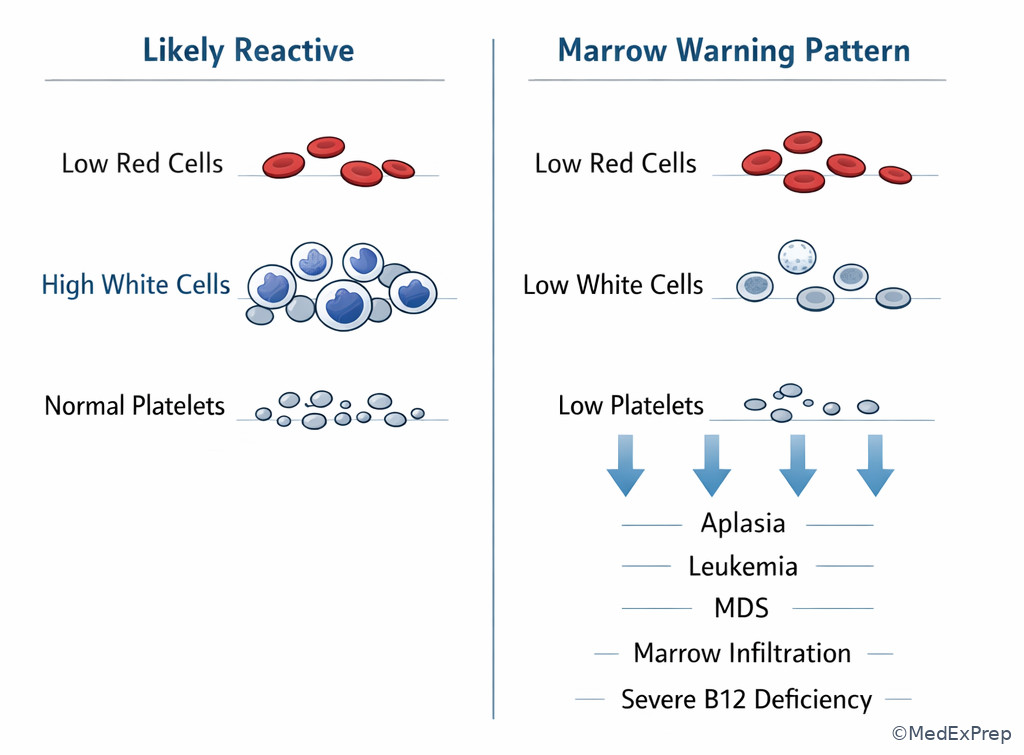
Once you can read RBC indices, the next conflict is whether an abnormal WBC or platelet count is isolated and reactive, or part of a broader marrow process. Board questions reward you for looking across all lines. An isolated mild neutrophilia with infection or steroid exposure usually behaves differently from leukocytosis paired with anemia, thrombocytopenia, constitutional symptoms, or circulating immature forms. Likewise, isolated thrombocytosis may be reactive—especially with iron deficiency, inflammation, infection, postsplenectomy state, or malignancy—but thrombocytosis with microvascular symptoms, splenomegaly, or other myeloproliferative clues raises concern for a clonal process.
The single most valuable rule: multilineage abnormalities are harder to dismiss. Leukopenia plus anemia, or thrombocytopenia plus leukocytosis, should push you beyond a reactive explanation. In cancer patients, chemotherapy, marrow infiltration, infection, hypersplenism, nutritional deficiency, and therapy-related myeloid neoplasms are all fair game. In non-cancer patients, marrow failure, aplasia, MDS, acute leukemia, and severe B12 deficiency become major considerations.
WHY THIS IS TESTED: The exam often presents a tempting benign explanation such as recent infection or iron deficiency, then inserts one second-line abnormality that should trigger concern for marrow pathology. See Fig 4.
Exam takeaway: Isolated mild abnormalities can be reactive, but paired or multilineage changes should trigger concern for marrow disease or malignancy.
flowchart TD
A[Abnormal WBC or platelet count] --> B{Other lines normal?}
B -->|Yes| C[Consider reactive causes first]
C --> C1[Infection or inflammation]
C --> C2[Steroids or stress]
C --> C3[Iron deficiency causing thrombocytosis]
C --> C4[Postsplenectomy]
B -->|No| D[Multilineage abnormality]
D --> D1[Marrow failure or aplasia]
D --> D2[MDS]
D --> D3[Acute leukemia]
D --> D4[Marrow infiltration]
D --> D5[Drug toxicity]
D --> D6[Severe nutritional deficiency] Exam takeaway: Simultaneous abnormalities in two or more cell lines should prompt a marrow-focused differential.
B Worked example
A 61-year-old man with metastatic colon cancer receiving capecitabine, prior splenectomy after trauma, and recent prednisone for COPD exacerbation presents for routine follow-up. CBC shows WBC 18,000/mcL, Hgb 12.4 g/dL, platelets 610,000/mcL, ANC 15,000/mcL. He feels well, has no fever, and the stem mentions chronic fatigue and low ferritin. The tempting wrong answer is “myeloproliferative neoplasm” because both WBCs and platelets are elevated. Identify the task: determine whether the pattern is most likely reactive or clonal. Extract key facts: recent steroids, postsplenectomy state, possible iron deficiency, no anemia progression, no thrombocytopenia, no constitutional symptoms, and no suggestion of left-shifted blasts. Apply the rule: multiple reactive explanations plus preserved other lines and a plausible context favor secondary changes first. Eliminate distractors: acute leukemia usually does not present with well appearance plus isolated steroid/postsplenectomy-compatible counts; marrow failure causes low counts, not thrombocytosis. Verify: trend the CBC, address iron deficiency, review smear if needed, and reserve clonal workup if counts persist without explanation.
C Exam trap
Common wrong answer: “Myeloproliferative neoplasm” for any thrombocytosis or leukocytosis.
Why it is tempting: Clonal diseases are high-yield, and exam writers often include dramatic count elevations.
Single clue that eliminates it: A strong reactive context such as iron deficiency, infection, steroid exposure, inflammation, or postsplenectomy state makes a secondary process more likely unless the pattern persists or other clonal clues emerge.
D Checkpoint
Question: Which CBC pattern most strongly argues against a purely reactive leukocytosis?
- WBC 14,500/mcL after high-dose prednisone, normal hemoglobin and platelets — tempting because the count is elevated, but steroids commonly cause this isolated pattern.
- WBC 16,000/mcL with fever and neutrophil predominance, normal platelets — tempting because leukocytosis can be marked, but infection readily explains it.
- WBC 22,000/mcL with anemia and thrombocytopenia — correct because multilineage abnormalities suggest marrow pathology rather than a simple reactive state.
- Platelets 550,000/mcL in iron deficiency with stable WBC count — tempting because thrombocytosis raises concern, but iron deficiency is a classic reactive cause.
1.1.1.6 Reticulocyte response and CBC kinetics: underproduction versus destruction or blood loss
A Explanation
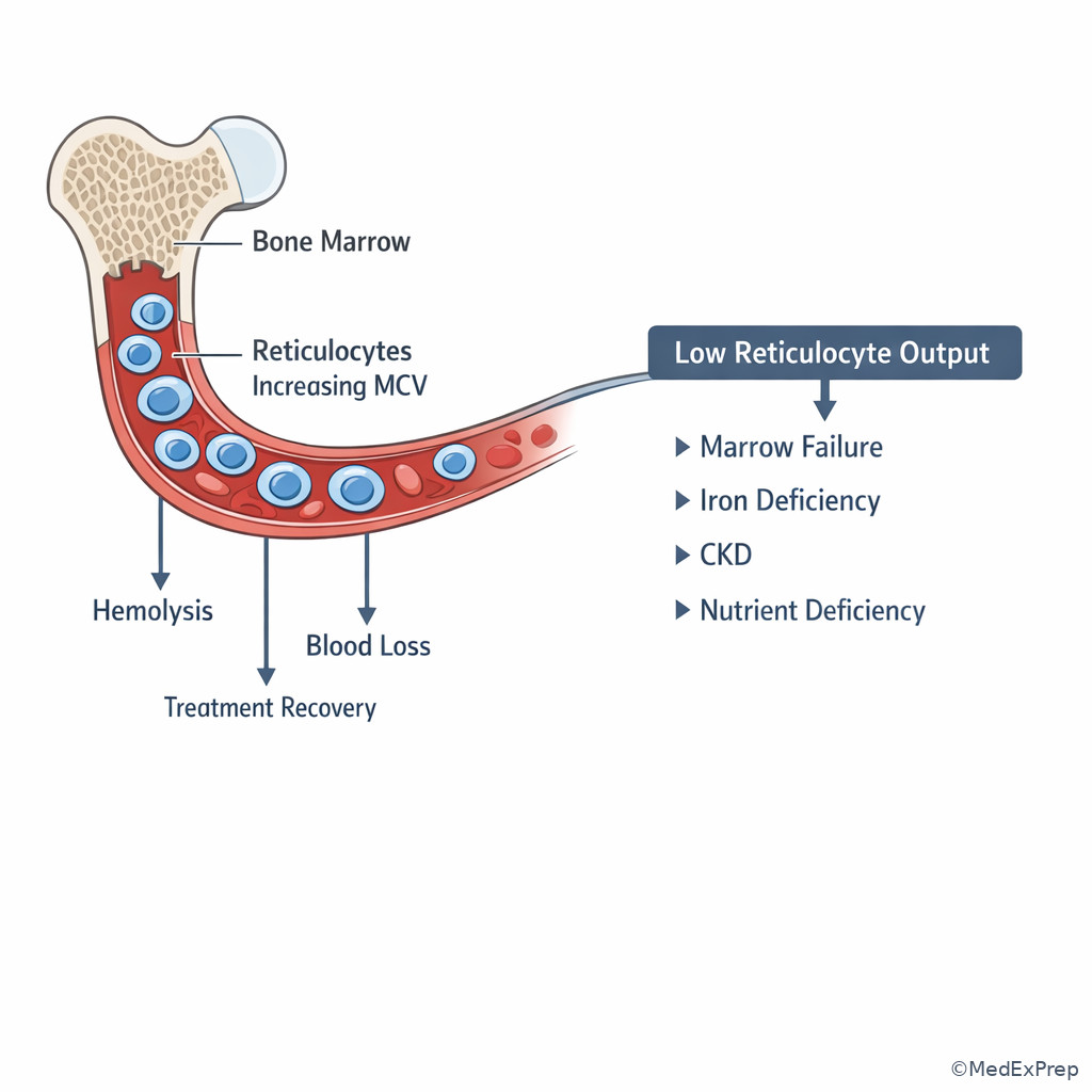
One of the highest-yield pattern questions is when to use the reticulocyte count to break a diagnostic tie. If anemia is present, the marrow should respond unless the problem is production failure. A low or inappropriately normal reticulocyte count supports underproduction: iron deficiency, inflammation, CKD, marrow suppression, marrow infiltration, aplasia, or severe nutrient deficiency. A high reticulocyte count points toward blood loss, hemolysis, or recovery after therapy. This is especially important in oncology patients where chemotherapy, marrow involvement, occult bleeding, hemolysis, and nutritional deficiency can coexist.
Reticulocytes also explain “false macrocytosis.” Young red cells are larger, so a strong reticulocyte response can raise MCV. That means macrocytosis is sometimes a kinetic clue rather than a maturation defect. On the exam, the stem may add elevated bilirubin, LDH, or recent transfusion/therapy to tempt hemolysis or recent bleeding. You still need to ask whether the reticulocyte response is appropriate for the degree of anemia.
WHY THIS IS TESTED: Many stems are unsolvable from MCV alone, but become easy once you choose whether the marrow is responding. See Fig 5.
Exam takeaway: The reticulocyte response is the fastest way to distinguish underproduction from loss or destruction when CBC patterns are ambiguous.
flowchart TD
A[Anemia present] --> B{Reticulocyte response}
B -->|Low or inappropriately normal| C[Underproduction]
C --> C1[Iron deficiency]
C --> C2[Inflammation]
C --> C3[CKD]
C --> C4[Marrow suppression or infiltration]
C --> C5[B12 or folate deficiency]
B -->|High| D[Loss or destruction]
D --> D1[Hemolysis]
D --> D2[Acute or occult blood loss]
D --> D3[Recovery after treatment] Exam takeaway: Larger reticulocytes can explain macrocytosis in hemolysis or recovery states.
B Worked example
A 54-year-old woman with diffuse large B-cell lymphoma in remission, type 2 diabetes, and chronic kidney disease presents with dyspnea on exertion. She recently completed trimethoprim-sulfamethoxazole for cellulitis and notes dark urine after a viral illness. CBC shows Hgb 8.6 g/dL, MCV 102 fL, WBC 7,100/mcL, platelets 280,000/mcL. The stem distracts with CKD and macrocytosis, making anemia of renal disease or B12 deficiency attractive. Identify the task: choose the best next interpretive step. Extract key facts: acute drop, dark urine, recent drug exposure, macrocytosis that could be reticulocytosis. Apply the rule: order reticulocyte count and hemolysis labs because a high reticulocyte response would reclassify this as destruction rather than underproduction. Eliminate distractors: CKD usually causes a more chronic normocytic pattern; isolated folate deficiency does not explain dark urine. Verify: if reticulocytes are high, pursue hemolysis rather than megaloblastic workup first.
C Exam trap
Common wrong answer: “Macrocytic anemia from B12/folate deficiency” in a hemolytic or recovery state.
Why it is tempting: The MCV is elevated and board learners anchor on nutrient deficiency.
Single clue that eliminates it: Evidence of increased turnover or recent recovery means the macrocytosis may be reticulocyte-driven.
D Checkpoint
Question: A patient with anemia has an elevated reticulocyte count. Which next diagnostic direction is most appropriate?
- Prioritize hemolysis or blood loss evaluation — correct because an elevated reticulocyte response signals preserved marrow production reacting to loss or destruction.
- Assume anemia of chronic kidney disease — tempting because CKD is common, but CKD usually causes an inadequate reticulocyte response.
- Assume marrow failure syndrome — tempting because anemia can be severe, but marrow failure suppresses reticulocytes.
- Assume iron deficiency until ferritin returns — tempting because iron deficiency is common, but high reticulocytes do not fit untreated iron-restricted erythropoiesis.
- True or False: A low reticulocyte count supports marrow underproduction. True.
- The most useful single test to separate underproduction from hemolysis/blood loss is the reticulocyte count.
- Macrocytosis from a high reticulocyte count is usually nonmegaloblastic.
1.1.1.7 Multilineage cytopenias, spurious results, and when the CBC requires urgent escalation
A Explanation
The final foundational integration step is deciding when the CBC pattern is dangerous, unreliable, or both. Not every abnormal CBC should trigger invasive workup, but some should never be dismissed. Urgent escalation is appropriate for severe neutropenia with infection risk, progressive thrombocytopenia with bleeding or hemolysis/DIC concern, leukocytosis suspicious for acute leukemia, and new pancytopenia suggesting marrow failure or infiltration. Equally important: a bizarre count may be spurious. EDTA-related platelet clumping can create pseudothrombocytopenia; cold agglutinins can distort MCV and MCHC; clotting in the tube can lower counts; recent transfusion or dilution can obscure the true pattern.
The board conflict is often: Do you diagnose a disease immediately, or do you first confirm the CBC is real? If the pattern is discordant with the clinical picture—such as severe thrombocytopenia in an asymptomatic patient with platelet clumping noted, or very high MCHC with agglutination—repeat the CBC and review the smear. But if the result is both plausible and high risk, do not delay escalation for perfect confirmation.
WHY THIS IS TESTED: The best answer is frequently “repeat and review smear” for likely artifact, but “urgent hematology evaluation” for real multilineage cytopenia or blast-suspicious leukocytosis. See Fig 6.
Exam takeaway: Repeat the test for likely artifact, but rapidly escalate real high-risk patterns such as pancytopenia, blasts, or severe neutropenia.
flowchart TD
A[Unexpected CBC abnormality] --> B{Pattern fits patient?}
B -->|No| C[Consider artifact]
C --> C1[Platelet clumping]
C --> C2[Cold agglutinins]
C --> C3[Clotted specimen]
C --> C4[Repeat CBC and smear review]
B -->|Yes| D{High-risk pattern?}
D -->|Yes| E[Urgent escalation]
E --> E1[Pancytopenia]
E --> E2[Blasts or leukemia concern]
E --> E3[Severe neutropenia]
E --> E4[Rapid platelet fall or bleeding]
D -->|No| F[Targeted outpatient workup] Exam takeaway: Pseudothrombocytopenia should be excluded before labeling destructive thrombocytopenia.
B Worked example
A 70-year-old woman with metastatic breast cancer on palbociclib, atrial fibrillation on apixaban, and hypothyroidism presents for routine labs. CBC shows WBC 2,100/mcL, Hgb 8.9 g/dL, platelets 74,000/mcL. She has mild fatigue but no active bleeding or fever. A tempting wrong answer is “immune thrombocytopenia” because the platelet count is low and she feels relatively well. Identify the task: determine the most appropriate next step. Extract key facts: all three lines are low, she is on a marrow-suppressive therapy, and the pattern is multilineage rather than isolated thrombocytopenia. Apply the rule: bicytopenia/pancytopenia requires marrow-suppression or marrow-infiltration thinking, not isolated immune destruction. Eliminate distractors: pseudothrombocytopenia would not explain anemia and leukopenia; iron deficiency does not cause leukopenia. Verify: review treatment timing, repeat CBC if needed, assess neutropenic risk, and escalate evaluation if counts are progressive or out of proportion to expected drug effect.
C Exam trap
Common wrong answer: Diagnosing isolated immune thrombocytopenia from a low platelet count without checking the other lines.
Why it is tempting: ITP is familiar and often tested.
Single clue that eliminates it: Concurrent anemia or leukopenia argues against isolated ITP and toward marrow, consumption, infiltration, treatment toxicity, or systemic disease.
D Checkpoint
Question: Which result most strongly requires immediate confirmation for possible artifact before assigning a diagnosis?
- Platelet count 24,000/mcL with platelet clumping reported on analyzer comment and no bleeding — correct because pseudothrombocytopenia must be excluded with repeat testing and smear review.
- Hgb 7.6 g/dL with reticulocytosis and jaundice — tempting because severe anemia needs prompt action, but this pattern is coherent and suggests real hemolysis.
- WBC 48,000/mcL with anemia and constitutional symptoms — tempting because lab error can occur, but the clinical pattern strongly supports real pathology requiring urgent evaluation.
- Pancytopenia in a patient receiving recent cytotoxic chemotherapy — tempting because treatment can suppress counts, but this is not an artifact pattern.
Exam Traps & Differentiators
Most common wrong answer and why: Calling every microcytic anemia “iron deficiency.” This is tempting because iron deficiency is common and often associated with thrombocytosis and fatigue. The differentiator is the RBC count and RDW: high RBC count with normal RDW pushes toward thalassemia trait.
Looks similar but isn’t: Macrocytosis from reticulocytosis versus megaloblastic anemia. Both have high MCV, but reticulocytosis means the marrow is responding; megaloblastic disease means ineffective production.
If the stem says ___, think ___:
- If the stem says marked microcytosis with relatively high RBC count, think thalassemia trait.
- If the stem says microcytosis plus thrombocytosis, think iron deficiency first, but verify with RBC count/RDW.
- If the stem says macrocytosis plus dark urine or jaundice, think hemolysis with reticulocytosis.
- If the stem says leukocytosis with anemia and thrombocytopenia, think marrow pathology or leukemia until proven otherwise.
- If the stem says isolated low platelets with platelet clumping noted, think pseudothrombocytopenia.
- If the stem says pancytopenia in an oncology patient, think therapy effect, marrow infiltration, MDS, or aplasia rather than isolated immune destruction.
| Pattern | Tempting wrong answer | Single discriminating clue | Better interpretation |
|---|---|---|---|
| Microcytosis + high RBC count | Iron deficiency | RBC count preserved/elevated | Thalassemia trait more likely |
| Macrocytosis + high reticulocytes | B12 deficiency | Reticulocyte response is brisk | Hemolysis, blood loss, or recovery state |
| Thrombocytopenia + platelet clumping | ITP | Analyzer/smear artifact noted | Pseudothrombocytopenia |
| Leukocytosis + anemia + thrombocytopenia | Infection alone | Multiple lines abnormal | Marrow pathology/leukemia concern |
| Normocytic anemia + low reticulocytes + CKD | Hemolysis | Inadequate marrow response | Low-EPO underproduction |
Common traps & distractors: Do not let a single symptom such as fatigue, jaundice, bleeding history, or cancer history override the CBC pattern. The board writer often inserts one vivid clue that is real but nonspecific. The winning move is to ask which single laboratory discriminator resolves the conflict: RBC count, RDW, reticulocyte count, number of affected cell lines, or evidence of artifact.
Tables
| Index / CBC feature | What it tells you | High-yield board use |
|---|---|---|
| Hemoglobin | Anemia severity | Gauge urgency and symptoms |
| MCV | Cell size classification | First anemia branch: microcytic, normocytic, macrocytic |
| RDW | Size variability | High in iron deficiency/mixed states; often normal in thalassemia trait |
| RBC count | Production pattern clue | High/preserved in thalassemia trait despite microcytosis |
| MCHC | Hemoglobin concentration | May rise with spherocytosis or artifact such as agglutination |
| Reticulocyte count | Marrow response | \(Low = underproduction\); \(high = loss/destruction/recovery\) |
| WBC differential | Lineage-specific shift | Reactive neutrophilia vs concerning immature/myeloid patterns |
| Platelet count | Bleeding/clotting context and marrow clue | Reactive thrombocytosis common; isolated thrombocytopenia may be artifactual |
| Feature | Iron deficiency anemia | Thalassemia trait | Anemia of inflammation |
|---|---|---|---|
| MCV | Low | Low, often markedly | Normal to low |
| RBC count | Often low/normal | Often normal/high | Usually low/normal |
| RDW | Often high | Often normal | Usually normal or mildly high |
| Platelets | May be high | Usually normal | Variable |
| Best next test | Iron studies | Hemoglobin electrophoresis after iron assessment as appropriate | Inflammatory context plus iron-restricted pattern assessment |
| Board discriminator | High RDW, low iron stores | Microcytosis out of proportion to anemia, high RBC count | Chronic inflammatory/renal disease context |
| CBC pattern | Likely mechanism/classification | Immediate next step |
|---|---|---|
| Isolated microcytic anemia | Deficiency or thalassemia-pattern production issue | Iron studies; use RBC count/RDW to refine |
| Normocytic anemia + low reticulocytes | Underproduction | Assess CKD, inflammation, marrow suppression/infiltration |
| Macrocytic anemia + high reticulocytes | Loss/destruction or recovery | Hemolysis or bleeding evaluation |
| Pancytopenia | Marrow failure, infiltration, therapy effect, severe deficiency, hypersplenism | Urgent smear review and marrow-focused workup |
| Isolated thrombocytopenia + clumping | Artifact | Repeat CBC in alternate tube, smear review |
| Leukocytosis + anemia + thrombocytopenia | Leukemia/marrow pathology concern | Urgent smear and hematology evaluation |
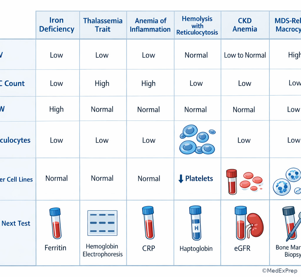
Exam takeaway: A compact side-by-side comparison prevents overcalling iron deficiency or megaloblastic disease from MCV alone.
Algorithm / Approach
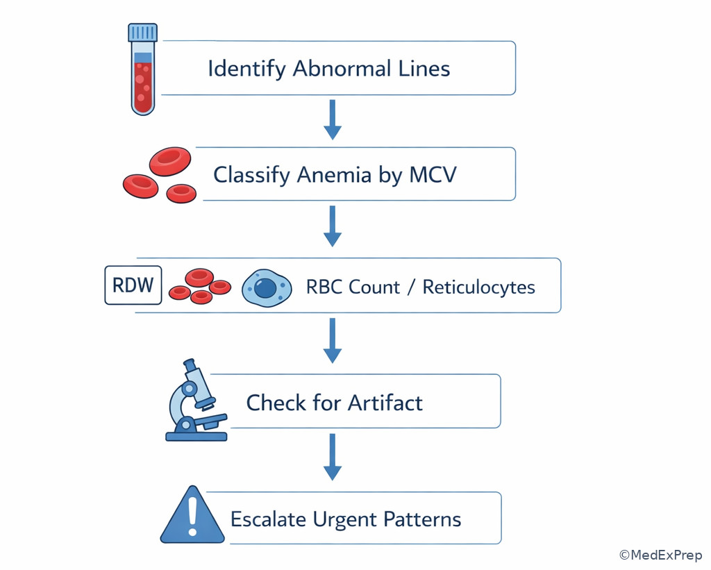
Exam takeaway: A fixed sequence—classify, tie-break, confirm, escalate—reduces common board mistakes.
Rapid Review
- Microcytosis + high RBC count → thalassemia trait over iron deficiency
- Microcytosis + high RDW → evolving or mixed deficiency pattern
- Normocytic anemia + low reticulocytes → underproduction until proven otherwise
- Normocytic anemia + high reticulocytes → blood loss or hemolysis
- Macrocytosis + isolated high MCV → think alcohol, liver disease, hypothyroidism, medications, MDS, B12/folate
- Macrocytosis + high reticulocytes → reticulocyte effect, not automatically megaloblastic disease
- Leukocytosis + anemia + thrombocytopenia → marrow pathology warning
- Thrombocytosis + iron deficiency → reactive until proven otherwise
- Isolated thrombocytopenia + platelet clumps → pseudothrombocytopenia
- Pancytopenia → marrow failure, infiltration, therapy effect, severe deficiency, hypersplenism
- Very abnormal CBC discordant with patient appearance → consider specimen artifact and repeat
- High MCHC with odd indices → consider spherocytosis or agglutination artifact
Memory anchor: For CBC interpretation, think “BARS” — Bucket by MCV, Affected lines, Reticulocytes, Spurious vs serious. If you do those four steps in order, you will avoid most board traps.
Self-check quiz
1. A 31-year-old woman with chronic heavy menses and no family history of anemia has Hgb 10.1 g/dL, MCV 71 fL, RBC count 3.8 million/mcL, and RDW 18%. Which interpretation is most likely?
- Iron deficiency anemia
- Thalassemia trait
- Anemia of chronic inflammation
- Acute blood loss anemia
2. A 66-year-old man with remote alcohol overuse, hypothyroidism, and stable coronary disease has Hgb 11.4 g/dL, MCV 107 fL, normal WBC count, and normal platelets. Which additional finding most strongly favors hemolysis or recovery rather than a primary megaloblastic process?
- Elevated reticulocyte count
- Low folate level
- Elevated thyroid-stimulating hormone
- Prior methotrexate exposure
3. A 48-year-old man with ulcerative colitis, prior iron deficiency, and no active bleeding presents with fatigue. CBC shows Hgb 11.0 g/dL, MCV 67 fL, RBC count 5.9 million/mcL, RDW 12.6%, platelets 330,000/mcL. Which is the most appropriate next step?
- Start empiric oral iron therapy without further testing
- Order iron studies and consider hemoglobin electrophoresis
- Diagnose anemia of inflammation and observe
- Order bone marrow biopsy as the initial test
4. A 59-year-old woman with metastatic ovarian cancer on carboplatin, chronic kidney disease, and recent epistaxis has Hgb 8.7 g/dL, MCV 101 fL, WBC 6,900/mcL, platelets 280,000/mcL. She notes dark stools after NSAID use. Which test best separates blood loss/hemolysis from underproduction in this scenario?
- Reticulocyte count
- Serum ferritin
- Vitamin B12 level
- Erythropoietin level
5. A 72-year-old man with COPD treated with prednisone, recent pneumonia, and no weight loss has WBC 17,800/mcL, Hgb 13.5 g/dL, platelets 290,000/mcL, and neutrophil predominance. Which interpretation is most appropriate?
- Reactive leukocytosis is more likely than marrow malignancy
- Acute leukemia is the most likely diagnosis
- Myelodysplastic syndrome is the most likely diagnosis
- Aplastic anemia is the most likely diagnosis
6. A 64-year-old woman with rheumatoid arthritis, CKD stage 4, and prior gastric bypass has Hgb 8.9 g/dL, MCV 90 fL, reticulocyte count low, WBC 5,400/mcL, and platelets 410,000/mcL. She reports fatigue but no bleeding. Which mechanism is most likely driving this CBC pattern?
- Underproduction anemia from chronic disease/renal insufficiency
- Hemolysis with compensatory marrow response
- Acute occult blood loss with appropriate reticulocytosis
- Pseudodilution from volume overload as the sole explanation
7. A 69-year-old man with prostate cancer on androgen deprivation therapy, recent trimethoprim-sulfamethoxazole exposure, and mild splenomegaly has WBC 3,000/mcL, Hgb 9.2 g/dL, platelets 82,000/mcL, and MCV 104 fL. He has no bleeding and no fever. Which interpretation best fits the CBC pattern?
- Isolated immune thrombocytopenia
- Multilineage process requiring marrow-focused differential
- Pseudothrombocytopenia from EDTA effect alone
- Simple iron deficiency anemia with reactive changes
8. A 58-year-old woman with cold-induced acrocyanosis, lymphoma history, and fatigue has Hgb 9.5 g/dL, MCV 116 fL, MCHC elevated, platelets normal, and analyzer flags agglutination. Which is the most appropriate next step?
- Diagnose megaloblastic anemia and give folate
- Repeat the CBC with attention to possible cold agglutinin artifact
- Diagnose myelodysplastic syndrome based on macrocytosis alone
- Start iron supplementation for mixed deficiency
Answer key
1.1.1.8 1. Answer: A — Iron deficiency anemia
A. Iron deficiency anemia: Tempting because heavy menses and fatigue are classic, and it is correct here because low RBC count with high RDW and microcytosis fits iron-restricted erythropoiesis. The single stem clue is MCV 71 with RBC count 3.8 and RDW 18%. Source: Harrison’s Principles of Internal Medicine; UpToDate.
B. Thalassemia trait: Tempting because thalassemia also causes marked microcytosis, but it is incorrect because the RBC count is not preserved/high and the RDW is elevated. The discriminating clue is low RBC count rather than high RBC count. Source: Harrison’s; UpToDate.
C. Anemia of chronic inflammation: Tempting because chronic inflammatory states can cause anemia, but it is incorrect because this pattern is more strongly microcytic with high RDW and bleeding history, not the typical normocytic or mildly microcytic inflammatory pattern. The clue is heavy menses and pronounced RDW elevation. Source: Harrison’s; Robbins & Cotran.
D. Acute blood loss anemia: Tempting because menstrual bleeding is present, but it is incorrect because acute blood loss is usually normocytic early and would not explain chronic microcytosis. The clue is microcytosis rather than a recent normocytic fall. Source: Harrison’s.
1.1.1.9 2. Answer: A — Elevated reticulocyte count
A. Elevated reticulocyte count: Tempting because it directly addresses marrow kinetics, and it is correct because high reticulocytes indicate response to loss, destruction, or recovery and can themselves raise MCV. The clue is macrocytosis with a question asking for recovery/hemolysis versus megaloblastic disease. Source: Harrison’s; UpToDate.
B. Low folate level: Tempting because folate deficiency is a classic cause of macrocytosis, but it is incorrect because it supports megaloblastic disease rather than arguing against it. The clue is the stem asks what favors hemolysis or recovery. Source: Harrison’s.
C. Elevated thyroid-stimulating hormone: Tempting because hypothyroidism can cause macrocytosis, but it is incorrect because it suggests another underproduction cause, not marrow response. The clue is thyroid disease does not explain reticulocyte-driven macrocytosis. Source: Harrison’s.
D. Prior methotrexate exposure: Tempting because medications can cause macrocytosis, but it is incorrect because it points toward impaired DNA synthesis rather than recovery or hemolysis. The clue is drug effect is not a kinetic response marker. Source: UpToDate; Harrison’s.
1.1.1.10 3. Answer: B — Order iron studies and consider hemoglobin electrophoresis
A. Start empiric oral iron therapy without further testing: Tempting because he has prior iron deficiency and microcytosis, but it is incorrect because the preserved-high RBC count and normal RDW make thalassemia trait a serious competing explanation that should not be ignored. The clue is RBC count 5.9 with RDW 12.6%. Source: Harrison’s; UpToDate.
B. Order iron studies and consider hemoglobin electrophoresis: Tempting because it is more deliberate, and it is correct because the pattern is not classic iron deficiency and requires confirmation of iron status plus evaluation for thalassemia-pattern disease. The clue is microcytosis out of proportion to anemia with high RBC count. Source: Harrison’s; UpToDate.
C. Diagnose anemia of inflammation and observe: Tempting because ulcerative colitis can cause inflammatory anemia, but it is incorrect because this degree of microcytosis with high RBC count is not the most typical inflammatory pattern. The clue is markedly low MCV with preserved RBC count. Source: Robbins & Cotran; Harrison’s.
D. Order bone marrow biopsy as the initial test: Tempting because persistent unexplained anemia can require marrow evaluation, but it is incorrect because targeted noninvasive tests are the appropriate first step in this CBC pattern. The clue is isolated microcytic anemia without multilineage abnormalities. Source: UpToDate; Harrison’s.
1.1.1.11 4. Answer: A — Reticulocyte count
A. Reticulocyte count: Tempting because it is often the forgotten test, and it is correct because it immediately separates underproduction from blood loss or hemolysis in a patient with mixed possible causes of anemia and borderline macrocytosis. The clue is several plausible etiologies with a need to distinguish kinetics. Source: Harrison’s; UpToDate.
B. Serum ferritin: Tempting because NSAID-associated bleeding and cancer raise iron deficiency concern, but it is incorrect because ferritin does not directly distinguish underproduction from hemolysis/blood loss in the moment and may be confounded by inflammation/malignancy. The clue is the stem asks specifically to separate mechanisms. Source: Harrison’s; UpToDate.
C. Vitamin B12 level: Tempting because MCV is 101 fL, but it is incorrect because mild macrocytosis in a patient with possible bleeding may be reticulocyte-related, and B12 does not resolve the key initial branch. The clue is recent blood loss clues and only slight macrocytosis. Source: Harrison’s.
D. Erythropoietin level: Tempting because CKD is present, but it is incorrect because EPO level rarely drives the next board-style step and does not sort loss/destruction from underproduction as efficiently as reticulocytes. The clue is best test to separate mechanisms. Source: UpToDate; Harrison’s.
1.1.1.12 5. Answer: A — Reactive leukocytosis is more likely than marrow malignancy
A. Reactive leukocytosis is more likely than marrow malignancy: Tempting because steroids and infection are common benign explanations, and it is correct because the leukocytosis is isolated, neutrophil-predominant, and occurs in the setting of prednisone and recent pneumonia without other line abnormalities. The clue is normal hemoglobin and platelets with clear reactive triggers. Source: Harrison’s; UpToDate.
B. Acute leukemia is the most likely diagnosis: Tempting because high WBC counts can signal leukemia, but it is incorrect because acute leukemia usually produces broader marrow disruption or immature forms rather than an isolated steroid/infection-compatible neutrophilia. The clue is absence of anemia, thrombocytopenia, or blast concern. Source: Robbins & Cotran; Harrison’s.
C. Myelodysplastic syndrome is the most likely diagnosis: Tempting in older adults with abnormal counts, but it is incorrect because MDS typically causes cytopenias, not isolated neutrophilic leukocytosis from an obvious trigger. The clue is count elevation rather than ineffective hematopoiesis with low lines. Source: Harrison’s.
D. Aplastic anemia is the most likely diagnosis: Tempting only if one focuses on marrow disorders generally, but it is incorrect because aplastic anemia causes pancytopenia or multilineage cytopenia, not isolated leukocytosis. The clue is elevated WBC count with normal other lines. Source: Harrison’s.
1.1.1.13 6. Answer: A — Underproduction anemia from chronic disease/renal insufficiency
A. Underproduction anemia from chronic disease/renal insufficiency: Tempting because CKD and inflammation are both present, and it is correct because the anemia is normocytic with low reticulocytes, the classic underproduction signature in renal/inflammatory states. The clue is reticulocyte count low. Source: Harrison’s; UpToDate.
B. Hemolysis with compensatory marrow response: Tempting because anemia can be severe, but it is incorrect because hemolysis should produce a reticulocyte response unless the marrow cannot respond. The clue is reticulocytes are low, not high. Source: Harrison’s.
C. Acute occult blood loss with appropriate reticulocytosis: Tempting because gastric bypass may predispose to deficiency and occult GI issues, but it is incorrect because the stem explicitly says reticulocytes are low rather than appropriately elevated. The clue is inadequate marrow response. Source: UpToDate; Harrison’s.
D. Pseudodilution from volume overload as the sole explanation: Tempting because CKD can cause fluid retention, but it is incorrect because dilution does not explain the persistent normocytic underproduction pattern as well as CKD/inflammation. The clue is low reticulocyte count indicating true reduced production. Source: Harrison’s.
1.1.1.14 7. Answer: B — Multilineage process requiring marrow-focused differential
A. Isolated immune thrombocytopenia: Tempting because the platelet count is low, but it is incorrect because anemia and leukopenia are also present, arguing strongly against isolated ITP. The clue is three cell lines affected. Source: Harrison’s; UpToDate.
B. Multilineage process requiring marrow-focused differential: Tempting because drug effect, marrow infiltration, and myeloid disease are all possible, and it is correct because leukopenia, anemia, and thrombocytopenia together define a broad marrow or systemic process rather than isolated peripheral destruction. The clue is bicytopenia/pancytopenia-type pattern with macrocytosis. Source: Harrison’s; Robbins & Cotran.
C. Pseudothrombocytopenia from EDTA effect alone: Tempting because platelet artifacts are board-tested, but it is incorrect because artifact would not account for concurrent leukopenia and anemia. The clue is other lines are abnormal too. Source: UpToDate.
D. Simple iron deficiency anemia with reactive changes: Tempting because iron deficiency is common, but it is incorrect because macrocytosis and leukopenia do not fit simple iron deficiency. The clue is MCV 104 with low WBC and platelets. Source: Harrison’s.
1.1.1.15 8. Answer: B — Repeat the CBC with attention to possible cold agglutinin artifact
A. Diagnose megaloblastic anemia and give folate: Tempting because MCV is very high, but it is incorrect because analyzer agglutination and cold-induced symptoms suggest an artifact or hemolytic process rather than isolated folate deficiency. The clue is agglutination flag with cold symptoms. Source: Harrison’s; UpToDate.
B. Repeat the CBC with attention to possible cold agglutinin artifact: Tempting because it seems cautious, and it is correct because cold agglutinins can distort indices, especially MCV and MCHC, and should be confirmed before assigning a diagnosis. The clue is elevated MCHC plus analyzer flags agglutination. Source: UpToDate; Harrison’s.
C. Diagnose myelodysplastic syndrome based on macrocytosis alone: Tempting because older patients with macrocytosis can have MDS, but it is incorrect because the immediate issue is a potentially spurious or hemolysis-related index abnormality. The clue is agglutination artifact signal. Source: Harrison’s.
D. Start iron supplementation for mixed deficiency: Tempting because fatigue and anemia are present, but it is incorrect because neither the macrocytosis nor the agglutination clue supports iron deficiency as the immediate explanation. The clue is cold-related agglutination rather than microcytic deficiency pattern. Source: Harrison’s; UpToDate.
Keep going with the full ABIM Hematology & Oncology Prep Course (Hem/Onc)
You just studied the way members do, in the same lesson interface. Full access unlocks every lesson, the question bank and flashcards for a full year.
One payment · 1 year of access · No auto-renewal