
Why it matters
- ABIM-style stems often hide the diagnosis inside hemodynamics: the loop tells you whether the ventricle cannot fill, cannot eject, cannot relax, or leaks across a valve.
- Pressure-volume reasoning converts memorized facts into decision logic: if stroke volume falls, the next question is whether preload, contractility, afterload, or valvular regurgitation explains it.
- Many “next best step” cardiology questions depend on knowing what therapy should move the loop left, down, wider, or narrower.
- This topic is a foundation for the next lessons on filling pressures and shock profiles; if the loop is unclear, catheter data will feel harder than it should. See Fig 1.
Key Terms & Must-Know Facts
1.1.1.1 Key terms
- EDV: end-diastolic volume
- ESV: end-systolic volume
- Stroke volume: \(SV = EDV - ESV\)
- Ejection fraction: \(EF = SV/EDV\)
- Preload: ventricular wall stress at end diastole; clinically tracks filling
- Afterload: load the ventricle must overcome during ejection
- Contractility: intrinsic inotropic state independent of load
- Compliance: change in volume per change in pressure; low compliance means stiff ventricle
- ESPVR: end-systolic pressure-volume relationship; slope reflects contractility
- EDPVR: end-diastolic pressure-volume relationship; reflects passive filling properties/compliance
- Isovolumic contraction: pressure rises with no volume change
- Isovolumic relaxation: pressure falls with no volume change
- Loop width: stroke volume
- Loop left border/right border: ESV/EDV landmarks
1.1.1.2 Must-know facts
- A wider loop means higher stroke volume; a narrower loop means lower stroke volume.
- Increased preload shifts the right border rightward and usually increases loop width if contractility is intact.
- Increased afterload raises systolic pressure and increases ESV, usually narrowing the loop.
- Increased contractility shifts the end-systolic point up and left, lowering ESV and increasing stroke volume.
- Decreased compliance shifts the diastolic filling curve up and left: small volume creates high filling pressure.
- Aortic stenosis increases afterload; aortic regurgitation abolishes isovolumic phases.
- Mitral regurgitation also abolishes isovolumic phases because volume changes while the ventricle should be closed.
- Mitral stenosis lowers LV preload and produces a small loop with reduced EDV.
- Loop interpretation is easier if you first ask: where are the borders, how tall is the loop, and are the vertical isovolumic limbs present?
- On exams, the single most useful discriminator is often whether ESV rose, EDV rose, or both.
| Parameter | What to look for on the loop | Board interpretation |
|---|---|---|
| EDV | Rightmost point | Higher EDV usually means higher preload |
| ESV | Leftmost point | Higher ESV suggests impaired ejection or higher afterload |
| Stroke volume | Horizontal width | Narrow loop = low forward output |
| Systolic pressure | Top of loop | Tall loop often implies high afterload or pressure generation |
| Compliance | Diastolic filling curve position | Up/left shift = stiff ventricle |
| Contractility | ESPVR slope | Steeper slope = stronger ventricle |
Core content
1.1.1.3 Foundational map of the normal pressure-volume loop
A Explanation
The decision conflict in a loop question is usually simple but easy to overcomplicate: are you being asked to identify a phase or a perturbation? For a normal loop, choose the phase first. The resolving rule is this: vertical limbs are isovolumic, horizontal movement reflects filling or ejection. Start at the bottom right: the mitral valve closes at end diastole, isovolumic contraction moves up, the aortic valve opens and ejection moves leftward, the aortic valve closes at end systole, and isovolumic relaxation moves down until the mitral valve opens and filling resumes. WHY THIS IS TESTED: exam items often give one valve event, one pressure change, or one border point and ask you to infer the rest of the cycle.
If you do not know where EDV and ESV live, every disease loop becomes harder. EDV is the rightmost point. ESV is the leftmost point. Loop width is stroke volume. The upper left corner is where ejection ends and the aortic valve closes. The lower left corner is where relaxation ends and the mitral valve opens. See Fig 1 for the core phase framework and Fig 2 for the geometric landmarks.
Exam takeaway: Anchor the loop by valve opening and closing events before judging preload, afterload, or contractility.
flowchart LR
A[Mitral valve opens
ventricular filling begins] --> B[Mitral valve closes
EDV reached]
B --> C[Isovolumic contraction
pressure rises volume fixed]
C --> D[Aortic valve opens
ejection begins]
D --> E[Aortic valve closes
ESV reached]
E --> F[Isovolumic relaxation
pressure falls volume fixed]
F --> A
Exam takeaway: Right border marks EDV, left border marks ESV, and loop width equals stroke volume.
{"v":1,"bbox":[-10,160,170,-10],"axis":true,"grid":false,"w":720,"h":360,"objects":[{"type":"point","name":"A","x":120,"y":10},{"type":"point","name":"B","x":120,"y":110},{"type":"point","name":"C","x":50,"y":130},{"type":"point","name":"D","x":50,"y":10},{"type":"segment","a":"A","b":"B"},{"type":"segment","a":"B","b":"C"},{"type":"segment","a":"C","b":"D"},{"type":"segment","a":"D","b":"A"},{"type":"text","x":125,"y":8,"text":"EDV"},{"type":"text","x":42,"y":8,"text":"ESV"},{"type":"text","x":75,"y":145,"text":"Ejection"},{"type":"text","x":123,"y":65,"text":"Iso contraction"},{"type":"text","x":20,"y":70,"text":"Iso relaxation"},{"type":"text","x":75,"y":3,"text":"Filling"}]} Exam takeaway: Use borders first, then height, then presence or absence of isovolumic phases.
B Worked example
A 62-year-old man with hypertension, type 2 diabetes, and obstructive sleep apnea undergoes evaluation for exertional dyspnea. He has mild anemia on recent labs, which is mentioned prominently in the stem, and a soft S4 on examination. A schematic loop question asks which point represents aortic valve closure. The tempting wrong clue is the dyspnea and anemia, which invite clinical overthinking, but the task is phase identification, not diagnosis.
Reasoning chain: Identify task — locate the valve event. Extract key facts — this is a normal schematic rather than a disease comparison. Apply rule — aortic valve closure occurs at the end of ejection and start of isovolumic relaxation, which is the upper left corner. Eliminate distractors — the upper right corner is aortic valve opening; lower right is mitral valve closure; lower left is mitral valve opening. Verify — the upper left point is ESV, matching end systole.
C Exam trap
D Checkpoint
Question: On a normal left ventricular pressure-volume loop, which event occurs at the lower left corner?
- Mitral valve opening
- Mitral valve closure
- Aortic valve opening
- Aortic valve closure
Answer: A. A: Tempting because the lower left point follows isovolumic relaxation; correct because mitral opening begins ventricular filling. B: Tempting because mitral closure is a diastolic-systolic transition; wrong because closure occurs at the lower right corner at EDV. C: Tempting because aortic opening also marks a transition; wrong because it occurs at the upper right corner after isovolumic contraction. D: Tempting because aortic closure is another landmark corner; wrong because it is the upper left corner at end ejection.
1.1.1.4 How preload, afterload, contractility, and compliance reshape the loop
A Explanation
The classic board conflict here is choosing among four mechanisms that all reduce cardiac output. The resolving rule is to identify which border moved and whether the ESPVR or EDPVR changed. Increased preload moves the right border rightward: EDV rises, stroke volume often rises by Frank-Starling if systolic function is preserved. Increased afterload makes the ventricle end ejection at a larger volume, so the left border shifts rightward and the loop narrows while peak pressure rises. Increased contractility shifts the end-systolic point up and left, reducing ESV and widening the loop. Decreased compliance shifts the diastolic filling relation up and left: the same filling pressure produces less EDV, and a small increase in volume produces a large rise in diastolic pressure.
WHY THIS IS TESTED: many items describe a drug, a blood pressure change, or a lesion and ask which loop alteration follows. Choose-vs-reject framing helps: choose preload if EDV primarily changes; reject pure preload if ESV is the main abnormality. Choose afterload if pressure rises and ESV increases; reject afterload if ESV actually falls. Choose contractility if ESPVR steepens and ESV falls. Choose impaired compliance when filling pressure is high despite modest filling volume. See Fig 3 and Fig 4.
Exam takeaway: Border movement identifies the dominant mechanism faster than memorizing disease names.
flowchart TD
A[Loop abnormality] --> B{Main border shift?}
B -->|Right border right| C[Higher EDV -> increased preload]
B -->|Left border right| D[Higher ESV -> increased afterload or lower contractility]
B -->|Left border left| E[Lower ESV -> increased contractility]
B -->|Diastolic curve up/left| F[Reduced compliance]
D --> G{Pressure high?}
G -->|Yes| H[Afterload]
G -->|No or low| I[Reduced contractility] Exam takeaway: Compare loop overlays visually to separate changes in EDV, ESV, pressure, and diastolic stiffness.
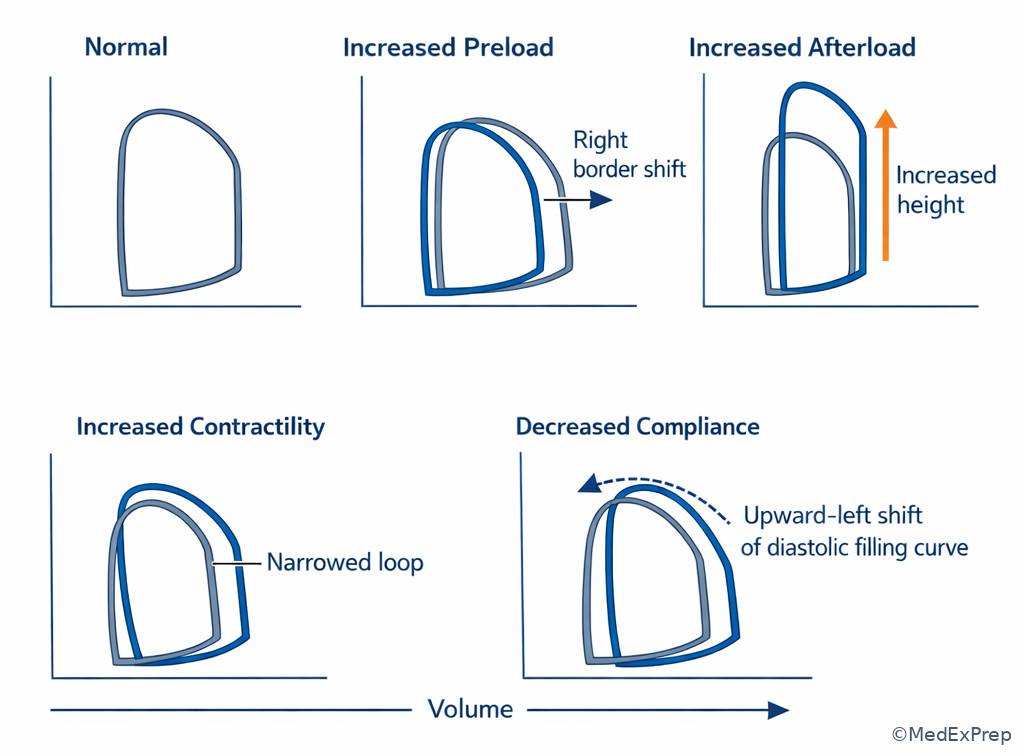
Exam takeaway: Ask which relationship changed before attaching a disease label.
B Worked example
A 71-year-old woman with long-standing hypertension, chronic kidney disease, and paroxysmal atrial fibrillation presents with acute pulmonary edema. Blood pressure is 198/102 mm Hg. Troponin is minimally elevated, creating a tempting ischemic distractor, but ECG shows only LVH with strain. A loop comparison asks which intervention would most immediately shift the end-systolic point leftward and increase stroke volume.
Reasoning chain: Identify task — determine the dominant variable and how therapy changes the loop. Extract key facts — hypertensive emergency physiology, marked afterload excess, acute pulmonary edema. Apply rule — excess afterload increases ESV and narrows the loop; reducing afterload should lower systolic pressure burden and shift the end-systolic point leftward. Eliminate distractors — pure fluid administration worsens congestion; slowing heart rate may help some patients but does not directly correct the high ESV from severe afterload; positive inotropy is not first-line for a markedly hypertensive wet patient. Verify — vasodilator therapy best matches the desired loop movement.
C Exam trap
D Checkpoint
Question: Which loop change is most consistent with an isolated increase in afterload?
- Rightward shift of EDV with preserved ESV
- Leftward shift of ESV with increased loop width
- Rightward shift of ESV with increased peak systolic pressure
- Up-left shift of the diastolic filling curve with unchanged ESPVR
Answer: C. A: Tempting because EDV often rises in compensation; wrong because isolated preload increase primarily changes EDV, not ESV. B: Tempting because improved performance widens the loop; wrong because this pattern fits increased contractility. C: Tempting because both pressure and ESV rise under a higher systolic load; correct because afterload makes ejection harder, leaving more blood behind. D: Tempting because diastolic changes alter filling; wrong because this is decreased compliance, not afterload.
- The loop width represents stroke volume.
- True or False: Increased contractility usually increases ESV. False.
- Name the hemodynamic variable most associated with an up-left shift of the diastolic curve: decreased compliance.
1.1.1.5 Systolic dysfunction and diastolic dysfunction: when low output comes from weak squeeze versus stiff filling
A Explanation
The decision conflict here is whether a small forward output reflects impaired contractility or impaired filling. The single finding that resolves it is the relationship between ESV, EDV, and diastolic pressure. In systolic dysfunction, contractility falls, ESPVR becomes less steep, ESV rises, and stroke volume falls; EDV may also rise over time because the ventricle dilates. In diastolic dysfunction, the ventricle is stiff, EDPVR shifts up and left, filling pressures rise, but EDV may be normal or even reduced. That means a patient can have pulmonary congestion with a non-dilated ventricle and relatively preserved EF because the filling pressure—not necessarily cavity size—is the problem.
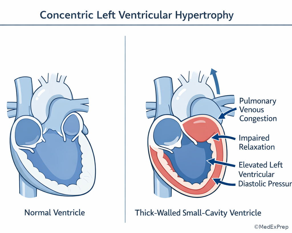
WHY THIS IS TESTED: board stems often present dyspnea plus elevated natriuretic peptides or left atrial enlargement and ask you to infer whether the loop abnormality is contractile failure or reduced compliance. Choose systolic dysfunction when ESV is the key abnormality and EF falls because the ventricle cannot empty. Choose diastolic dysfunction when small volume increments cause large pressure rises, especially in hypertensive LVH, infiltrative disease, or aging myocardium. Reject pure systolic dysfunction if the stem emphasizes concentric LVH, preserved EF, S4, and high filling pressures disproportionate to EDV. See Fig 5.
Exam takeaway: High ESV points to weak systolic emptying, whereas high filling pressure at low-normal volume points to diastolic stiffness.
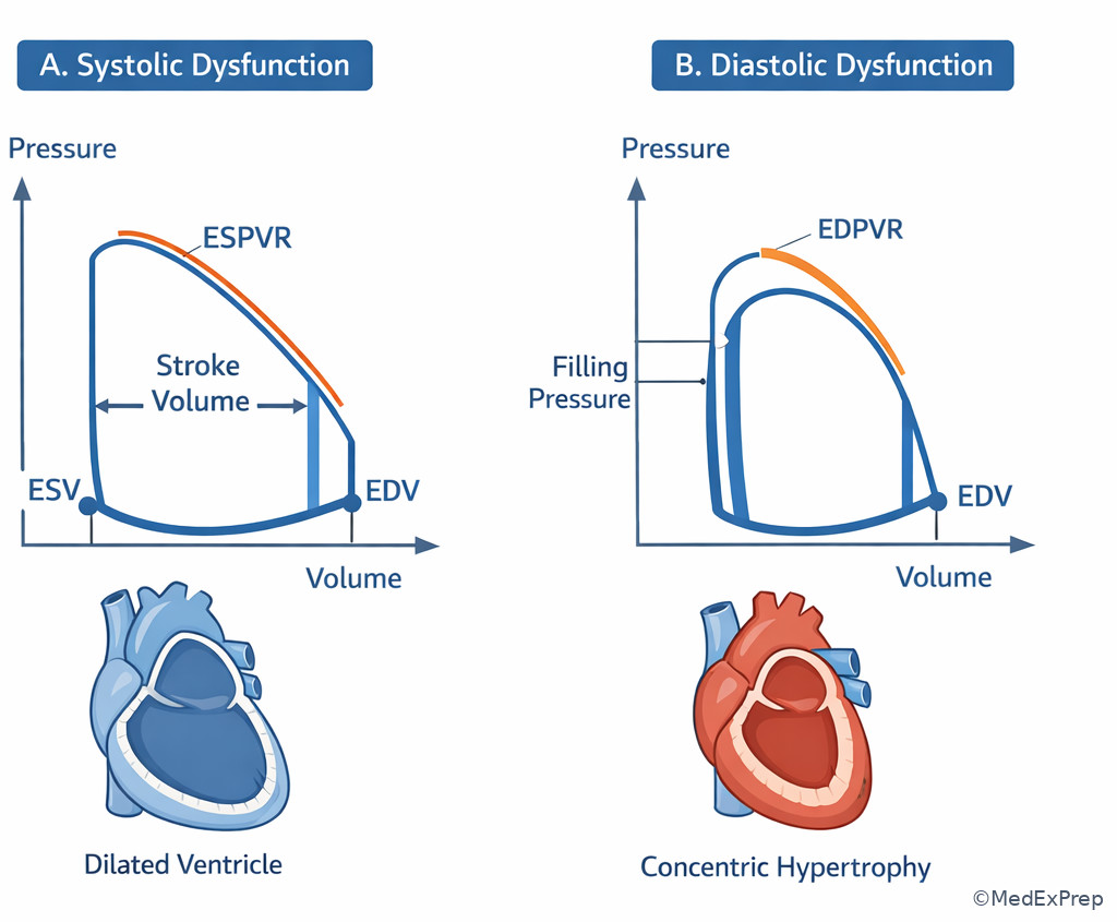
Exam takeaway: Concentric hypertrophy reduces compliance and raises filling pressure before EF must fall.
Exam takeaway: Separate failure to empty from failure to fill by locating the dominant abnormal relationship.
B Worked example
A 76-year-old woman with obesity, long-standing hypertension, chronic kidney disease, and no prior MI presents with exertional dyspnea and episodic orthopnea. She has an S4, bibasilar crackles, preserved blood pressure, and normal troponin. Echocardiography shows EF 62%, concentric LVH, and left atrial enlargement. A hemodynamic question asks which loop change is most likely present. The distracting clue is crackles, which may push the reader toward systolic failure, but the preserved EF and LVH pattern point elsewhere.
Reasoning chain: Identify task — choose loop physiology. Extract key facts — preserved EF, concentric hypertrophy, elevated filling phenotype, no infarct history. Apply rule — reduced compliance shifts the EDPVR upward and leftward, so diastolic pressure rises at relatively low volume. Eliminate distractors — a major rise in ESV with flattened ESPVR would fit systolic dysfunction; absent isovolumic phases would suggest regurgitation; isolated preload increase does not explain chronic stiff-ventricle features. Verify — the stem is classic HFpEF mechanics.
C Exam trap
D Checkpoint
Question: Which loop feature most strongly favors diastolic dysfunction over isolated systolic dysfunction?
- Marked increase in ESV due to flatter ESPVR
- Upward-left shift of the end-diastolic pressure-volume relationship
- Loss of isovolumic relaxation and isovolumic contraction
- Large rightward shift in EDV with increased loop width
Answer: B. A: Tempting because systolic failure is common; wrong because this is the hallmark of impaired contractility. B: Tempting because stiffness changes the passive filling relation; correct because decreased compliance raises diastolic pressure at lower volumes. C: Tempting because diastolic disease affects relaxation; wrong because complete loss of isovolumic phases points more to regurgitant valvular lesions. D: Tempting because congestion may coexist with higher volume; wrong because this suggests preload increase rather than a primary compliance abnormality.
1.1.1.6 Valvular lesions on loops: stenosis versus regurgitation patterns
A Explanation
The decision conflict is whether a valve lesion acts mainly as a pressure overload, a volume overload, or a filling obstruction. The single best rule: regurgitant lesions abolish isovolumic phases; stenotic lesions preserve them but distort pressure or filling. Aortic stenosis behaves like severe afterload excess: high systolic pressure, increased ESV, often compensatory LV hypertrophy, and a narrowed loop. Aortic regurgitation causes marked volume overload; because blood runs into the LV during diastole and some ejected blood returns during diastole, the classic loop is very wide with increased EDV and absent true isovolumic relaxation/contraction. Mitral regurgitation also abolishes isovolumic phases because during what should be isovolumic contraction, blood can move backward into the left atrium; forward stroke volume may be low even when total stroke volume is high. Mitral stenosis restricts LV filling, decreasing preload and EDV, producing a small loop.
WHY THIS IS TESTED: these are among the most recognizable pressure-volume patterns on board exams. Choose regurgitation when the vertical limbs disappear; reject stenosis if the question explicitly says isovolumic contraction is absent. Choose aortic stenosis when afterload physiology dominates. Choose mitral stenosis when the ventricle itself may contract normally but starts with too little volume. See Fig 6.
Exam takeaway: Absent isovolumic phases strongly favor regurgitation, whereas pressure overload or restricted filling favors stenosis.
flowchart TD
A[Abnormal valve loop] --> B{Are vertical isovolumic limbs present?}
B -->|No| C{Which chamber volume pattern?}
C -->|Very large EDV and wide loop| D[Aortic regurgitation]
C -->|Backward flow into LA with low forward output| E[Mitral regurgitation]
B -->|Yes| F{Main issue?}
F -->|High systolic pressure and higher ESV| G[Aortic stenosis]
F -->|Small EDV from impaired filling| H[Mitral stenosis] Exam takeaway: Side-by-side valve loop patterns are faster to recognize than isolated memorized descriptions.
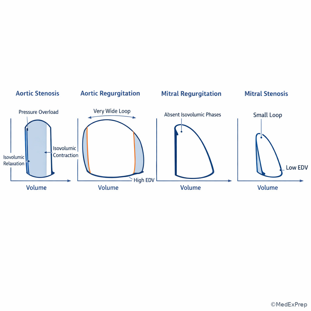
Exam takeaway: Pressure overload and volume overload create different loop signatures even when both involve the aortic valve.
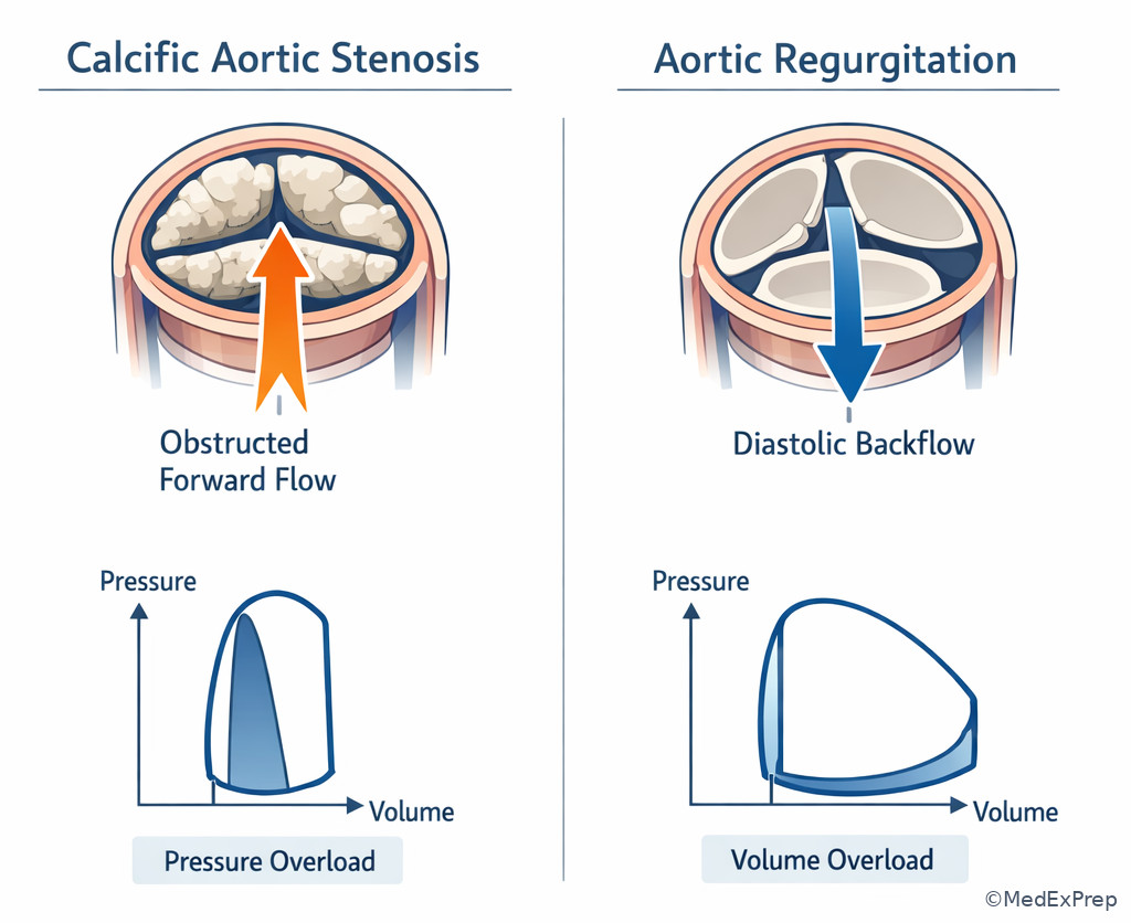
Exam takeaway: Use isovolumic phases first, then decide whether the problem is pressure overload, volume overload, or filling obstruction.
B Worked example
A 58-year-old man with bicuspid aortic valve history, hypertension, and hyperlipidemia reports progressive exertional dyspnea and exertional presyncope. Creatinine is mildly elevated, a distracting detail that may make catheterization feel central, but the current task is loop recognition. A diagram shows a tall loop with increased systolic pressure, preserved isovolumic phases, and increased ESV. Which lesion best fits?
Reasoning chain: Identify task — classify valve lesion by loop morphology. Extract key facts — pressure overload pattern, preserved vertical limbs, increased ESV, symptomatic outflow obstruction phenotype. Apply rule — stenotic lesions preserve isovolumic phases; aortic stenosis behaves like increased afterload. Eliminate distractors — aortic regurgitation should show a very wide loop and no true isovolumic phases; mitral regurgitation also loses isovolumic phases; mitral stenosis produces a small underfilled loop rather than a high-pressure ejection loop. Verify — symptomatic bicuspid valve disease supports severe aortic stenosis.
C Exam trap
D Checkpoint
Question: Which lesion most characteristically abolishes both isovolumic contraction and isovolumic relaxation while markedly increasing EDV?
- Aortic stenosis
- Aortic regurgitation
- Mitral stenosis
- Hypertrophic cardiomyopathy
Answer: B. A: Tempting because outflow valve disease changes systolic pressure; wrong because aortic stenosis preserves isovolumic phases. B: Tempting because severe regurgitant backflow creates continuous volume movement; correct because aortic regurgitation causes marked volume overload and no true closed-valve isovolumic segments. C: Tempting because a stenotic valve alters filling; wrong because mitral stenosis underfills the LV but does not abolish both isovolumic phases. D: Tempting because dynamic obstruction affects pressure generation; wrong because it is not the classic lesion causing absent isovolumic phases with very high EDV.
- The valve lesion most associated with a tall pressure-overload loop is aortic stenosis.
- True or False: Mitral regurgitation preserves a normal isovolumic contraction phase. False.
- Name the lesion that primarily reduces LV EDV by obstructing filling: mitral stenosis.
1.1.1.7 Integrating loops with bedside hemodynamics, therapy effects, and exam strategy
A Explanation
The final decision conflict is not “what does the loop look like?” but “what should I infer clinically?” The single resolving rule is to translate loop morphology into the expected bedside pressure and management direction. A stiff ventricle means high filling pressures and pulmonary congestion at modest volumes. A weak ventricle means high ESV, reduced forward output, and often chronic dilation. Increased afterload may improve by vasodilation if blood pressure allows. Pure preload deficiency produces a small loop with low EDV and low pressure. Regurgitation can produce a deceptively large total stroke volume with low forward stroke volume. On board questions, the best answer usually targets the dominant abnormality rather than a generic heart failure response.
WHY THIS IS TESTED: ABIM-style cardiology stems blend physiology with “most appropriate next step.” If the loop suggests acute pressure overload, afterload reduction matters. If the loop suggests severe valvular obstruction, definitive intervention and urgent echo matter. If the loop suggests low preload in shock, indiscriminate vasodilators are harmful. If the loop suggests high filling pressure from stiffness, aggressive fluids worsen symptoms. See Fig 1 again for the basic cycle, then connect it to the bedside through the algorithm below.
Exam takeaway: The loop predicts which hemodynamic number will be high or low and which therapy is most likely to help.
flowchart LR
A[Loop pattern] --> B[Main hemodynamic problem]
B --> C[Expected bedside finding]
C --> D[Best immediate management direction]
A1[High ESV + low SV] --> B1[Poor systolic emptying]
B1 --> C1[Low output signs]
C1 --> D1[HF therapy or inotropy if shock]
A2[Up-left EDPVR] --> B2[High filling pressure]
B2 --> C2[Pulmonary congestion]
C2 --> D2[Diuresis and BP control]
A3[Tall narrow loop] --> B3[High afterload]
B3 --> C3[Hypertension or outflow obstruction]
C3 --> D3[Afterload reduction or valve evaluation]
Exam takeaway: Pressure-volume abnormalities should predict filling pressures and output trends before you see the catheter numbers.
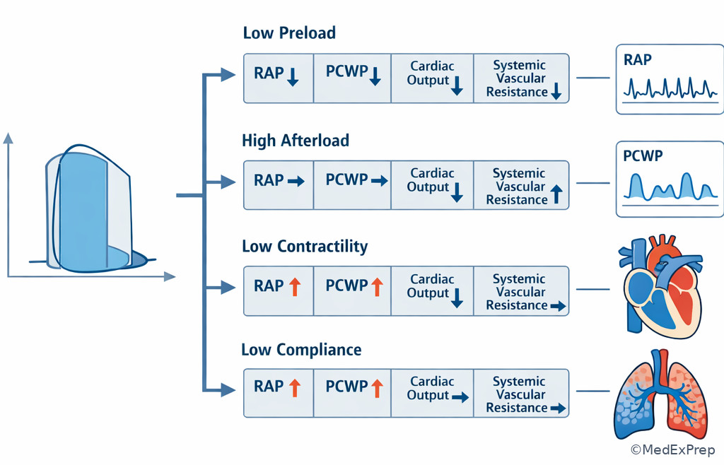
Exam takeaway: Identify the dominant hemodynamic error, then choose the therapy that reverses that specific loop change.
B Worked example
A 68-year-old woman with ischemic cardiomyopathy, CKD stage 3, and chronic atrial fibrillation presents with cold extremities, narrow pulse pressure, and worsening dyspnea. Blood pressure is 86/58 mm Hg. She received a liter of IV fluid in the ED because of a borderline lactate, a tempting but potentially harmful clue. A loop-based question asks which dominant abnormality best explains her low cardiac output. Exam reveals elevated JVP and pulmonary rales.
Reasoning chain: Identify task — determine which hemodynamic variable is limiting output. Extract key facts — low BP, congestion, elevated JVP, ischemic cardiomyopathy, not volume depletion. Apply rule — a failing systolic ventricle has high ESV and low stroke volume; giving more preload when filling pressures are already high will not solve the main problem. Eliminate distractors — low preload would not fit JVP elevation and rales; isolated increased afterload is less likely in hypotension; pure diastolic dysfunction is possible but the ischemic cardiomyopathy history plus cold profile favors systolic failure. Verify — the dominant loop abnormality is impaired contractility with elevated ESV.
C Exam trap
D Checkpoint
Question: A patient with acute hypertensive pulmonary edema has a loop showing high peak systolic pressure and increased ESV. Which intervention most directly targets the dominant abnormality?
- Rapid isotonic fluid bolus
- Afterload reduction with vasodilator therapy
- Immediate permanent pacemaker implantation
- Broad-spectrum antibiotics
Answer: B. A: Tempting because low forward output may suggest underfilling; wrong because congestion with afterload excess worsens with fluid. B: Tempting because the loop shows pressure-loaded impaired ejection; correct because reducing afterload lowers ESV and improves stroke volume if blood pressure permits. C: Tempting if pulmonary edema is misread as bradycardic low output; wrong because no rhythm clue identifies pacing as the main fix. D: Tempting if dyspnea is misattributed to infection; wrong because the loop directly indicates a cardiovascular loading problem.
Exam Traps & Differentiators
- Most common wrong answer and why: choosing “increased preload” whenever stroke volume rises or the loop shifts right. This is tempting because Frank-Starling is familiar, but it fails when ESV is the main abnormality or when a regurgitant lesion is present.
- Looks similar but isn't: aortic stenosis versus systolic dysfunction. Both may reduce stroke volume, but AS produces a high-pressure afterload loop; systolic dysfunction produces flatter ESPVR and poor emptying.
- Looks similar but isn't: HFpEF loop versus hypovolemia. Both may show relatively small volumes, but HFpEF has high diastolic pressure at low-normal volumes; hypovolemia has low volume and low filling pressure.
- Looks similar but isn't: mitral stenosis versus aortic stenosis. Both reduce output; mitral stenosis underfills the LV, whereas aortic stenosis overloads systolic pressure.
- Looks similar but isn't: aortic regurgitation versus mitral regurgitation. Both lose isovolumic phases, but AR usually produces more dramatic EDV increase and wide total stroke volume.
| Stem says... | Think... | Single discriminator |
|---|---|---|
| High systolic pressure, increased ESV, preserved isovolumic phases | Aortic stenosis or increased afterload | Pressure overload pattern |
| Absent isovolumic contraction and relaxation | Regurgitant valve lesion | Volume is changing while valves should be closed |
| Concentric LVH, S4, preserved EF, dyspnea | Diastolic dysfunction/HFpEF physiology | Up-left EDPVR shift |
| Cold, congested patient with ischemic cardiomyopathy | Reduced contractility | High ESV with low forward SV |
| Small loop, low EDV, low pressure | Low preload | Right border shifted left |
| Markedly wide loop with large EDV and regurgitant clue | Aortic regurgitation | Volume overload plus absent isovolumic phases |
| Comparison | Shared feature | Key difference | Board choice |
|---|---|---|---|
| Afterload increase vs reduced contractility | Both can increase ESV and reduce SV | Afterload raises peak systolic pressure; reduced contractility flattens ESPVR | Use pressure top and contractility line |
| HFpEF vs hypovolemia | Both may have smaller LV volume | HFpEF has high filling pressure; hypovolemia has low filling pressure | Use EDPVR and congestion clues |
| Aortic regurgitation vs mitral regurgitation | Both lose isovolumic phases | AR more dramatically increases EDV and total stroke volume | Use volume overload severity and valve context |
| Aortic stenosis vs mitral stenosis | Both can reduce forward output | AS is pressure overload; MS is underfilling | Use pressure burden versus low EDV |
If the stem says exertional syncope, late-peaking systolic murmur, and a tall narrow loop, think aortic stenosis.
If the stem says preserved EF, LVH, S4, pulmonary congestion, and high filling pressure, think diastolic dysfunction.
If the stem says absent isovolumic phases with a very wide loop, think regurgitant lesion, especially aortic regurgitation.
If the stem says hypotension plus elevated JVP and rales, think low output from poor emptying or stiff filling, not automatic volume depletion.
If the stem says hypertensive flash pulmonary edema with high ESV, think afterload reduction if clinically safe.
Algorithm / Approach
Rapid Review
- EDV → rightmost loop point
- ESV → leftmost loop point
- Stroke volume → horizontal width, not loop height
- Increased preload → right border rightward with larger SV if inotropy intact
- Increased afterload → higher pressure + higher ESV
- Increased contractility → lower ESV via steeper ESPVR
- Reduced compliance → high diastolic pressure at lower volume
- Systolic dysfunction → flatter ESPVR, increased ESV
- Diastolic dysfunction → up-left EDPVR, congestion despite preserved EF
- Aortic stenosis → pressure overload loop with preserved isovolumic phases
- Aortic regurgitation → very wide loop, increased EDV, absent isovolumic phases
- Mitral regurgitation → absent isovolumic phases, low forward SV despite possible large total SV
- Mitral stenosis → small underfilled LV loop with low EDV
- High loop top alone → think afterload before preload
- Elevated JVP + rales → reject routine fluid bolus unless the stem clearly proves low preload
Self-check quiz
1. A 64-year-old man with hypertension and hyperlipidemia is shown a normal left ventricular pressure-volume loop during a board review session. Which loop feature most directly represents stroke volume?
- The loop height
- The loop width
- The slope of the EDPVR
- The pressure at the upper right corner
2. A 70-year-old woman with obesity and longstanding hypertension has dyspnea, concentric LVH, and EF 60%. Which loop change best matches the dominant abnormality?
- Upward-left shift of the end-diastolic pressure-volume relationship
- Marked flattening of the ESPVR with large rise in ESV
- Loss of isovolumic contraction from regurgitant flow
- Large rightward shift of EDV from isolated volume loading
3. A 72-year-old man with diabetes, CKD, and severe hypertension presents with flash pulmonary edema. Blood pressure is 210/110 mm Hg, JVP is mildly elevated, and ECG shows LVH without STEMI. A loop would most likely show increased peak systolic pressure and increased ESV. What is the most appropriate next hemodynamic direction?
- Increase preload with rapid isotonic saline infusion
- Reduce afterload with vasodilator therapy if no contraindication
- Slow sinus rate with permanent pacing
- Increase contractility first with routine inotrope despite severe hypertension
4. A 59-year-old man with bicuspid aortic valve, exertional angina, and presyncope has a loop with preserved vertical isovolumic limbs, high systolic pressure, and increased ESV. Which diagnosis is most likely?
- Aortic stenosis
- Aortic regurgitation
- Mitral regurgitation
- Mitral stenosis
5. A 67-year-old woman with prior anterior MI, reduced EF, COPD, and CKD has worsening fatigue and cool extremities. JVP is elevated, lungs have crackles, and blood pressure is 88/60 mm Hg. Which loop abnormality best explains her low forward output?
- Low EDV from isolated hypovolemia
- High ESV from reduced contractility
- High EDV from isolated increased preload with preserved squeeze
- Absent isovolumic phases from mitral stenosis
6. A 61-year-old man with rheumatic heart disease, atrial fibrillation, and progressive exertional dyspnea has a diastolic rumble and pulmonary hypertension. A loop-based question asks which pattern best fits the dominant lesion. Which is most appropriate?
- Small loop with decreased EDV due to impaired LV filling
- Tall loop with increased systolic pressure due to outflow obstruction
- Very wide loop with markedly increased EDV and absent isovolumic phases
- Flattened ESPVR with large ESV rise from primary systolic failure
7. A 65-year-old woman with chronic hypertension, diabetes, and preserved EF develops acute dyspnea after receiving two liters of IV fluid for presumed sepsis in the ED. She has bibasilar crackles, S4, BP 168/92 mm Hg, and no ischemic ECG changes. Which interpretation best requires immediate clarification before more fluid is given?
- Her loop likely reflects reduced compliance with high filling pressures at modest volumes
- Her loop likely reflects isolated low preload and needs another fluid bolus
- Her loop likely reflects absent isovolumic phases from acute aortic regurgitation
- Her loop likely reflects fixed severe aortic stenosis as the sole abnormality
8. A 57-year-old man with endocarditis develops abrupt pulmonary edema and hypotension. A new holosystolic murmur is heard at the apex. A loop diagram shows no true isovolumic contraction and reduced effective forward output despite substantial total stroke volume. Which diagnosis is most likely?
- Acute mitral regurgitation
- Severe mitral stenosis
- Chronic aortic stenosis
- Restrictive cardiomyopathy
Answer key
1.1.1.8 1. Question 1
Correct answer: B. The loop width.
A: Tempting because a tall loop feels like “more work” or “more output.” Incorrect for this scenario because height reflects pressure generation, not directly the amount ejected. Single clue: stroke volume is EDV - ESV , a horizontal distance. Source: Braunwald’s Heart Disease; Costanzo Physiology.
B: Tempting because width literally spans EDV to ESV. Correct because stroke volume is the difference between rightmost and leftmost points. Single clue: the question asks what most directly represents ejected volume. Source: Costanzo Physiology; AHA/ACC hemodynamic principles.
C: Tempting because diastolic properties influence filling. Incorrect because the EDPVR slope reflects compliance, not the amount ejected per beat. Single clue: compliance is a pressure-volume relation during filling, not a beat-to-beat ejection measure. Source: Costanzo Physiology.
D: Tempting because upper-right pressure is a familiar landmark. Incorrect because a point pressure does not encode the entire ejected volume. Single clue: stroke volume requires two volume endpoints. Source: Braunwald’s Heart Disease.
1.1.1.9 2. Question 2
Correct answer: A. Upward-left shift of the end-diastolic pressure-volume relationship.
A: Tempting because HFpEF physiology centers on stiffness. Correct because concentric LVH with preserved EF and dyspnea implies reduced compliance and higher filling pressures at smaller volumes. Single clue: preserved EF plus concentric LVH. Source: ACC/AHA Heart Failure Guideline; Braunwald’s Heart Disease.
B: Tempting because dyspnea is often equated with systolic failure. Incorrect because a marked ESV rise from flattened ESPVR fits HFrEF, not the preserved EF phenotype given. Single clue: EF 60%. Source: ACC/AHA Heart Failure Guideline.
C: Tempting because symptoms can be severe. Incorrect because absent isovolumic contraction suggests regurgitation, which is not indicated by the stem. Single clue: hypertensive concentric LVH is a stiffness clue, not valve regurgitation. Source: Braunwald’s Heart Disease.
D: Tempting because volume loading can cause dyspnea. Incorrect because the stem describes chronic diastolic dysfunction, not isolated preload expansion. Single clue: long-standing hypertension with concentric LVH. Source: Costanzo Physiology.
1.1.1.10 3. Question 3
Correct answer: B. Reduce afterload with vasodilator therapy if no contraindication.
A: Tempting because pulmonary edema can coexist with low output and clinicians may fear underfilling. Incorrect because the loop clue is severe afterload excess with increased ESV, and more fluid worsens congestion. Single clue: BP 210/110 mm Hg with flash pulmonary edema. Source: ACC/AHA HF guidance; Braunwald’s Heart Disease.
B: Tempting because reducing systemic resistance improves ejection under a high-pressure load. Correct because afterload excess is the dominant hemodynamic problem, so vasodilator therapy can lower ESV and improve forward stroke volume if perfusion is adequate. Single clue: increased peak systolic pressure plus increased ESV. Source: Costanzo Physiology; ACC/AHA HF guidance.
C: Tempting because pacing helps select low-output bradyarrhythmias. Incorrect because no bradyarrhythmia is described; the stem is about hypertensive afterload excess. Single clue: LVH and severe hypertension, not conduction disease. Source: Braunwald’s Heart Disease.
D: Tempting because inotropes can raise output. Incorrect because routine inotropy is not the dominant fix in a markedly hypertensive patient whose main problem is excessive afterload. Single clue: very high blood pressure. Source: ACC/AHA HF guidance.
1.1.1.11 4. Question 4
Correct answer: A. Aortic stenosis.
A: Tempting because AS is the classic pressure overload valve lesion. Correct because preserved isovolumic limbs and increased systolic pressure with increased ESV fit severe outflow obstruction. Single clue: preserved vertical limbs plus pressure overload. Source: ACC/AHA Valvular Heart Disease Guideline; Braunwald’s Heart Disease.
B: Tempting because the aortic valve is involved. Incorrect because aortic regurgitation abolishes true isovolumic phases and typically causes large EDV with a wide loop. Single clue: vertical limbs are preserved. Source: ACC/AHA Valvular Guideline.
C: Tempting because MR also can cause dyspnea. Incorrect because mitral regurgitation causes absent isovolumic contraction/relaxation, not preserved limbs. Single clue: preserved isovolumic phases. Source: Braunwald’s Heart Disease.
D: Tempting because stenotic lesions lower output. Incorrect because mitral stenosis primarily reduces EDV from impaired filling rather than creating a tall pressure-overload ejection loop. Single clue: increased systolic pressure, not underfilling. Source: ACC/AHA Valvular Guideline.
1.1.1.12 5. Question 5
Correct answer: B. High ESV from reduced contractility.
A: Tempting because hypotension may suggest low preload. Incorrect because elevated JVP and crackles indicate congestion, arguing against isolated hypovolemia. Single clue: elevated JVP with pulmonary congestion. Source: ACC/AHA HF guidance; Braunwald’s Heart Disease.
B: Tempting because prior anterior MI and reduced EF suggest systolic failure. Correct because impaired contractility raises ESV and lowers forward output, matching a cold and wet phenotype. Single clue: prior MI with reduced EF and congestion. Source: ACC/AHA HF guidance.
C: Tempting because high EDV may occur in chronic HF. Incorrect because the question asks for the dominant abnormality explaining low forward output, which is poor emptying, not merely more filling. Single clue: cool extremities indicate low output from pump failure. Source: Braunwald’s Heart Disease.
D: Tempting because valvular lesions can reduce forward output. Incorrect because mitral stenosis does not abolish isovolumic phases, and the history strongly favors ischemic systolic dysfunction. Single clue: ischemic cardiomyopathy history, not rheumatic MS. Source: ACC/AHA Valvular Guideline.
1.1.1.13 6. Question 6
Correct answer: A. Small loop with decreased EDV due to impaired LV filling.
A: Tempting because rheumatic disease and AF strongly suggest mitral stenosis. Correct because mitral stenosis limits flow into the LV, reducing preload and EDV. Single clue: diastolic rumble with pulmonary hypertension and AF. Source: ACC/AHA Valvular Guideline; Braunwald’s Heart Disease.
B: Tempting because exertional dyspnea can occur in AS. Incorrect because the stem gives rheumatic mitral clues, not outflow obstruction; tall pressure-overload loop fits AS. Single clue: diastolic rumble, not systolic ejection murmur. Source: ACC/AHA Valvular Guideline.
C: Tempting because severe regurgitation can cause dyspnea and pulmonary edema. Incorrect because very wide loop with absent isovolumic phases is regurgitation, not mitral stenosis. Single clue: rheumatic MS usually underfills the LV. Source: Braunwald’s Heart Disease.
D: Tempting because dyspnea and low output can accompany systolic failure. Incorrect because the stem is dominated by a filling obstruction lesion, not primary cardiomyopathy. Single clue: classic MS auscultatory profile. Source: ACC/AHA Valvular Guideline.
1.1.1.14 7. Question 7
Correct answer: A. Her loop likely reflects reduced compliance with high filling pressures at modest volumes.
A: Tempting because hypertension, S4, and preserved EF signal a stiff ventricle. Correct because extra fluid in a noncompliant ventricle sharply raises filling pressure and worsens pulmonary edema. Single clue: S4 with preserved EF and fluid-triggered dyspnea. Source: ACC/AHA Heart Failure Guideline; Costanzo Physiology.
B: Tempting because prior fluid administration sometimes implies sepsis-related underfilling. Incorrect because crackles, hypertension, and S4 argue against isolated low preload; more fluid requires clarification and is likely harmful. Single clue: pulmonary congestion after fluids. Source: Braunwald’s Heart Disease.
C: Tempting because acute AR can cause pulmonary edema. Incorrect because no diastolic murmur, widened pulse pressure, or valve-injury clue is given; preserved EF hypertensive stiffness is more consistent. Single clue: chronic hypertensive HFpEF phenotype. Source: ACC/AHA Valvular Guideline.
D: Tempting because severe AS can cause dyspnea. Incorrect because the stem lacks syncope/angina/ejection murmur clues and instead emphasizes fluid-sensitive stiff physiology. Single clue: S4 and preserved EF after fluid loading. Source: ACC/AHA Valvular Guideline.
1.1.1.15 8. Question 8
Correct answer: A. Acute mitral regurgitation.
A: Tempting because acute endocarditis can rupture leaflet support and cause abrupt MR. Correct because absence of true isovolumic contraction with low effective forward output despite sizable total stroke volume is classic for MR. Single clue: new apical holosystolic murmur with acute pulmonary edema. Source: ACC/AHA Valvular Guideline; Braunwald’s Heart Disease.
B: Tempting because mitral valve disease can cause dyspnea. Incorrect because severe MS causes underfilling and a diastolic rumble, not loss of isovolumic contraction with large regurgitant stroke. Single clue: holosystolic apical murmur. Source: ACC/AHA Valvular Guideline.
C: Tempting because chronic AS causes dyspnea and low output. Incorrect because AS preserves isovolumic phases and produces a pressure-overload loop rather than regurgitant backflow. Single clue: no true isovolumic contraction. Source: Braunwald’s Heart Disease.
D: Tempting because restrictive physiology causes pulmonary congestion. Incorrect because restrictive cardiomyopathy changes compliance, not via an abrupt new holosystolic murmur and regurgitant loss of isovolumic phases. Single clue: endocarditis with sudden apical murmur. Source: Braunwald’s Heart Disease.