1.1.1 - Primary Lesion Morphology
Why it matters
- The ABD exam often tests diagnosis indirectly: the lesion term is the hidden first question, and the final diagnosis follows from that choice.
- One wrong morphology label can push you into the wrong disease family, wrong biopsy type, or unsafe empiric treatment.
- Primary morphology is most valuable early, before excoriation, crust, scale, ulceration, or lichenification obscure the original process.
- Dermatology stems reward the dermatologist who identifies the dominant primary lesion rather than cataloging every visible change.
Key Terms & Must-Know Facts
1.1.1.1 Key terms
- Macule: Flat, circumscribed color change, less than 1 cm.
- Patch: Flat color change, 1 cm or more.
- Papule: Solid, elevated lesion, less than 1 cm.
- Plaque: Elevated, broad-topped lesion, 1 cm or more; often a confluence of papules.
- Nodule: Solid, palpable lesion with depth, often extending into dermis or subcutis.
- Tumor: Large nodule or mass; board stems may use this term descriptively, not necessarily to imply malignancy.
- Vesicle: Small fluid-filled blister, less than 1 cm.
- Bulla: Fluid-filled blister, 1 cm or more.
- Pustule: Pus-filled superficial lesion.
- Wheal: Evanescent edematous papule or plaque caused by dermal edema.
- Cyst: Encapsulated cavity with liquid or semisolid contents, usually deeper and not a blister.
- Telangiectasia: Visible dilated superficial blood vessel.
- Primary lesion: The original morphologic expression of disease before secondary alteration.
1.1.1.2 Must-know facts
- Flat vs palpable is the first major split.
- Solid vs fluid-filled is the second major split.
- Less than 1 cm vs 1 cm or more remains a classic board threshold for several lesion pairs.
- Nodule implies depth; plaque implies surface breadth.
- Wheals are transient; persistence should make you reconsider the label.
- Pustules contain inflammatory cells; vesicles and bullae contain clear or serosanguinous fluid.
- Macule and patch are color changes only; if you can feel elevation, they are no longer macules or patches.
- A cyst is not just a large vesicle; it is deeper and structurally distinct.
- When multiple findings exist, identify the dominant primary lesion that best explains the disease family.
- Primary morphology must be paired with distribution and configuration in later diagnostic steps.
| Lesion | Key feature | Size cue | High-yield example |
|---|---|---|---|
| Macule | Flat color change | < 1 cm | Freckle, early drug exanthem spot |
| Patch | Flat color change | ≥ 1 cm | Vitiligo patch, melasma patch |
| Papule | Solid elevation | < 1 cm | Verruca papule, lichen planus papule |
| Plaque | Broad elevated lesion | ≥ 1 cm | Psoriasis plaque |
| Nodule | Solid, deeper palpable lesion | Variable | Dermatofibroma, rheumatoid nodule mimic |
| Vesicle | Clear fluid-filled blister | < 1 cm | Allergic contact dermatitis vesicle |
| Bulla | Large fluid-filled blister | ≥ 1 cm | Bullous pemphigoid bulla |
| Pustule | Pus-filled lesion | Usually small | Folliculitis pustule |
| Wheal | Transient edematous lesion | Variable | Urticaria |
Exam takeaway: Start with flat versus elevated, then solid versus fluid-filled, then size and depth to avoid the most common morphology errors.
flowchart TD
A[Skin lesion on exam] --> B{Flat or palpable?}
B -->|Flat| C{Color change only?}
C -->|Yes| D{Size}
D -->|Under 1 cm| E[Macule]
D -->|1 cm or larger| F[Patch]
B -->|Palpable| G{Fluid-filled?}
G -->|No| H{Broad surface or depth?}
H -->|Small elevated under 1 cm| I[Papule]
H -->|Broad raised 1 cm or larger| J[Plaque]
H -->|Deep solid lesion| K[Nodule or tumor]
G -->|Yes| L{Clear fluid or pus?}
L -->|Clear fluid under 1 cm| M[Vesicle]
L -->|Clear fluid 1 cm or larger| N[Bulla]
L -->|Purulent content| O[Pustule]
A --> P{Evanescent edema?}
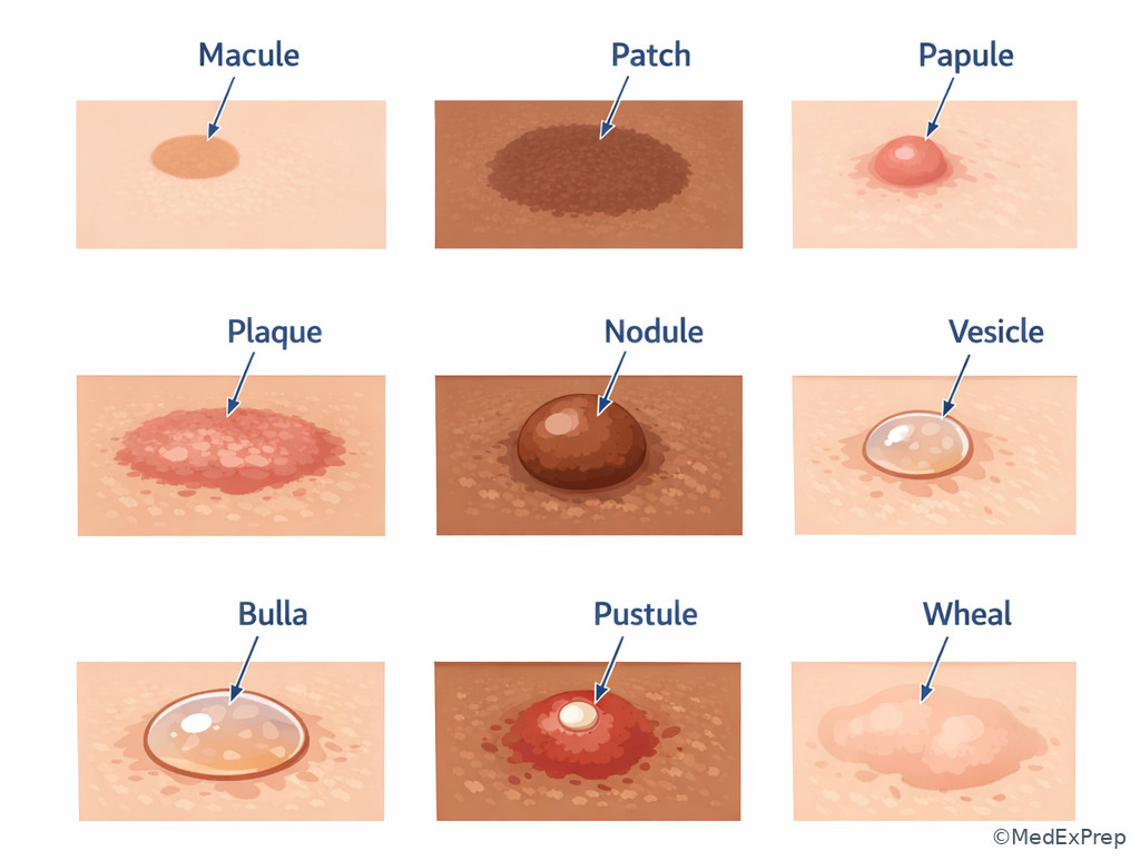
P -->|Yes| Q[Wheal] Exam takeaway: Visual side-by-side comparison strengthens rapid recognition of lesion categories before secondary change appears.
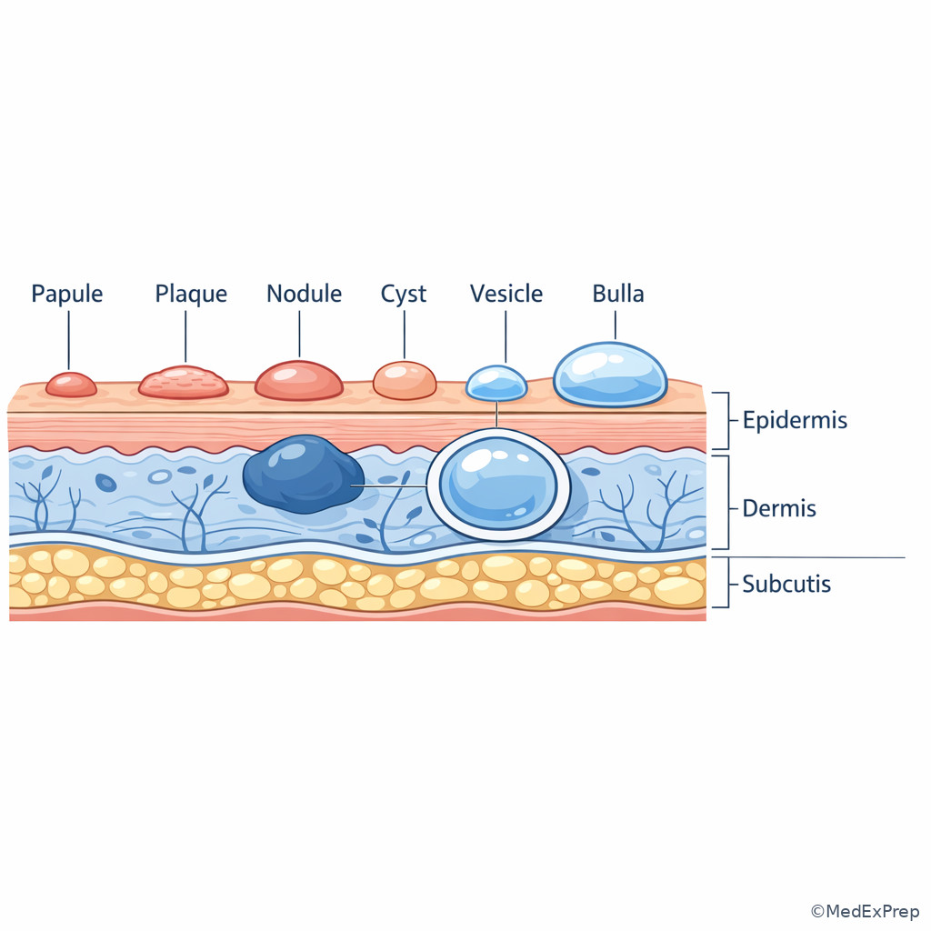
Exam takeaway: Depth is the key discriminator between superficial plaques and deeper nodules or cysts.
Core content
1.1.1.3 Foundational split: flat lesions versus palpable lesions
A Explanation
The first board-style decision conflict is simple but decisive: Do you choose a flat color change or a palpable lesion term? The single resolving rule is whether the lesion is elevated on inspection or palpation. If it is flat, choose macule or patch; if it is elevated, reject those terms immediately. On exam stems, learners overvalue color and underweight palpability. A red lesion can still be a papule, plaque, or wheal if it is raised. See Fig 1.
WHY THIS IS TESTED: Many diagnoses differ first by whether the process is only a color change or a true tissue elevation. Vitiligo, melasma, and lentigines begin in the flat bucket; psoriasis, lichen planus, and urticaria do not. The stem may add scale or excoriation later, but your first cut remains primary morphology.
Use classic size thresholds after you decide flatness. Macule is a flat circumscribed color change less than 1 cm; patch is the same but 1 cm or more. The board does not usually punish tiny deviations in exact measurement if the lesion is obviously broad and flat, but it will punish calling a palpable lesion a patch.
Choose-vs-reject framing matters: choose macule/patch when the lesion is seen but not felt; reject them when there is any true elevation, induration, edema, or depth. If uncertain, re-read the stem for verbs such as “palpable,” “raised,” “firm,” “dome-shaped,” or “edematous,” which all push you out of the flat category.
B Worked example
A 34-year-old woman with systemic lupus erythematosus and iron deficiency presents with multiple light-brown lesions on the cheeks that became more noticeable after summer travel. She uses hydroxychloroquine and reports mild pruritus from a recent cosmetic peel. Examination shows several tan to brown, sharply demarcated, nonpalpable lesions, most 3–6 mm, without scale. A resident calls them “thin papules” because they are easy to see. What is the most appropriate morphology term?
Reasoning chain: Identify task: morphology labeling, not final diagnosis. Extract key facts: visible color change, nonpalpable, 3–6 mm, no scale or induration. Apply rule: flat and under 1 cm equals macules. Eliminate distractors: papules are palpable; patches would be larger; plaques are elevated and broad. Verify: the cosmetic peel causing itch is distracting noise and does not alter the primary morphology described.
C Exam trap
D Checkpoint
Question: A 52-year-old man with diabetes and chronic venous insufficiency has several well-demarcated brown lesions on the lower legs. They are flat, nonpalpable, and each measures 1.5–2 cm. Which term is most appropriate?
- A. Macules — tempting because they are flat color changes, but wrong because each lesion is larger than 1 cm.
- B. Patches — tempting because pigmentary lesions are often called patches, and right because they are flat and at least 1 cm.
- C. Papules — tempting if one assumes visible means raised, but wrong because the stem says nonpalpable.
- D. Plaques — tempting because the lesions are broad, but wrong because plaques are elevated, not simply flat discolorations.
1.1.1.4 Solid elevated lesions: papule, plaque, nodule, and tumor
A Explanation
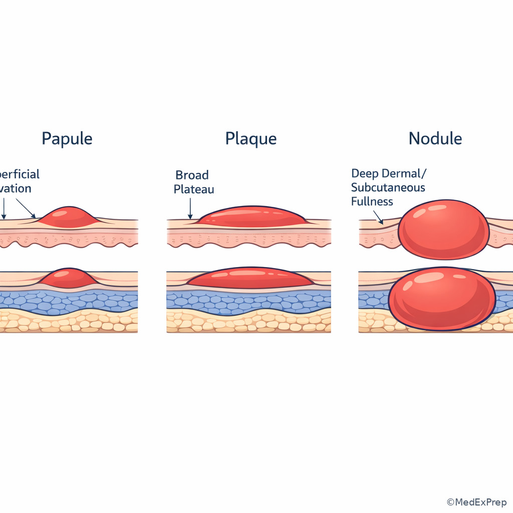
The second major conflict is surface elevation versus deeper mass. Students often choose between papule, plaque, and nodule based only on size; the better resolving rule is the combination of breadth, height, and depth. A papule is a small solid elevated lesion, usually less than 1 cm. A plaque is a raised lesion with a broad plateau-like surface, typically 1 cm or more and often formed by coalescent papules. A nodule is solid and palpable with significant depth into the dermis or subcutis. A tumor is a large mass-like lesion; on modern boards it is used less often as the best answer unless the stem emphasizes a bulky mass.
WHY THIS IS TESTED: Papulosquamous disease, infiltrative tumors, granulomatous disorders, and adnexal lesions are separated by whether the process is superficial and broad or deep and firm. A superficial broad scaly lesion is a plaque; a deeper firm mobile lesion is a nodule even if the surface looks smooth.
Choose papule when the lesion is small and elevated but not deeply seated. Choose plaque when the surface area dominates and the lesion reads as a broad tabletop rather than a dome. Choose nodule when palpation and depth are emphasized by words like “firm,” “deep,” “subcutaneous,” “mobile,” or “extends into the dermis.” See Fig 2.
Reject the common shortcut that “anything larger than 1 cm is a plaque.” A large deep lesion may still be a nodule. Likewise, not every papule cluster should be called a plaque unless the lesions truly coalesce into a broad raised area.
Exam takeaway: Surface breadth points to plaque, whereas palpable depth points to nodule even when size overlaps.
flowchart LR
A[Solid elevated lesion] --> B{Main feature}
B -->|Small superficial bump| C[Papule]
B -->|Broad raised plateau| D[Plaque]
B -->|Deep palpable mass| E[Nodule]
E --> F[May extend into dermis or subcutis]
D --> G[Often formed by coalescing papules]
C --> H[Usually under 1 cm] Exam takeaway: The visual difference between tabletop breadth and deeper dermal fullness is easier to remember than size cutoffs alone.
B Worked example
A 61-year-old man with obesity, hypertension, and longstanding psoriasis presents for evaluation of a 2.2-cm tender lesion on the forearm. He says it “looks like a raised patch.” Examination shows a firm, dome-shaped, deeply palpable lesion with overlying smooth skin and minimal scale. He also has unrelated erythematous scaly plaques on the elbows. Which morphology term best describes the forearm lesion?
Reasoning chain: Identify task: morphology of one lesion, not classification of all skin findings. Extract key facts: firm, dome-shaped, deeply palpable, 2.2 cm, smooth surface. Apply rule: depth dominates, so this is a nodule. Eliminate distractors: plaque is broad and superficial; papule is too small and not deep; patch is flat. Verify: his true psoriatic plaques on the elbows are distracting comparison data.
C Exam trap
D Checkpoint
Question: A 28-year-old woman with celiac disease and atopic dermatitis has multiple violaceous, flat-topped, 4-mm, pruritic lesions on the flexor wrists. Which primary lesion term is most appropriate?
- A. Papules — tempting because the lesions are small and elevated, and right because they are under 1 cm and solid.
- B. Plaques — tempting because lichen planus can coalesce, but wrong because the stem describes discrete 4-mm lesions.
- C. Nodules — tempting because the lesions are palpable, but wrong because there is no deep dermal or subcutaneous component.
- D. Patches — tempting if one focuses on color rather than texture, but wrong because the lesions are elevated.
- The most useful discriminator between plaque and nodule is depth.
- True or False: Any elevated lesion larger than 1 cm is a plaque. False.
- Name the 3 key features of a papule: solid, elevated, under 1 cm.
1.1.1.5 Fluid-filled lesions: vesicle, bulla, pustule, and cyst
A Explanation
This section tests whether you can tell what is inside the lesion and how superficial it is. A vesicle is a small, clear fluid-filled blister less than 1 cm. A bulla is the same process at 1 cm or more. A pustule contains purulent material. A cyst is not a blister at all; it is a deeper epithelial-lined or encapsulated cavity with liquid or semisolid contents. In practice, the exam often asks you to reject “cyst” when a lesion is clearly superficial and blistering, or reject “bulla” when the lesion is a deeper dome-shaped sac.
WHY THIS IS TESTED: Vesiculobullous disease, pustular eruptions, acneiform processes, infections, and cystic adnexal lesions diverge immediately once you classify fluid content correctly. Choosing vesicle instead of pustule changes the disease family. Choosing bulla instead of cyst changes the likely tissue level and workup.
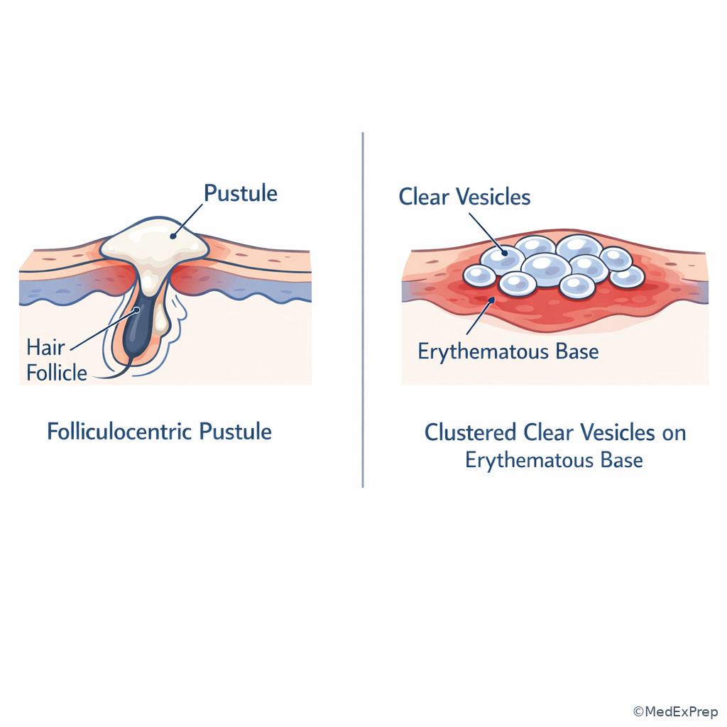
Use three cues: content, size, and plane. Clear superficial blister under 1 cm equals vesicle. Same but larger equals bulla. Yellow-white inflammatory content favors pustule. Deeper, round, often mobile lesion with punctum or semisolid keratinous material favors cyst. See Fig 3.
Choose-vs-reject framing: choose pustule when purulence is named or implied. Reject vesicle/bulla if the lesion is centered on a hair follicle with pus. Reject cyst when the lesion is fragile, tense, or ruptures like a blister. Reject bulla when the lesion has a punctum and expresses keratinous debris.
Exam takeaway: Clear fluid, pus, and deeper cystic contents belong to different lesion families and should not be used interchangeably.
flowchart TD
A[Palpable lesion with fluid] --> B{Superficial blister or deeper sac?}
B -->|Superficial blister| C{Content}
C -->|Clear fluid| D{Size}
D -->|Under 1 cm| E[Vesicle]
D -->|1 cm or larger| F[Bulla]
C -->|Purulent| G[Pustule]
B -->|Deeper saclike lesion| H[Cyst]
H --> I[Often punctum or keratinous material] Exam takeaway: Superficial clear blisters, purulent pustules, and deeper cysts should be recognized as distinct lesion types at a glance.
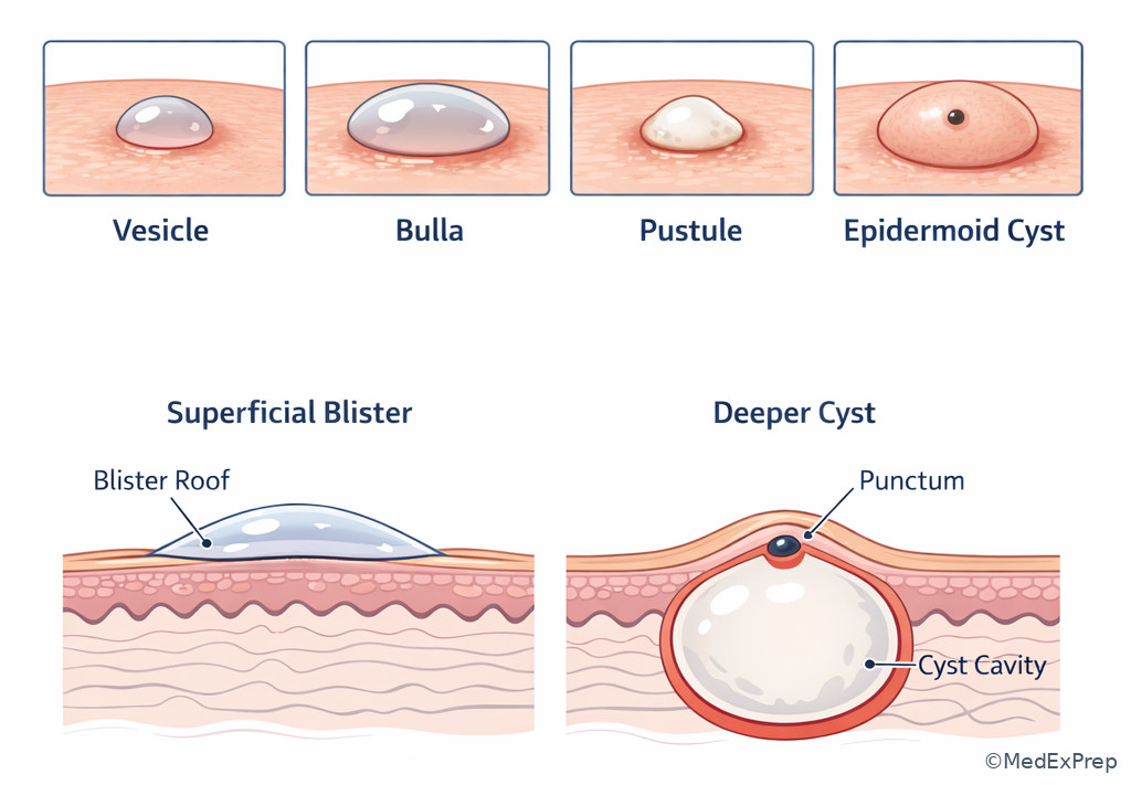
Exam takeaway: Follicle-centered purulence is a powerful clue that the primary lesion is a pustule, not a vesicle.
B Worked example
A 73-year-old woman with hypertension, type 2 diabetes, and chronic kidney disease presents with tense pruritic lesions on the abdomen and thighs. She recently completed doxycycline for rosacea and worries these are “infected boils.” Examination shows several tense, clear, 1.5- to 3-cm fluid-filled lesions on urticarial plaques; no follicular centering or purulent heads are present. What is the most appropriate primary lesion term?
Reasoning chain: Identify task: lesion type, not final autoimmune diagnosis. Extract key facts: clear fluid, tense blister roof, 1.5–3 cm, not follicular, not purulent. Apply rule: clear fluid and at least 1 cm means bullae. Eliminate distractors: pustules require pus; cysts are deeper and saclike; vesicles are too small. Verify: “infected boils” is a patient interpretation and a tempting but wrong framing.
C Exam trap
D Checkpoint
Question: A 19-year-old wrestler with asthma and acne develops multiple 2–4 mm yellow-white follicle-centered lesions on the upper back after hot-tub use. Which term is most appropriate?
- A. Vesicles — tempting because they are small superficial lesions, but wrong because the content is purulent and folliculocentric.
- B. Bullae — tempting if one only thinks “fluid-filled,” but wrong because these lesions are tiny and contain pus, not clear fluid.
- C. Pustules — tempting because folliculitis commonly presents this way, and right because they are pus-filled superficial lesions.
- D. Cysts — tempting because acne can involve cystic lesions, but wrong because these are small superficial follicular lesions, not deep nodulocystic structures.
1.1.1.6 Edematous and vascular primary lesions: wheal and telangiectasia
A Explanation
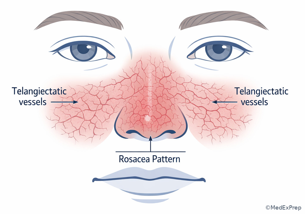
Not every raised lesion is solid or fluid-filled. Some are vascular or edematous. A wheal is an evanescent edematous papule or plaque caused by superficial dermal edema; it often changes shape or location over hours and may have central pallor. Telangiectasias are visibly dilated superficial blood vessels; they are not papules unless there is a separate raised component. The board frequently tests the mistake of calling urticaria a plaque or calling telangiectasia a macule just because it is red.
WHY THIS IS TESTED: Wheals push you toward urticaria, arthropod reactions, or edema-mediated processes; telangiectasias raise vascular, photodamage, rosacea, connective tissue disease, or neoplastic clues. These labels carry different implications for duration, pathophysiology, and management.
The single rule for wheal is transience from edema. If the lesion appears and fades within hours, migrates, or leaves no residual surface change, think wheal. The single rule for telangiectasia is visible vessel morphology—fine linear, arborizing, or mat-like red vessels that blanch. See Fig 4.
Reject plaque when the stem emphasizes fleeting duration. Reject macule when individual vessels are visibly delineated. Reject purpura when blanching is present. These are common exam pivots because the visible color can mislead.
Exam takeaway: A transient edematous swelling is a wheal, whereas a persistent visible superficial vessel is telangiectasia.
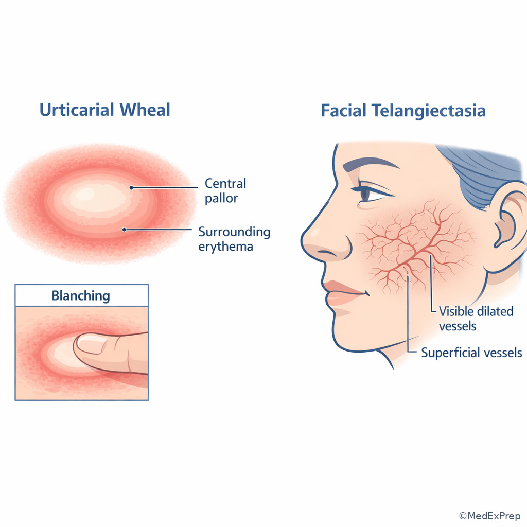
Exam takeaway: When individual superficial vessels are seen directly, think vascular morphology rather than inflammatory papules alone.
B Worked example
A 42-year-old woman with Hashimoto thyroiditis and chronic spontaneous urticaria presents with intensely pruritic “plaques” that appear after stress and fade by morning. She started a new vitamin supplement and worries about a drug eruption. Examination during the visit shows several blanchable edematous pink lesions with central pallor on the trunk and arms, none with scale or residual hyperpigmentation. What is the best primary lesion term?
Reasoning chain: Identify task: classify the lesion causing recurrent episodes. Extract key facts: pruritic, blanchable, edematous, central pallor, appears and fades within hours. Apply rule: transient edema equals wheal. Eliminate distractors: plaque would not be so evanescent; patch is flat; pustule contains pus. Verify: the recent vitamin supplement is distracting noise that may suggest a fixed eruption or drug rash, but the time course resolves the conflict.
C Exam trap
D Checkpoint
Question: A 67-year-old man with rosacea and prior nonmelanoma skin cancer has fine linear red blanchable vessels on the nose and medial cheeks without scale or palpable substance. Which primary lesion term is best?
- A. Macules — tempting because the lesions are flat and red, but wrong because the stem specifically describes visible superficial vessels.
- B. Papules — tempting because rosacea can include papules, but wrong because these lesions are not palpable.
- C. Telangiectasias — tempting because vascular morphology is explicit, and right because the lesions are visible dilated superficial vessels.
- D. Wheals — tempting because they are blanchable, but wrong because there is no edema or transient course.
- The most common primary lesion in urticaria is a wheal.
- True or False: Telangiectasias are usually described as palpable lesions. False.
- Name the one-word discriminator for wheal: transient.
1.1.1.7 Integration: choosing the dominant primary lesion in mixed eruptions
A Explanation
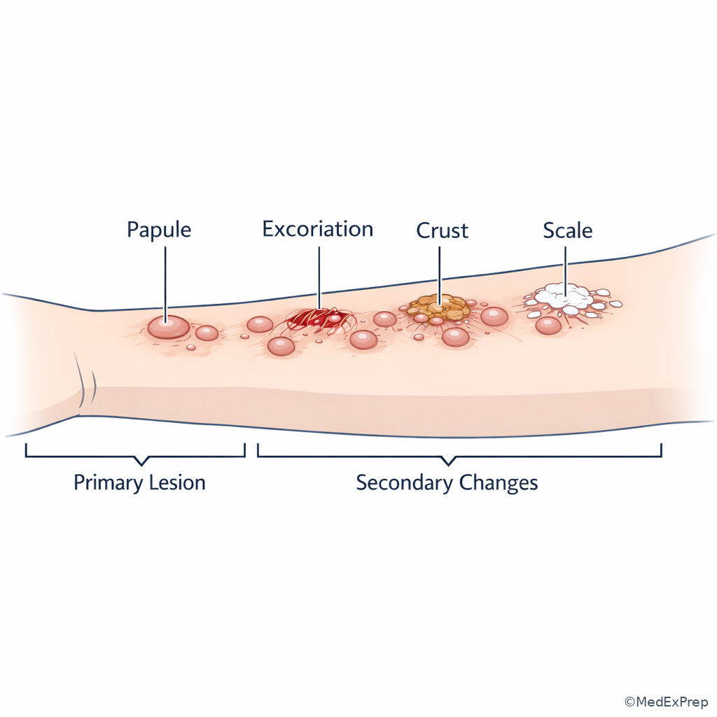
Real patients rarely present with perfect textbook lesions. The board therefore tests a higher-level conflict: When multiple lesion types are present, which one is the dominant primary lesion that best organizes the differential? The resolving rule is to prioritize the lesion that appears earliest in the disease process and best reflects the underlying mechanism, while downgrading secondary changes such as scale, crust, excoriation, erosion, or lichenification. This is where primary morphology connects forward to the next topics.
WHY THIS IS TESTED: The exam wants dermatologist-level reasoning, not vocabulary recitation. A pruritic eruption with excoriations may still be primarily papular. A blistering disease with ruptured surfaces still began as vesicles or bullae. A scaly red eruption may be papules coalescing into plaques. If you classify by the wrong visible late change, you enter the wrong differential family.
Choose the dominant primary lesion by asking three questions: What came first? What is repeated across most lesions? What single term best predicts the diagnostic category? In a mixed papule-plus-plaque eruption, the disease may still be described as papulosquamous if discrete papules and broad plaques coexist. In a mixed vesicle-plus-erosion process, choose vesicle if the stem says the lesions rupture easily. See Fig 5.
This is also where lesion morphology begins to link with distribution and configuration. A morphology label alone rarely ends the differential, but it creates the correct lane. The next topics teach how annular, grouped, dermatomal, flexural, acral, or photodistributed patterns refine the diagnosis after morphology is correct.
Exam takeaway: When several findings coexist, choose the lesion that came first and best explains the disease family, then treat later changes as modifiers.
flowchart TD
A[Mixed eruption] --> B[List visible findings]
B --> C{Primary or secondary?}
C -->|Primary| D[Keep in differential lane]
C -->|Secondary| E[Use as modifier only]
D --> F{Which lesion came first and repeats most?}
F --> G[Choose dominant primary lesion]
G --> H[Add distribution and configuration next]
E --> H B Worked example
A 46-year-old man with seasonal allergies, generalized anxiety disorder, and chronic scratching presents with a 3-week pruritic eruption on the forearms and neck. He recently changed laundry detergent. Examination shows multiple erythematous 2–5 mm raised lesions, some grouped, with overlying excoriations and scattered crust. One note in the chart describes “crusted plaques.” What is the best dominant primary lesion term?
Reasoning chain: Identify task: dominant primary lesion, not every change. Extract key facts: multiple 2–5 mm raised lesions are repeated across the eruption; excoriations and crust are overlying changes. Apply rule: choose the earlier repeated lesion type, so papules. Eliminate distractors: crust is secondary, plaque would require broader surface-dominant elevation, patch is flat. Verify: the chart phrase “crusted plaques” is tempting but imprecise because it collapses primary and secondary morphology.
C Exam trap
D Checkpoint
Question: A 31-year-old woman with ulcerative colitis and a new fragrance exposure develops grouped tiny clear lesions on an erythematous base of the fingers; several have ruptured, leaving superficial raw areas. Which is the best dominant primary lesion term?
- A. Erosions — tempting because ruptured lesions leave raw surfaces, but wrong because erosion is a secondary change.
- B. Vesicles — tempting because the lesions began as tiny clear blisters, and right because that is the primary morphology.
- C. Pustules — tempting because inflamed finger lesions may look yellow later, but wrong because the original content is clear fluid.
- D. Plaques — tempting if grouped lesions are confluent, but wrong because the repeated earliest lesions are small blisters.
Exam Traps & Differentiators
Most common wrong answer and why: Calling any visible lesion a patch or any large raised lesion a plaque. These errors happen when size is used before palpability and depth.
| Looks similar | Choose this when... | Do not choose this when... | Single discriminator |
|---|---|---|---|
| Macule vs patch | Both are flat color changes | Do not ignore size | < 1 cm vs ≥ 1 cm |
| Papule vs plaque | Both are solid and elevated | Do not use size alone | Small discrete bump vs broad plateau |
| Plaque vs nodule | Both may be larger than 1 cm | Do not miss deep palpability | Surface breadth vs depth |
| Vesicle vs pustule | Both are small superficial lesions | Do not ignore content | Clear fluid vs pus |
| Bulla vs cyst | Both may be dome-shaped and fluid-containing | Do not confuse superficial blister with deeper sac | Blister roof vs punctum/deeper cavity |
| Wheal vs plaque | Both can be broad raised lesions | Do not miss time course | Evanescent edema |
| Telangiectasia vs erythematous macule | Both can appear red and flat | Do not ignore vessel architecture | Visible superficial vessels |
| If the stem says... | Think... |
|---|---|
| “Nonpalpable” | Macule or patch, not papule or plaque |
| “Firm” and “deeply palpable” | Nodule |
| “Tense clear blister 2 cm” | Bulla |
| “Follicle-centered yellow-white lesions” | Pustules |
| “Appears and fades within hours” | Wheal |
| “Visible branching red vessels” | Telangiectasia |
Exam takeaway: Boards often hide the correct diagnosis by showing secondary change overlying a more informative primary lesion.
Tables
| Reference summary | Definition core | Exam pivot |
|---|---|---|
| Macule | Flat color change < 1 cm | Reject if palpable |
| Patch | Flat color change ≥ 1 cm | Reject if raised |
| Papule | Solid elevated lesion < 1 cm | Reject if broad plateau or deep mass |
| Plaque | Broad elevated lesion ≥ 1 cm | Reject if depth dominates |
| Nodule | Solid deeper palpable lesion | Depth matters more than diameter alone |
| Vesicle | Clear-fluid blister < 1 cm | Reject if purulent |
| Bulla | Clear-fluid blister ≥ 1 cm | Reject if deeper cystic sac |
| Pustule | Pus-filled superficial lesion | Follicular pattern is a common clue |
| Wheal | Edematous transient papule/plaque | Time course is the discriminator |
| Telangiectasia | Visible superficial dilated vessel | Look for vessel architecture, not just redness |
| Comparison | Superficiality | Contents/feel | Best board discriminator |
|---|---|---|---|
| Papule | Superficial | Solid | Small raised bump |
| Plaque | Superficial to mid-dermal | Solid broad surface | Tabletop breadth |
| Nodule | Dermal/subcutaneous | Solid deep mass | Palpable depth |
| Vesicle | Superficial | Clear fluid | Small blister |
| Pustule | Superficial | Purulent | Pus, often follicular |
| Cyst | Deeper | Liquid or semisolid cavity | Punctum or keratinous material |
| Differential/classification problem | Tempting wrong choice | Why it tempts | One clue that separates them |
|---|---|---|---|
| Vitiligo lesion | Macule | Flat color change is correct family | Usually broad depigmented areas, so patch is often better |
| Psoriasis lesion | Papule | Plaques can begin as papules | Coalescent broad raised scaly surface favors plaque |
| Bullous pemphigoid lesion | Cyst | Dome-shaped fluid lesion | Tense superficial blister, no punctum |
| Folliculitis lesion | Vesicle | Small superficial fluid lesion | Pus and follicular centering favor pustule |
| Urticaria lesion | Plaque | Broad raised erythematous area | Fades or migrates within hours |
| Rosacea vascular lesion | Macule | Flat red lesion | Individual superficial vessels are visible |
Algorithm / Approach
Exam takeaway: The safest exam approach is to classify lesion plane and contents first, then use size and time course as tie-breakers.
flowchart TD
A[Board stem] --> B[Identify dominant lesion]
B --> C{Flat?}
C -->|Yes| D[Macule or patch by size]
C -->|No| E{Raised lesion type}
E -->|Solid superficial| F[Papule or plaque]
E -->|Deep solid| G[Nodule]
E -->|Clear fluid| H[Vesicle or bulla]
E -->|Purulent| I[Pustule]
E -->|Transient edema| J[Wheal]
E -->|Visible vessel| K[Telangiectasia]
F --> L[Then add distribution and secondary change]
G --> L
H --> L
I --> L
J --> L
K --> L
See Fig 6 for the compact exam-day algorithm. The key operational rule is sequencing: flatness and contents come before size. Size alone is a weak classifier when learners have not yet determined whether the lesion is broad, deep, blistering, or edematous.
Rapid Review
- Macule → flat color change under 1 cm; if palpable, reject it.
- Patch → flat color change 1 cm or larger; breadth without elevation.
- Papule → small solid bump; think discrete elevation.
- Plaque → broad raised surface; think tabletop rather than dome.
- Nodule → depth beats diameter; firm deep palpability is the clue.
- Vesicle → small clear blister; content matters more than redness.
- Bulla → large blister; do not confuse with cyst.
- Pustule → purulence, often follicular; not just a tiny blister.
- Wheal → transient edema; time course is the discriminator.
- Telangiectasia → visible superficial vessel pattern; not just a red spot.
- Cyst → deeper cavity with punctum or keratinous material; not a blister.
- Mixed eruption → choose what came first; later crust or erosion is secondary.
- Size rule → useful only after flat vs raised and content vs depth are settled.
Self-check quiz
1.1.1.8 Question 1
A 26-year-old woman with migraine and oral contraceptive use presents with symmetric tan facial discoloration after sun exposure. The lesions are flat, nonpalpable, and each involved area is larger than 1 cm. Which primary lesion term is most appropriate?
- A. Macules
- B. Patches
- C. Papules
- D. Plaques
1.1.1.9 Question 2
A 17-year-old male with asthma and acne develops multiple 3-mm follicle-centered yellow-white lesions on the forehead during football season. Which primary lesion term is most appropriate?
- A. Vesicles
- B. Pustules
- C. Papules
- D. Cysts
1.1.1.10 Question 3
A 64-year-old woman with hypertension, type 2 diabetes, and pruritus presents with several tense clear 2-cm lesions on the lower abdomen over urticarial erythema. She also has a few excoriations from scratching. Which description best identifies the dominant primary lesion?
- A. Erosions
- B. Bullae
- C. Cysts
- D. Plaques
1.1.1.11 Question 4
A 39-year-old man with allergic rhinitis and chronic urticaria reports recurrent itchy lesions that appear after pressure and resolve within 6 hours. During the visit, examination shows blanchable edematous pink raised areas with central pallor on the trunk. Which primary lesion term is most appropriate?
- A. Wheals
- B. Plaques
- C. Patches
- D. Nodules
1.1.1.12 Question 5
A 55-year-old man with obesity and psoriasis presents for evaluation of a 2.5-cm firm dome-shaped lesion on the upper arm. The overlying skin is smooth, and palpation shows clear dermal depth. He also has unrelated scaly plaques on the elbows. Which morphology term best fits the arm lesion?
- A. Plaque
- B. Nodule
- C. Papule
- D. Patch
1.1.1.13 Question 6
A 33-year-old woman with hand dermatitis, allergic rhinitis, and recent detergent exposure develops grouped 2-mm clear lesions on the lateral fingers. Several have ruptured, leaving superficial raw areas, and one area has mild crust from scratching. Which is the most likely dominant primary lesion?
- A. Erosions
- B. Vesicles
- C. Pustules
- D. Plaques
1.1.1.14 Question 7
A 71-year-old man with rosacea, actinic damage, and coronary artery disease has persistent fine branching red lines across the nasal sidewalls and cheeks. They blanch with pressure and are not palpable. Which term best describes these lesions?
- A. Telangiectasias
- B. Macules
- C. Wheals
- D. Purpura
1.1.1.15 Question 8
A 48-year-old woman with obesity, hypothyroidism, and chronic pruritus presents with a 4-week eruption on the extensor forearms. Exam shows dozens of 4-mm erythematous raised lesions, many with overlying scale and excoriation; several neighboring lesions merge into larger rough areas. Which description best captures the dominant primary morphology?
- A. Scaly plaques
- B. Excoriated papules
- C. Crusted erosions
- D. Hyperkeratotic patches
Answer key
1.1.1.16 Question 1: B. Patches
Correct-answer reasoning: These lesions are flat, nonpalpable color changes larger than 1 cm, which makes them patches. Mechanistically, the stem tests the flat-versus-raised split first and then size. This is classic morphology logic for pigmentary disorders. Sources: Bolognia, Schaffer, and Cerroni; Habif.
- A. Macules — tempting because the lesions are flat pigment changes, but incorrect because each involved area is larger than 1 cm; the single discriminating clue is size.
- B. Patches — tempting and correct because they are flat and broad; the single discriminating clue is nonpalpable lesions larger than 1 cm.
- C. Papules — tempting if visible lesions are assumed to be raised, but incorrect because the stem explicitly says nonpalpable; the clue is absence of elevation.
- D. Plaques — tempting because facial lesions can be broad, but incorrect because plaques require elevation; the clue is flatness.
1.1.1.17 Question 2: B. Pustules
Correct-answer reasoning: Follicle-centered yellow-white lesions indicate purulence within or around follicles, so the primary lesion is a pustule. This is a content-based classification, not just a size-based one. Sources: Bolognia, Schaffer, and Cerroni; Andrews' Diseases of the Skin.
- A. Vesicles — tempting because the lesions are small and superficial, but incorrect because vesicles contain clear fluid; the clue is yellow-white purulence.
- B. Pustules — tempting and correct because folliculitis commonly produces folliculocentric pus-filled lesions; the clue is purulent content.
- C. Papules — tempting because inflamed acne lesions can be papules, but incorrect because the stem specifically highlights pus; the clue is lesion content.
- D. Cysts — tempting because acne can include cysts, but incorrect because these are tiny superficial follicular lesions rather than deep cavities; the clue is superficial folliculocentric morphology.
1.1.1.18 Question 3: B. Bullae
Correct-answer reasoning: Tense clear 2-cm lesions are bullae. The excoriations are secondary changes from scratching and should not replace the dominant blister morphology. Mechanism-to-treatment logic matters because blistering diseases and excoriated eruptions have different differentials and management pathways. Sources: Bolognia, Schaffer, and Cerroni; Wolff et al., Fitzpatrick's Dermatology.
- A. Erosions — tempting because excoriations and ruptured lesions can leave raw areas, but incorrect because erosion is secondary; the clue is intact tense clear 2-cm blisters.
- B. Bullae — tempting and correct because clear fluid-filled lesions at least 1 cm meet the definition; the clue is size plus blister content.
- C. Cysts — tempting because the lesions are dome-shaped and fluid-containing, but incorrect because they are superficial tense blisters rather than deeper sacs; the clue is blister roof on erythematous skin.
- D. Plaques — tempting because the blisters sit on urticarial erythema, but incorrect because the lesion being classified is the fluid-filled blister; the clue is dominant primary lesion selection.
1.1.1.19 Question 4: A. Wheals
Correct-answer reasoning: Recurrent pruritic blanchable edematous lesions that resolve within hours are wheals. The key mechanism is transient superficial dermal edema, which distinguishes urticaria from fixed plaques or nodules. Sources: Bolognia, Schaffer, and Cerroni; Habif.
- A. Wheals — tempting and correct because the lesions are transient, edematous, and blanchable; the clue is resolution within 6 hours.
- B. Plaques — tempting because wheals may be broad and raised, but incorrect because plaques are not classically migratory and fleeting; the clue is time course.
- C. Patches — tempting because erythematous areas can appear broad, but incorrect because patches are flat; the clue is edema and elevation.
- D. Nodules — tempting if firmness is assumed from palpability, but incorrect because nodules are deep and persistent; the clue is evanescence.
1.1.1.20 Question 5: B. Nodule
Correct-answer reasoning: A firm dome-shaped lesion with clear dermal depth is a nodule. Although it is larger than 1 cm, the decisive feature is depth rather than surface breadth. This is a classic plaque-versus-nodule trap. Sources: Bolognia, Schaffer, and Cerroni; Andrews' Diseases of the Skin.
- A. Plaque — tempting because the lesion is raised and larger than 1 cm, but incorrect because plaques are surface-dominant rather than deeply palpable; the clue is dermal depth.
- B. Nodule — tempting and correct because deep palpable solidity defines a nodule; the clue is firm dome-shaped lesion with depth.
- C. Papule — tempting because it is a discrete bump, but incorrect because it is too large and too deep; the clue is 2.5 cm with dermal fullness.
- D. Patch — tempting only if one overlooks palpability, but incorrect because the lesion is clearly raised; the clue is elevation.
1.1.1.21 Question 6: B. Vesicles
Correct-answer reasoning: The earliest repeated lesions are grouped 2-mm clear blisters, so vesicles are the dominant primary morphology. Rupture leading to raw areas creates erosions, but those are secondary changes. This is a direct test of dominant lesion selection in a mixed eruption. Sources: Bolognia, Schaffer, and Cerroni; Fitzpatrick's Dermatology.
- A. Erosions — tempting because several lesions have ruptured, but incorrect because erosion is secondary; the clue is grouped clear intact 2-mm lesions.
- B. Vesicles — tempting and correct because the original lesions are small clear blisters under 1 cm; the clue is tiny clear grouped lesions.
- C. Pustules — tempting because inflamed hand lesions may look yellow after rupture, but incorrect because the original content is clear, not purulent; the clue is clear fluid.
- D. Plaques — tempting if grouped lesions blend into an erythematous field, but incorrect because the repeated discrete lesion type is vesicular; the clue is lesion size and blister content.
1.1.1.22 Question 7: A. Telangiectasias
Correct-answer reasoning: Persistent fine branching blanchable visible superficial vessels are telangiectasias. The mechanism is vascular dilation, not dermal edema, hemorrhage, or simple erythematous discoloration. Sources: Bolognia, Schaffer, and Cerroni; Habif.
- A. Telangiectasias — tempting and correct because the lesions are visible branching superficial vessels; the clue is vessel architecture.
- B. Macules — tempting because the lesions are flat and red, but incorrect because the stem highlights visible vascular lines rather than a homogeneous color spot; the clue is branching vessels.
- C. Wheals — tempting because they can be red and blanchable, but incorrect because wheals are edematous and transient; the clue is persistence without swelling.
- D. Purpura — tempting because vascular lesions can look red-purple, but incorrect because purpura typically does not blanch; the clue is blanching.
1.1.1.23 Question 8: B. Excoriated papules
Correct-answer reasoning: The dominant repeated lesions are dozens of 4-mm erythematous raised lesions, which are papules. Scale and excoriation are modifiers, so the best full descriptor is excoriated papules. This answer preserves the primary lesion and adds the secondary change without letting the secondary change take over. Sources: Bolognia, Schaffer, and Cerroni; Andrews' Diseases of the Skin.
- A. Scaly plaques — tempting because some neighboring lesions merge into larger rough areas, but incorrect because the dominant repeated lesion described across the eruption remains 4-mm papules; the clue is lesion repetition.
- B. Excoriated papules — tempting and correct because it captures the primary lesion plus a common secondary change; the clue is multiple 4-mm raised lesions with scratching.
- C. Crusted erosions — tempting because crust and surface damage are present, but incorrect because those are not the earliest repeated lesions; the clue is baseline papular morphology.
- D. Hyperkeratotic patches — tempting because rough scale may suggest a flat keratotic process, but incorrect because the lesions are raised and discrete; the clue is palpable 4-mm elevation.
Keep going with the full ABD Dermatology Prep Course (Derm)
You just studied the way members do, in the same lesson interface. Full access unlocks every lesson, the question bank and flashcards for a full year.
One payment · 1 year of access · No auto-renewal