1.1.1 - Anal Canal Landmarks
Why it matters
- Board questions often hinge on one landmark: above vs below the dentate line changes pain, innervation, lymphatic drainage, and differential diagnosis.
- Operative safety depends on knowing where anoderm ends, where internal sphincter is at risk, and where the anorectal ring marks the upper surgical boundary of the anal canal.
- Precise landmark language improves interpretation of anoscopy, pathology, MRI, and operative reports.
- Many common anorectal distractors look similar clinically but separate cleanly when localized to the verge, anoderm, dentate line, or ring.
Key Terms & Must-Know Facts
1.1.1.1 Key terms
- Anal verge: external junction where anal canal meets perianal skin
- Anoderm: hairless squamous lining distal to the dentate line
- Perianal skin: keratinized skin outside the verge
- Dentate line: mucocutaneous transition created by anal valves between columns
- Anal columns of Morgagni: longitudinal mucosal folds in upper anal canal
- Anal valves: folds joining inferior ends of columns; together form the dentate line
- Anal sinuses/crypts: recesses above valves; source of cryptoglandular sepsis
- Intersphincteric groove: palpable groove between internal and external sphincter distally
- Internal anal sphincter: continuation of circular smooth muscle
- External anal sphincter: striated voluntary sphincter complex
- Anorectal ring: palpable ring formed by puborectalis plus upper external and internal sphincter contributions
- Pectinate line: commonly used synonym for dentate line in exam settings
- White line of Hilton: intersphincteric line demarcating subcutaneous external sphincter from internal sphincter/anoderm region
1.1.1.2 Must-know facts
- Above the dentate line: columnar/transitional mucosa, visceral innervation, less pain sensitivity.
- Below the dentate line: anoderm, somatic innervation via inferior rectal nerves, marked pain sensitivity.
- Internal hemorrhoids arise above the dentate line; external hemorrhoids arise distal to it.
- Most cryptoglandular infections start in anal glands draining into anal crypts at the dentate line.
- The anal canal is typically described from anal verge to anorectal ring.
- Fissures classically involve distal anoderm, usually posterior midline.
- Lymphatic drainage above the dentate line favors internal iliac/inferior mesenteric pathways; below favors superficial inguinal nodes.
- Venous drainage differs across the dentate line and explains mixed internal-external hemorrhoidal disease.
- The anorectal ring is a crucial operative landmark for transanal dissection and continence preservation.
Exam takeaway: Localizing a lesion relative to the verge, dentate line, and anorectal ring immediately narrows pain pattern, pathology, and procedure choice.
flowchart TD
A[Anal verge] --> B[Anoderm below dentate line]
B --> C[Dentate line]
C --> D[Anal columns and crypts]
D --> E[Upper anal canal]
E --> F[Anorectal ring]
B --> G[Somatic pain]
C --> H[Internal hemorrhoids originate above]
D --> I[Crypt glands start sepsis]
F --> J[Transition to rectum and pelvic floor influence]
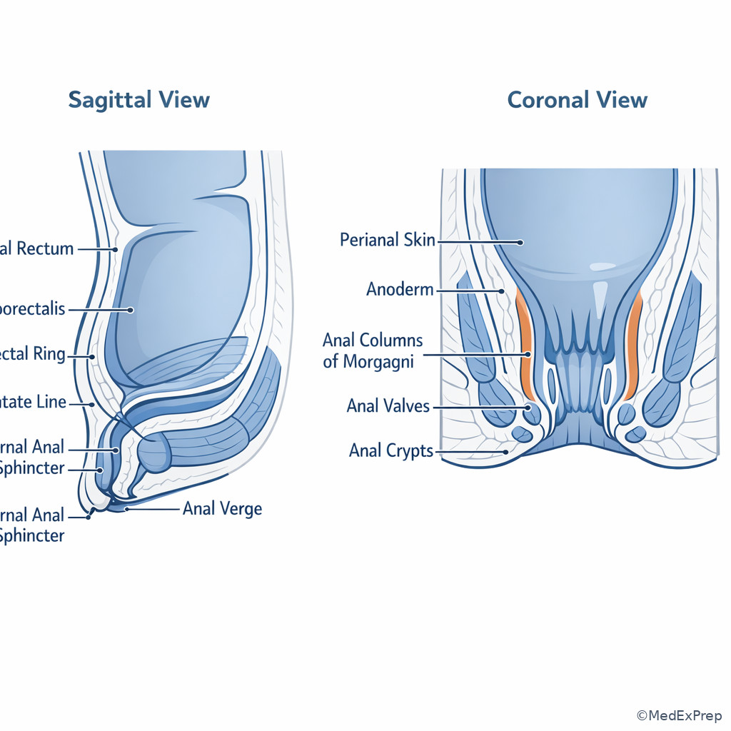
Exam takeaway: Use a labeled orientation image to anchor where the verge, dentate line, columns, crypts, and anorectal ring sit in relation to each other.
Core content
1.1.1.3 Foundational orientation: where the anal canal begins and ends
A Explanation
The first decision conflict on exam stems is simple but heavily tested: Which landmark should you use to orient the lesion? If the question asks about external inspection, pain at the skin edge, or distal lesions, anchor yourself to the anal verge. If it asks about mucosal transition, hemorrhoid type, sensory pattern, or lymphatic implications, anchor to the dentate line. If it asks about the proximal limit of the anal canal, continence-preserving dissection, or the point where rectum becomes functionally tethered by the pelvic floor, anchor to the anorectal ring. See Fig 1.
WHY THIS IS TESTED: many board questions hide the real task behind clinical noise, but the single rule that resolves them is whether the pathology lies at the verge, below the dentate line, above the dentate line, or near the ring. The anal canal is generally described from the anal verge to the anorectal ring. The verge is a visible external landmark; the ring is a palpable functional/surgical landmark. The dentate line sits in between and acts as the key biologic divider.
Choose the verge when the stem describes visible perianal skin or a lesion “at the opening.” Reject the dentate line if the lesion is clearly on external keratinized skin. Choose the dentate line when the stem asks about pain, hemorrhoidal category, epithelial type, or nodal drainage. Reject the anorectal ring if the question is about mucosal sensitivity or hemorrhoids; it is too proximal for that distinction. Choose the anorectal ring when describing the upper end of the surgical anal canal or the palpable high point reached on digital exam before entering the rectum.
Key rule: \(Verge = external opening\); dentate line = biologic divider; anorectal ring = proximal functional/surgical limit.
Clinician Action: Diagnose by explicitly localizing lesions relative to verge, dentate line, and ring on exam. Order anoscopy or EUA when office localization is uncertain. Treat with anatomy-driven planning, especially for hemorrhoids, fissures, and fistula surgery. Refer/Escalate to multidisciplinary pelvic imaging or subspecialty input when a proximal lesion may extend beyond the surgical anal canal.
B Worked example
A 58-year-old man with obesity, atrial fibrillation on apixaban, and type 2 diabetes presents with “a painful lump at the anus” after constipation. He reports bright red blood on wiping and says a prior colonoscopy showed “internal hemorrhoids,” which tempts an examinee toward the same diagnosis. On inspection, there is a tense bluish nodule at the anal margin; anoscopy is poorly tolerated because of pain. His hemoglobin is normal, and a mildly elevated glucose distracts from the local anatomy question.
Reasoning chain: Identify task: localize the lesion and infer the best category. Extract key facts: severe pain, visible anal margin lesion, bluish thrombosed appearance, poor tolerance of manipulation. Apply rule: a visible lesion at the margin is referenced to the anal verge, and painful thrombosis distal to the dentate line favors external hemorrhoid, not internal hemorrhoid. Eliminate distractors: internal hemorrhoids are above the dentate line and usually painless unless prolapsed/strangulated; fissure causes severe pain but is a linear tear rather than a tense nodule; perianal abscess is fluctuant and inflammatory. Verify: distal somatic innervation explains the marked pain.
C Exam trap
Common wrong answer: Dentate line as the “beginning” of the anal canal. Why it is tempting: it is the most famous landmark and separates internal from external disease, so learners overuse it as the main boundary for everything. Single clue that eliminates it: when the stem asks for the external starting point seen on inspection, the correct landmark is the anal verge.
D Checkpoint
Question: During operative dictation, which landmark most appropriately defines the proximal limit of the anal canal?
- Anal verge
- Dentate line
- Anorectal ring
- White line of Hilton
Answer: C. Anorectal ring.
- A: Tempting because it is the obvious external boundary, but it marks the distal opening rather than the proximal limit.
- B: Tempting because it is the key biologic transition, but it lies within the canal rather than defining its upper end.
- C: Correct because the anal canal is commonly described from the verge to the anorectal ring, the palpable functional upper boundary.
- D: Tempting because it is another named line in the region, but it is not the standard proximal endpoint of the canal.
1.1.1.4 Dentate line as the master divider: epithelium, innervation, lymphatics, venous drainage
A Explanation
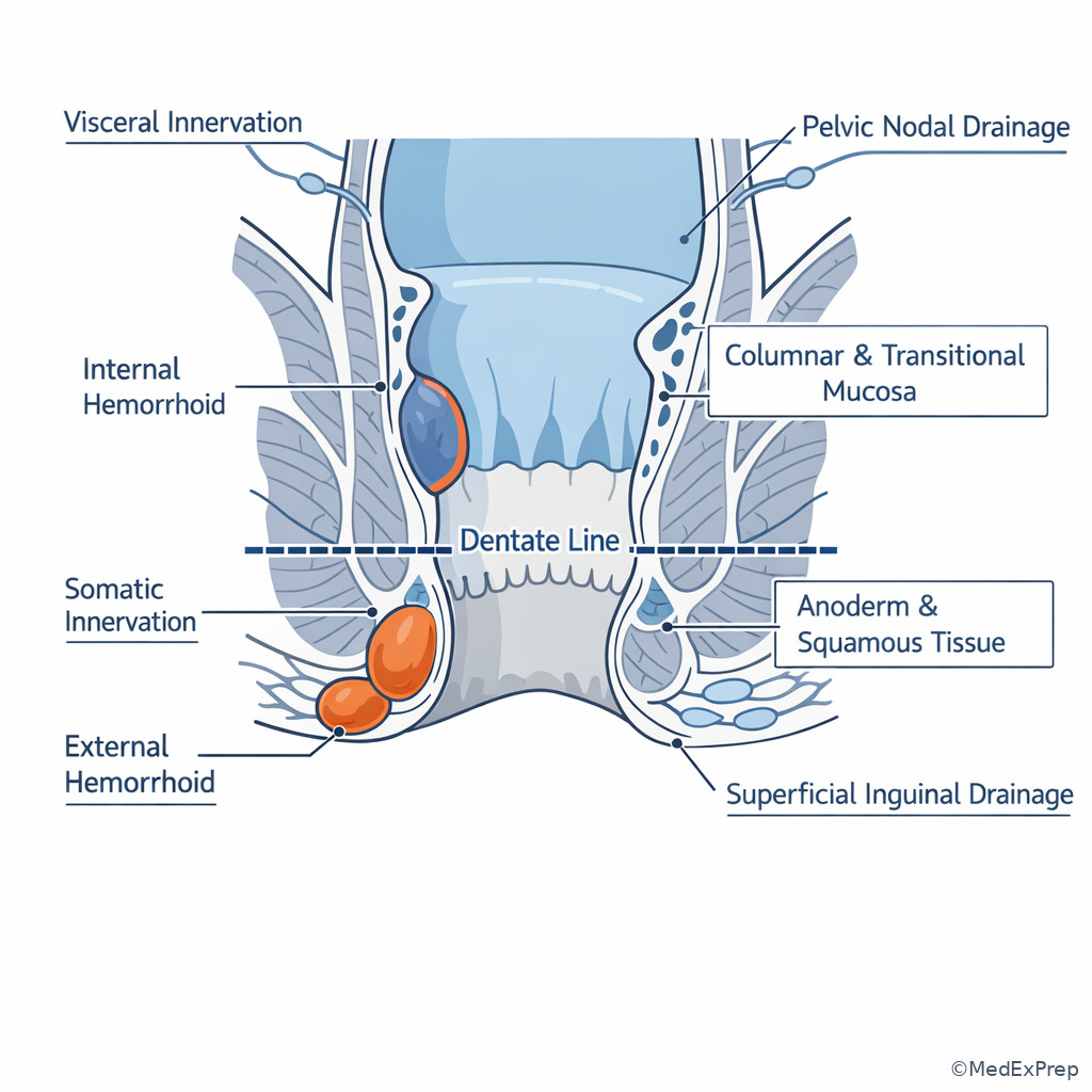
The second foundational decision conflict is: When the stem gives pain, pathology, or nodal spread, should you think above or below the dentate line? This is the single highest-yield rule in anal canal anatomy. Above the dentate line, mucosa is columnar/transitional and innervated viscerally, so office instrumentation and internal hemorrhoidal disease are usually less painful. Below it, the lining is anoderm and then perianal skin, innervated somatically through the inferior rectal branches, so fissures, thrombosed external hemorrhoids, and distal wounds are acutely painful.
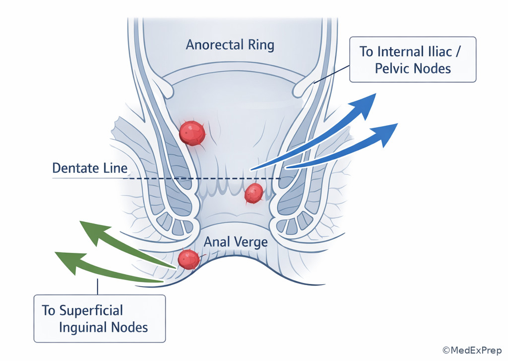
This same landmark predicts lymphatic drainage and often exam wording about malignancy. Lesions above the dentate line preferentially drain to internal iliac and related pelvic nodal pathways; lesions below the line drain toward superficial inguinal nodes. Venous drainage also differs across the line and contributes to the internal-external hemorrhoid distinction. Choose the dentate line whenever the stem asks which one finding best explains severe pain, nodal basin, epithelial type, or why an office procedure is tolerated differently above versus below. Reject the anal verge if the question is not about external inspection. Reject the anorectal ring if the issue is sensory biology rather than proximal canal support.
WHY THIS IS TESTED: exam writers repeatedly convert anatomy into decisions about symptoms, exam tolerance, hemorrhoid class, and cancer staging implications. See Fig 2 and Fig 3.
Memory anchor: DENTATE = Drainage changes, Epithelium changes, Nerve supply changes, Thrombosed external pain is below, Anal crypts sit here, Transition point, Exam stems love it.
High-Yield Connection: This concept is tested alongside hemorrhoid grading/treatment and anal squamous lesions. Know both dentate-line anatomy and pain/lymphatic consequences for exam day — they are frequently paired in stems.
Clinician Action: Diagnose by relating symptom intensity and lesion level to the dentate line. Order targeted anoscopy, biopsy, or nodal assessment based on location. Treat internal versus external pathology differently because pain expectations and tissue type differ. Refer/Escalate to oncology and cross-sectional staging when malignant lesions imply pelvic or inguinal nodal risk.
B Worked example
A 67-year-old woman with COPD, prior hysterectomy, and chronic constipation presents with intermittent bright red bleeding and tissue prolapse during defecation. She reports surprisingly little pain, though she is anxious because a cousin had anal cancer. On anoscopy, there are engorged cushions in the left lateral and right anterior positions. A superficial tender skin tag at the verge is also present and acts as a tempting distractor. The question asks which anatomic landmark best explains the relative lack of pain from the primary lesion.
Reasoning chain: Identify task: explain symptom pattern by anatomy. Extract key facts: bleeding, prolapse, minimal pain, anoscopic cushions above the margin, distracting external skin tag. Apply rule: painless bleeding/prolapsing cushions are internal hemorrhoids, which arise above the dentate line. Eliminate distractors: anal verge localizes the skin tag but does not explain the major lesion’s symptom pattern; anorectal ring is too proximal and not the sensory divider; white line of Hilton is not the main rule tested here. Verify: visceral innervation above the dentate line explains why the internal hemorrhoids bleed and prolapse more than hurt.
C Exam trap
Common wrong answer: Superficial inguinal drainage for every anal canal lesion. Why it is tempting: learners remember “anal cancer goes inguinal” and overgeneralize. Single clue that eliminates it: if the lesion is above the dentate line, expect pelvic/internal iliac-pattern drainage rather than superficial inguinal as the primary route.
D Checkpoint
Question: A lesion below the dentate line is most likely associated with which feature?
- Columnar mucosa and visceral afferents
- Marked pain with manipulation and superficial inguinal drainage
- Painless prolapse due to internal hemorrhoidal cushions
- Primary drainage to inferior mesenteric nodes
Answer: B. Marked pain with manipulation and superficial inguinal drainage.
- A: Tempting because it describes upper anal canal mucosa, but those are above the dentate line.
- B: Correct because below the dentate line tissue is somatically innervated and drains toward superficial inguinal nodes.
- C: Tempting because prolapse is common in anorectal disease, but painless internal cushions arise above the line.
- D: Tempting because colorectal nodal pathways can involve mesenteric nodes, but distal anal canal below the line does not primarily drain there.
- The most common landmark separating internal from external hemorrhoidal disease is the dentate line.
- True or False: Severe pain with light touch suggests tissue below the dentate line. True.
- Name the 3 key systems that change across the dentate line: epithelium, innervation, lymphatic drainage.
Exam takeaway: If you know whether the lesion is above or below the dentate line, you can predict pain, epithelium, hemorrhoid type, and nodal drainage.
flowchart LR
A[Dentate line] --> B[Above]
A --> C[Below]
B --> B1[Columnar or transitional]
B --> B2[Visceral sensation]
B --> B3[Internal hemorrhoids]
B --> B4[Pelvic nodal drainage]
C --> C1[Anoderm and squamous tissue]
C --> C2[Somatic sensation]
C --> C3[External hemorrhoids and fissure pain]
C --> C4[Superficial inguinal drainage]
Exam takeaway: The dentate line is not just a visual line; it marks a change in mucosa, pain fibers, and nodal pathways that drives board-style answers.
1.1.1.5 Anal columns, valves, crypts, and glands: why infection starts where it does
A Explanation
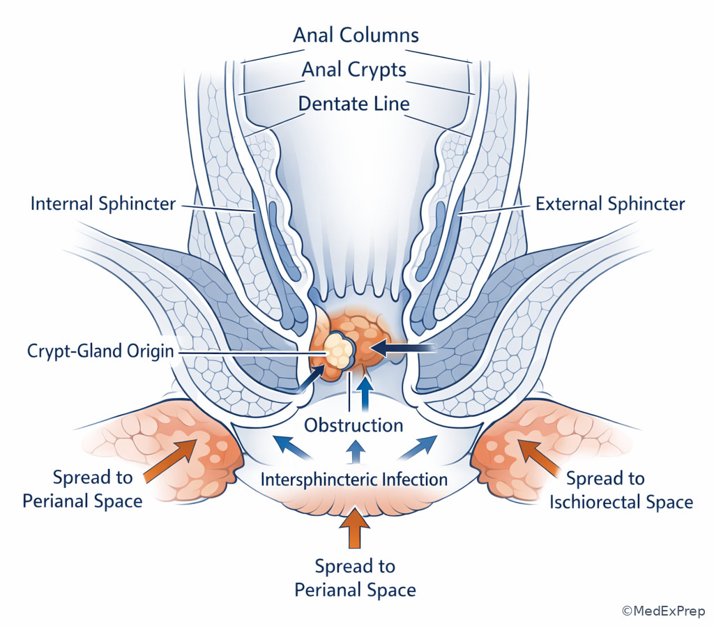
The next decision conflict is: Where do cryptoglandular abscesses and fistulas begin? The answer is not the verge and not the anorectal ring. It is the anal crypt-gland complex at the level of the dentate line. The anal columns are vertical folds; the anal valves connect their inferior ends, creating the dentate line. Just above these valves are the anal crypts, into which anal glands drain. Obstruction and infection of these glands seed the intersphincteric plane and then spread into perianal, ischiorectal, supralevator, or other spaces depending on the path of extension.
WHY THIS IS TESTED: anorectal sepsis questions repeatedly ask for the source anatomy, and distractors often include fissure, hemorrhoid, or skin-based infection. The single finding that resolves the conflict is whether the stem points to a crypt origin near the dentate line. Choose cryptoglandular origin when there is an anorectal abscess or fistula-in-ano without another specific cause such as Crohn disease, trauma, radiation, or malignancy. Reject the anal verge if the stem asks where infection starts internally rather than where it points externally. Reject “external hemorrhoid thrombosis” when the problem is fever, fluctuance, drainage, or recurrent fistula.
Recall: This is the first topic in the course, so there is no prior anatomy lesson to pull from. Build a habit now: whenever you read “abscess” or “fistula,” ask first whether the process started in the crypt-gland unit at the dentate line. That framework will carry directly into the later topic on anorectal abscess patterns.
Forward reference: In Pelvic Floor Compartments, you will extend this logic proximally and learn how levators and puborectalis alter the direction of spread for supralevator and complex sepsis.
Clinician Action: Diagnose abscess/fistula with the assumption of crypt-gland origin unless the history points elsewhere. Order EUA or imaging for complex, recurrent, or supralevator concern. Treat sepsis promptly with drainage; do not mistake cryptoglandular infection for hemorrhoidal disease. Refer/Escalate for MRI/pelvic floor correlation or multidisciplinary care when Crohn disease or complex sphincter involvement is suspected.
B Worked example
A 41-year-old man with smoking history, obesity, and poorly controlled diabetes presents with throbbing anal pain, fever, and purulent drainage after two days of worsening symptoms. He also reports a remote history of “hemorrhoids,” a classic distracting clue. On exam he has tenderness at the left posterolateral perianal region and induration; DRE is limited by pain. White blood cell count is 15,000/µL. The question asks which anatomic structure is the most likely site of origin of this process.
Reasoning chain: Identify task: site of origin, not final cavity location. Extract key facts: acute sepsis, pain, drainage, localized inflammatory findings, no data favoring thrombosis or fissure. Apply rule: cryptoglandular anorectal sepsis typically begins in the anal glands opening into crypts at the dentate line. Eliminate distractors: the anal verge is where external findings may be seen but not the usual origin; internal hemorrhoids bleed/prolapse rather than suppurate; the anorectal ring is too proximal and tied to pelvic floor mechanics, not crypt origin. Verify: the clinical pattern matches a cryptoglandular abscess.
C Exam trap
Common wrong answer: Thrombosed external hemorrhoid for any very painful anal complaint. Why it is tempting: both produce severe pain near the anal opening. Single clue that eliminates it: fever or purulent drainage points to sepsis arising from the crypt-gland system, not venous thrombosis.
D Checkpoint
Question: The usual anatomic source of cryptoglandular fistula-in-ano is which structure?
- Anal crypts and glands at the dentate line
- Perianal sebaceous glands at the verge
- Subcutaneous external sphincter at the white line of Hilton
- Puborectalis fibers at the anorectal ring
Answer: A. Anal crypts and glands at the dentate line.
- A: Correct because cryptoglandular infection originates in glands that drain into crypts near the dentate line.
- B: Tempting because external skin infection can cause local inflammation, but it is not the classic source of fistula-in-ano.
- C: Tempting because fistula surgery involves sphincters, but the source is glandular, not the muscle itself.
- D: Tempting because complex sepsis may extend superiorly, but the usual origin is far more distal.
Exam takeaway: Most cryptoglandular abscesses begin at glands draining into anal crypts near the dentate line before tracking into adjacent spaces.
Exam takeaway: Distinguish painful thrombosis, fissure, and abscess by lesion form, associated findings, and relation to the dentate line.
flowchart TD
A[Painful anal complaint] --> B{Main finding}
B --> C[Linear tear]
B --> D[Bluish tense nodule]
B --> E[Fluctuance fever drainage]
C --> F[Fissure in distal anoderm]
D --> G[Thrombosed external hemorrhoid below dentate line]
E --> H[Cryptoglandular abscess from dentate-line glands]
1.1.1.6 Sphincter complex, intersphincteric groove, and anorectal ring: what matters for continence and surgery
A Explanation
The first integration step is recognizing that not every landmark is mucosal. Some are muscular and surgical. The intersphincteric groove marks the distal plane between the internal and external sphincters. The internal sphincter is smooth muscle and a continuation of rectal circular muscle; the external sphincter is striated voluntary muscle. The anorectal ring is a palpable composite landmark formed by puborectalis wrapping the anorectal junction together with upper sphincter contributions. On exam stems, choose muscular landmarks when the issue is continence preservation, fistula tract course, or proximal dissection.
WHY THIS IS TESTED: surgeons are expected to reason beyond mucosal anatomy. If the stem asks what structure is at risk during lateral internal sphincterotomy, fistulotomy, intersphincteric dissection, transanal excision, or low pelvic dissection, you must know which muscle and plane is being referenced. Choose the internal sphincter when the question is about smooth muscle hypertonicity or division for chronic fissure. Choose the external sphincter or puborectalis/anorectal ring when the question is about continence risk, high tracts, or upper anal canal support. Reject dentate line if the stem is about muscle-preserving surgery rather than mucosal transition.
Key rule: Dentate line answers sensory and epithelial questions; anorectal ring answers high canal and continence questions.
High-Yield Connection: This concept is tested alongside pelvic floor disorders and rectal dissection planes. Know both anorectal ring anatomy and puborectalis/levator support; upcoming topics build directly on this relationship.
Clinician Action: Diagnose sphincter-related disease by defining whether the process is in the internal sphincter, intersphincteric space, or external complex. Order EUA, endoanal ultrasound, or MRI when anatomy affects operative choice. Treat with sphincter-preserving intent whenever feasible. Refer/Escalate when continence risk is high, anatomy is recurrent/complex, or pelvic floor dysfunction may coexist.
B Worked example
A 36-year-old woman with prior vaginal deliveries, irritable bowel syndrome, and hypothyroidism has chronic posterior midline anal pain with defecation, a sentinel tag, and exposed internal sphincter fibers. Topical therapy has failed. She asks what muscle is targeted by the most common operative treatment. A mild iron-deficiency anemia from menorrhagia is included as distracting data but does not alter the anorectal decision.
Reasoning chain: Identify task: name the muscle relevant to chronic fissure surgery. Extract key facts: chronic fissure signs, posterior midline, failed medical therapy. Apply rule: standard operative treatment when indicated is lateral internal sphincterotomy, so the targeted muscle is the internal anal sphincter. Eliminate distractors: external sphincter division would threaten continence; puborectalis belongs to the anorectal ring and is not divided for routine fissure surgery; dentate line is a mucosal landmark, not a muscle. Verify: fissure pathophysiology includes internal sphincter hypertonicity and reduced perfusion.
C Exam trap
Common wrong answer: External sphincter as the muscle divided in fissure surgery. Why it is tempting: learners remember “sphincterotomy” but forget which sphincter is targeted. Single clue that eliminates it: the operation is specifically lateral internal sphincterotomy because the pathology centers on internal sphincter hypertonicity.
D Checkpoint
Question: Which landmark best represents the palpable upper boundary of the surgical anal canal and is closely related to continence preservation?
- Dentate line
- Anorectal ring
- Anal verge
- Anal crypt
Answer: B. Anorectal ring.
- A: Tempting because it is the best-known internal landmark, but it does not define the upper surgical boundary.
- B: Correct because it is the palpable composite landmark at the upper anal canal related to puborectalis and continence.
- C: Tempting because it marks the distal opening, but the question asks for the upper boundary.
- D: Tempting because crypts matter in sepsis, but they are not the proximal continence landmark.
- The muscle divided in classic operative treatment of chronic fissure is the internal anal sphincter.
- True or False: The anorectal ring is primarily a biologic mucosal transition line. False.
- Name the 2 main structures you should think of when a stem says “continence risk”: external sphincter complex and puborectalis/anorectal ring.
Exam takeaway: Distinguish mucosal landmarks from muscular ones so you can answer continence, fissure surgery, and fistula-plane questions correctly.
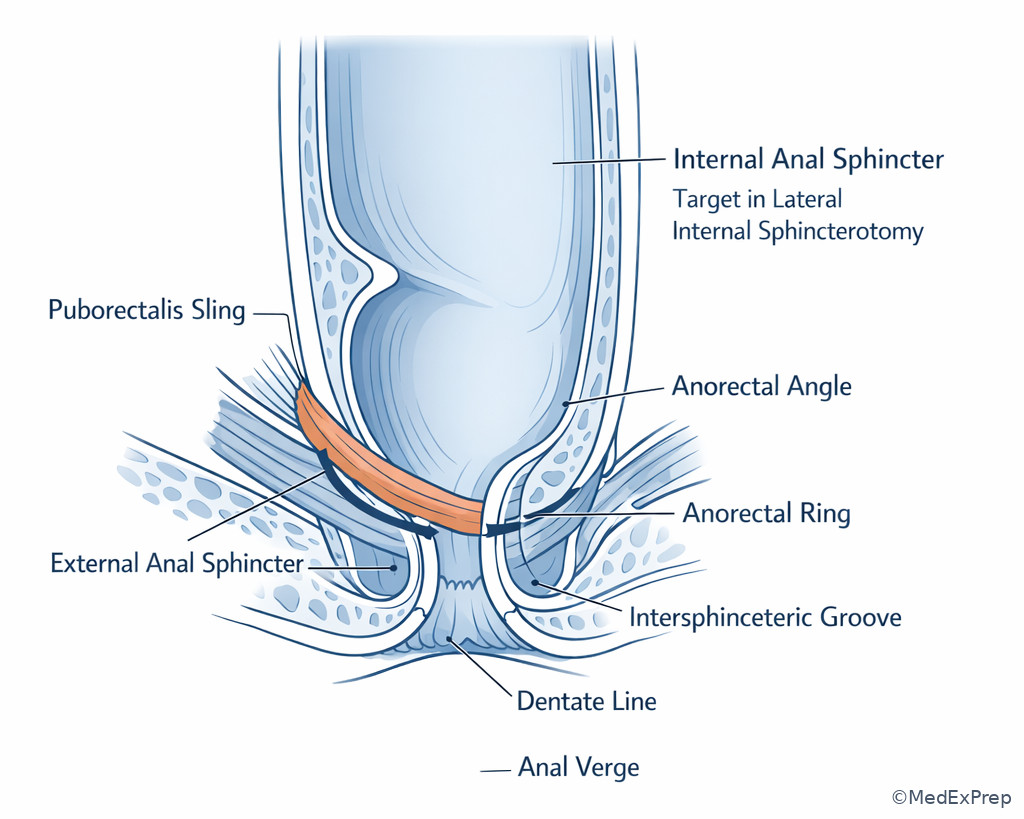
Exam takeaway: Surface and palpable landmarks help translate external inspection and digital examination into operative orientation.
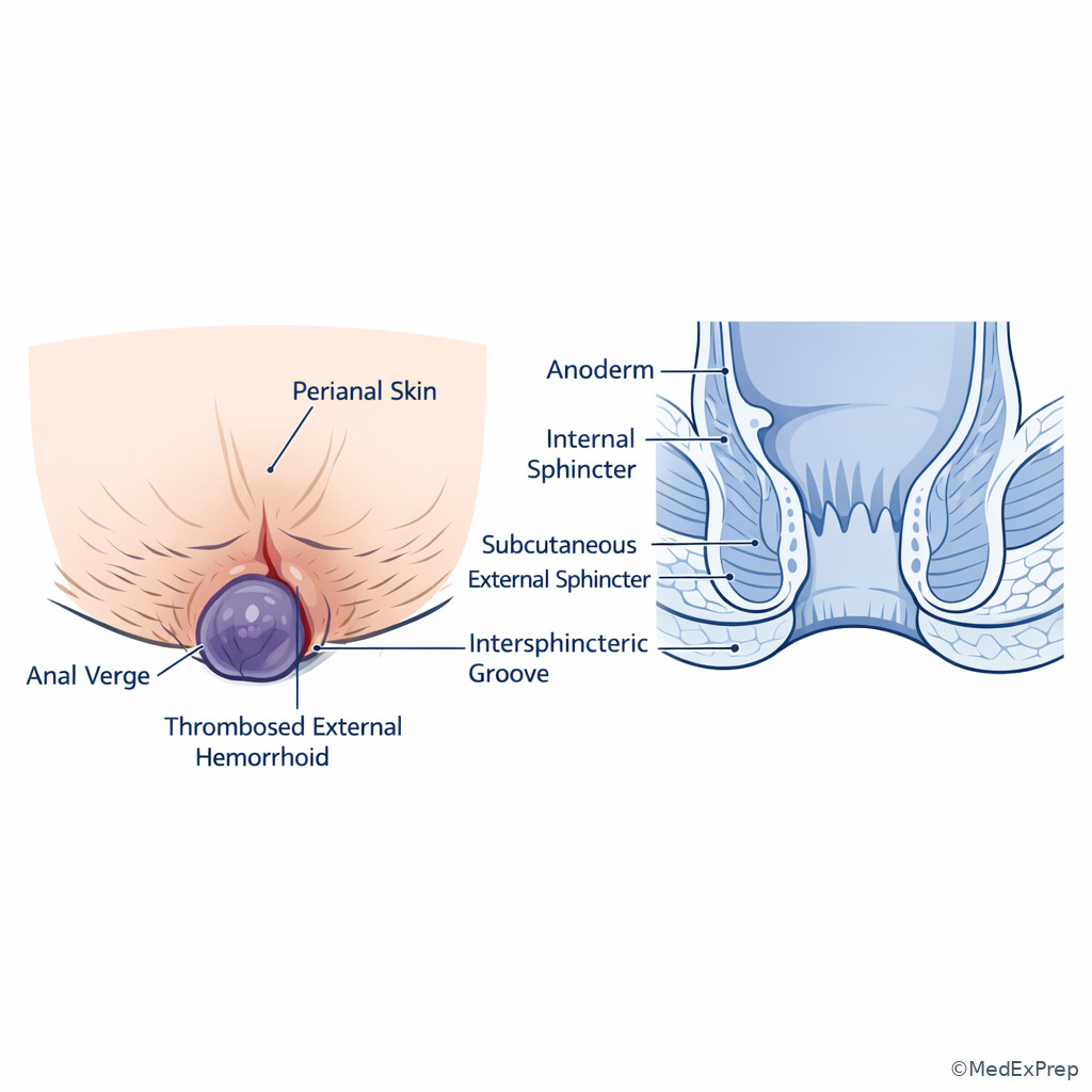
1.1.1.7 Applying landmarks to board-style disease localization
A Explanation
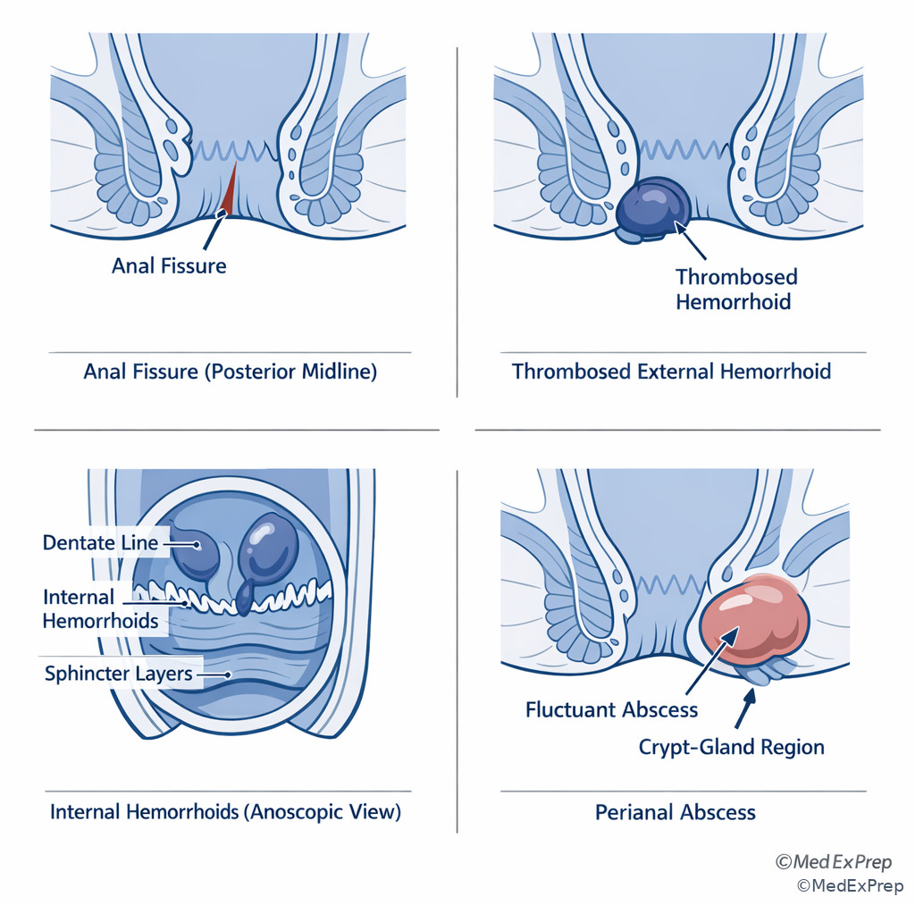
The final integration step is using landmarks as a differential diagnosis engine. In ABCRS stems, landmark anatomy is rarely asked in isolation; it is embedded in disease selection. If the lesion is painful, distal, and visible, think below the dentate line: fissure, thrombosed external hemorrhoid, painful skin lesion, or distal anal neoplasia. If the lesion is bleeding/prolapsing with minimal pain, think above the dentate line: internal hemorrhoids or proximal anal canal mucosal disease. If the process is suppurative, think crypt-gland unit at the dentate line. If the concern is sphincter preservation, orient to the intersphincteric plane and anorectal ring. See Fig 6.
WHY THIS IS TESTED: candidates must make definitive specialty-level choices under imperfect information. The correct answer often comes from one discriminating clue: severe touch pain, inguinal nodes, visible verge lesion, crypt origin, or continence-related high tract. Choose the diagnosis or next step that matches the landmark consequences; reject alternatives that fit only if the lesion were on the other side of the dentate line or at a different structural level.
Memory anchor: V-D-R = Verge for visible lesions, Dentate for drainage and pain rules, Ring for resection plane and continence questions.
Clinician Action: Diagnose by translating symptom pattern into landmark level before naming the disease. Order anoscopy, EUA, biopsy, or imaging based on that level. Treat the disease that fits the landmark logic, not the loudest symptom alone. Refer/Escalate for oncology, pelvic floor, or advanced imaging when lesion level changes nodal staging or continence risk.
B Worked example
A 72-year-old man with coronary artery disease, chronic kidney disease, and prior pelvic radiation for prostate cancer presents with a 2-cm ulcerated lesion just distal to the dentate line and exquisite pain during exam. He has one enlarged right superficial inguinal node. A distractor in the stem notes intermittent bright red bleeding, which may push test-takers toward internal hemorrhoids. The question asks which nodal basin is most concerning for regional spread.
Reasoning chain: Identify task: match lesion level to nodal drainage. Extract key facts: lesion is distal to the dentate line, severe pain, inguinal node already enlarged, bleeding is nonspecific noise. Apply rule: lesions below the dentate line drain primarily to superficial inguinal nodes. Eliminate distractors: internal iliac drainage is more relevant above the line; inferior mesenteric nodes fit more proximal rectal/colonic disease; para-aortic spread is too remote for first-echelon reasoning. Verify: severe pain and distal location both reinforce below-line anatomy.
C Exam trap
Common wrong answer: Internal hemorrhoids for any bright red blood per rectum. Why it is tempting: bleeding is common and familiar. Single clue that eliminates it: exquisite pain in a lesion distal to the dentate line should push you away from painless internal hemorrhoids and toward distal anal pathology.
D Checkpoint
Question: Which finding most strongly indicates a process below the dentate line?
- Painless prolapsing tissue during defecation
- Primary pelvic nodal drainage
- Exquisite pain with light touch of the lesion
- Engorged hemorrhoidal cushions seen above anal valves
Answer: C. Exquisite pain with light touch of the lesion.
- A: Tempting because prolapse is anorectal, but painless prolapsing tissue suggests internal hemorrhoids above the line.
- B: Tempting because pelvic nodes are relevant to the region, but they imply a more proximal location above the line.
- C: Correct because somatic innervation below the dentate line produces marked pain with manipulation.
- D: Tempting because this describes a true lesion pattern, but it localizes above the dentate line.
Exam takeaway: Convert symptom pattern into lesion level first, then choose the diagnosis and nodal or operative consequence.
flowchart TD
A[Anorectal stem] --> B{Main clue}
B --> C[Visible painful distal lesion]
B --> D[Bleeding prolapse minimal pain]
B --> E[Fever drainage fluctuance]
B --> F[Continence or high tract concern]
C --> C1[Below dentate line pathology]
D --> D1[Above dentate line pathology]
E --> E1[Crypt-gland origin near dentate line]
F --> F1[Sphincter complex and anorectal ring] Exam takeaway: Side-by-side lesion localization makes it easier to separate fissure, external hemorrhoid, internal hemorrhoid, and abscess on exam stems.
Exam takeaway: Remember that nodal drainage on anal cancer stems follows the lesion’s relation to the dentate line, not the symptom alone.
Exam Traps & Differentiators
Most common wrong answer and why: choosing the dentate line for every anatomic question. It is tempting because it is the best-known landmark, but many questions are really asking about the anal verge for external inspection or the anorectal ring for proximal functional anatomy.
| Looks similar | Key discriminator | Think this instead |
|---|---|---|
| Anal verge vs dentate line | External visible opening vs internal mucosal transition | Verge for inspection; dentate line for pain/lymphatics/hemorrhoid type |
| Dentate line vs anorectal ring | Biologic divider vs proximal functional boundary | Dentate for mucosa and sensation; ring for continence and surgery |
| External hemorrhoid vs internal hemorrhoid | Severe pain and distal location vs bleeding/prolapse with less pain | External below dentate line; internal above dentate line |
| Abscess vs thrombosed external hemorrhoid | Fever/drainage/fluctuance vs tense bluish thrombosis | Abscess originates from crypt-gland infection |
| Distal anal lesion nodal basin vs rectal lesion nodal basin | Relation to dentate line | Below line favors superficial inguinal; above favors pelvic pathways |
If the stem says ... think ...
- “Visible lesion at the opening” → think anal verge
- “Painless bleeding/prolapse” → think above the dentate line
- “Exquisite pain with light touch” → think below the dentate line/anoderm
- “Abscess/fistula originates from” → think anal crypt-gland unit at the dentate line
- “Palpable upper limit of anal canal” → think anorectal ring
- “Inguinal node concern for anal lesion” → think distal lesion below dentate line
Related traps will recur in upcoming lessons. In Pelvic Floor Compartments, the distractor pair will shift from dentate line vs verge to anorectal ring vs levator compartments. In Rectal Mesorectal Planes, a common confusion is using anal canal landmarks when the correct oncologic frame is mesorectal fascia and circumferential margin. Do not let a familiar landmark override the actual anatomic level being tested.
Tables
| Landmark | What it is | Highest-yield exam use | Common trap |
|---|---|---|---|
| Anal verge | External mucocutaneous opening to the perianal skin | Visible lesion localization, thrombosed external hemorrhoid, skin lesions | Mistaken for dentate line |
| Anoderm | Hairless squamous distal canal lining | Fissure pain, somatic sensitivity | Confused with perianal keratinized skin |
| Dentate line | Line formed by anal valves between columns | Pain, epithelium, hemorrhoid class, lymphatic drainage | Used incorrectly as proximal canal limit |
| Anal crypts/glands | Recesses and gland openings near dentate line | Cryptoglandular abscess/fistula origin | Confused with skin-origin infection |
| Intersphincteric groove | Plane between internal and external sphincters distally | Sphincter-plane orientation in surgery | Treated as a mucosal line |
| Anorectal ring | Palpable ring from puborectalis and sphincter complex | Upper anal canal boundary, continence, high tracts | Confused with dentate line |
| Feature | Above dentate line | Below dentate line |
|---|---|---|
| Epithelium | Columnar/transitional | Squamous anoderm/distal skin transition |
| Innervation | Visceral | Somatic |
| Pain sensitivity | Lower | High |
| Typical lesion clue | Painless bleeding or prolapse | Exquisite pain with touch |
| Hemorrhoid type | Internal | External |
| Primary nodal pattern | Pelvic/internal iliac-related | Superficial inguinal |
| Office tolerance | Better tolerated | Poorly tolerated if inflamed or incised |
| Presentation | Most likely landmark level | Best leading diagnosis | Single discriminator |
|---|---|---|---|
| Painful bluish anal margin lump | At/just distal to verge, below dentate line | Thrombosed external hemorrhoid | Tense nodule, no sepsis |
| Bleeding prolapse with little pain | Above dentate line | Internal hemorrhoids | Painless cushion disease |
| Posterior midline tearing pain | Distal anoderm | Anal fissure | Linear tear with severe defecatory pain |
| Fever, drainage, fluctuance | Crypt origin at dentate line with outward spread | Cryptoglandular abscess | Sepsis clues |
| High tract/continence concern | Intersphincteric or near anorectal ring | Complex fistula/sphincter-related process | Muscle plane matters more than mucosa |
| Distal anal lesion with inguinal nodes | Below dentate line | Distal anal canal/perianal malignancy pattern | Somatic pain plus inguinal drainage |
| Structure | Surgical relevance | When to choose it on an exam |
|---|---|---|
| Internal anal sphincter | Target in lateral internal sphincterotomy; continence contributor | Chronic fissure, hypertonicity, smooth muscle question |
| External anal sphincter | Voluntary continence mechanism; avoid injury in fistula surgery | Continence-risk procedures and muscle-preserving decisions |
| Puborectalis | Maintains anorectal angle; part of anorectal ring | Upper anal canal support and pelvic floor linkage |
| Anorectal ring | Upper surgical boundary; palpated on DRE/EUA | Question asks about proximal anal canal limit |
Algorithm / Approach
Rapid Review
- Anal verge → external visible starting point, not the sensory master divider
- Dentate line → single best landmark for pain, epithelium, lymphatics, hemorrhoid type
- Anorectal ring → proximal functional/surgical limit, not hemorrhoid classifier
- Internal hemorrhoids → above dentate line, bleeding/prolapse outweigh pain
- External hemorrhoids → distal to dentate line, pain dominates when thrombosed
- Anal fissure → distal anoderm lesion, usually posterior midline, severe pain
- Cryptoglandular abscess → starts at anal glands opening into crypts near dentate line
- Below dentate line malignancy → superficial inguinal drainage more likely
- Above dentate line lesion → pelvic/internal iliac nodal pattern more likely
- Internal sphincter → smooth muscle target in lateral internal sphincterotomy
- External sphincter/puborectalis → continence-preserving structures, high-cost injury
- Intersphincteric groove → muscle-plane landmark, not mucosal transition line
Self-check quiz
1.1.1.8 Question 1
A 63-year-old man with hypertension and chronic constipation has painless bright red bleeding and prolapsing tissue during bowel movements. Anoscopy shows enlarged vascular cushions above the anal valves. Which landmark most directly separates this lesion from a painful thrombosed external hemorrhoid?
- Anal verge
- Dentate line
- Anorectal ring
- Intersphincteric groove
1.1.1.9 Question 2
A 45-year-old woman with obesity and tobacco use presents with a tender ulcerated lesion just distal to the dentate line. She has no abdominal symptoms, but one superficial groin node is enlarged. Which nodal basin is most likely involved first?
- Inferior mesenteric nodes
- Paraaortic nodes
- Superficial inguinal nodes
- Pericolic nodes
1.1.1.10 Question 3
A 39-year-old man with diabetes and obesity presents with fever, severe anal pain, and a fluctuant perianal mass. He also reports a remote history of hemorrhoids, which makes thrombosis seem plausible. The most appropriate anatomic origin of this process is which structure?
- Anal crypts and glands at the dentate line
- Internal hemorrhoidal plexus above the anorectal ring
- Perianal epidermis at the anal verge
- Puborectalis fibers at the anorectal angle
1.1.1.11 Question 4
A 34-year-old woman with hypothyroidism, prior vaginal deliveries, and chronic constipation has a chronic posterior midline fissure refractory to topical calcium channel blocker therapy. The best operative target, if surgery is chosen, is which structure?
- External anal sphincter
- Internal anal sphincter
- Puborectalis sling
- Anal columns of Morgagni
1.1.1.12 Question 5
A 71-year-old man with coronary artery disease and chronic kidney disease undergoes examination under anesthesia for a low anal canal lesion. The surgeon needs the landmark that marks the upper boundary of the anal canal for operative orientation. Which is the most appropriate structure?
- Anal verge
- Dentate line
- Anorectal ring
- Anal crypt
1.1.1.13 Question 6
A 58-year-old woman with COPD, anticoagulation for atrial fibrillation, and chronic constipation presents with bright red blood per rectum and a painful anal mass. Examination shows a tense bluish nodule at the anal margin and separate noninflamed internal cushions on anoscopy. Which single clue most strongly identifies the painful lesion as below the dentate line?
- Presence of bright red bleeding
- Visualization on anoscopy
- Exquisite pain with light touch at the anal margin
- History of chronic constipation
1.1.1.14 Question 7
A 66-year-old man with prior pelvic radiation, smoking history, and weight loss has an ulcerated anal lesion. Biopsy confirms malignancy. The lesion is described as extending from the anal verge to just distal to the dentate line, and it is very painful on examination. Which regional drainage pattern is the best match for this specific lesion location?
- Primarily superficial inguinal drainage
- Primarily pericolic drainage along the sigmoid mesentery
- Primarily middle colic drainage
- Primarily paraesophageal drainage
1.1.1.15 Question 8
A 47-year-old woman with Crohn disease, prior seton placement, and mild urgency undergoes EUA for recurrent drainage. The stem asks for the landmark that is most relevant if the tract extends high enough to threaten continence mechanisms and the upper limit of the surgical anal canal. Which landmark best answers that question?
- Dentate line
- Anal verge
- Anorectal ring
- Anal valve
Answer key
1.1.1.16 Question 1 Answer: B. Dentate line
Mechanism first: Internal hemorrhoids arise above the dentate line and are typically associated with bleeding and prolapse with relatively less pain because of visceral innervation. Thrombosed external hemorrhoids occur distal to the dentate line and are acutely painful because of somatic innervation. This single landmark separates the two entities. Sources: ASCRS clinical practice patterns; Beck et al., The ASCRS Textbook of Colon and Rectal Surgery.
- A. Anal verge: Tempting because it marks the visible opening and external anatomy; incorrect because the distinguishing rule between internal and external hemorrhoids is the dentate line, not the verge. Discriminating clue: the question asks what separates painless internal cushions from painful external thrombosis.
- B. Dentate line: Correct because it marks the transition in innervation, mucosa, and hemorrhoidal classification. Discriminating clue: enlarged cushions above the anal valves with little pain.
- C. Anorectal ring: Tempting because it is another major landmark; incorrect because it defines the upper anal canal, not hemorrhoid class. Discriminating clue: this is a symptom-location question, not a proximal boundary question.
- D. Intersphincteric groove: Tempting because it is surgically relevant; incorrect because it is a muscle-plane landmark, not the primary divider for hemorrhoids. Discriminating clue: the stem centers on bleeding/prolapse versus pain.
1.1.1.17 Question 2 Answer: C. Superficial inguinal nodes
Mechanism first: Lesions below the dentate line drain primarily to the superficial inguinal nodal basin. The clue “just distal to the dentate line” plus marked tenderness localizes the lesion below the line. Sources: NCCN Anal Carcinoma guideline framework; Beck et al., The ASCRS Textbook of Colon and Rectal Surgery.
- A. Inferior mesenteric nodes: Tempting because colorectal lesions can drain proximally; incorrect for a distal anal lesion below the dentate line. Discriminating clue: distal painful lesion, not proximal rectal/colonic disease.
- B. Paraaortic nodes: Tempting as an advanced metastatic site; incorrect because the question asks the first regional basin. Discriminating clue: regional, not distant spread.
- C. Superficial inguinal nodes: Correct because below-line anal lesions commonly drain here. Discriminating clue: superficial groin node enlargement with a lesion distal to the dentate line.
- D. Pericolic nodes: Tempting because they are standard for colonic tumors; incorrect in anal canal anatomy. Discriminating clue: anal lesion rather than colonic lesion.
1.1.1.18 Question 3 Answer: A. Anal crypts and glands at the dentate line
Mechanism first: Most anorectal abscesses and fistulas are cryptoglandular. The anal glands open into crypts near the dentate line; obstruction and infection seed the intersphincteric plane and then spread. Fever and fluctuance distinguish abscess from hemorrhoidal thrombosis. Sources: Steele et al., ASCRS Clinical Practice Guidelines for Anorectal Abscess, Fistula-in-Ano, and Rectovaginal Fistula; Beck et al.
- A. Anal crypts and glands at the dentate line: Correct because this is the classic origin of cryptoglandular sepsis. Discriminating clue: fever plus fluctuant mass.
- B. Internal hemorrhoidal plexus above the anorectal ring: Tempting because hemorrhoids are common and the patient mentions them; incorrect because hemorrhoids do not usually cause febrile fluctuance, and “above the anorectal ring” is anatomically off-target. Discriminating clue: suppuration rather than bleeding/prolapse.
- C. Perianal epidermis at the anal verge: Tempting because the external manifestation is perianal; incorrect because the usual origin is deeper crypt-gland infection. Discriminating clue: asks origin, not where the abscess points.
- D. Puborectalis fibers at the anorectal angle: Tempting because deep pelvic structures matter in complex sepsis; incorrect because routine cryptoglandular disease starts distally. Discriminating clue: common rather than supralevator origin.
1.1.1.19 Question 4 Answer: B. Internal anal sphincter
Mechanism first: Chronic anal fissure is linked to internal sphincter hypertonicity and local ischemia, especially in posterior midline disease. When medical therapy fails and surgery is selected, the classic operative target is the internal sphincter via lateral internal sphincterotomy. Sources: ASCRS anal fissure guidelines; Beck et al.
- A. External anal sphincter: Tempting because it is a named sphincter; incorrect because dividing it would increase continence risk and does not address the standard mechanism. Discriminating clue: operative treatment for fissure is lateral internal sphincterotomy.
- B. Internal anal sphincter: Correct because it is the hypertonic smooth muscle targeted when surgery is indicated. Discriminating clue: refractory chronic posterior midline fissure.
- C. Puborectalis sling: Tempting because it affects anorectal angle; incorrect because it is not the routine target in fissure surgery. Discriminating clue: fissure, not defecatory floor dysfunction.
- D. Anal columns of Morgagni: Tempting because they are real landmarks within the canal; incorrect because they are mucosal folds, not the operative muscle target. Discriminating clue: asks for the structure targeted by surgery.
1.1.1.20 Question 5 Answer: C. Anorectal ring
Mechanism first: The anorectal ring is the palpable composite upper boundary of the surgical anal canal, reflecting the puborectalis and sphincteric complex. It is the correct orientation point when the surgeon needs the proximal canal limit. Sources: Beck et al.; standard colorectal anatomy texts.
- A. Anal verge: Tempting because it is the distal visible starting point; incorrect because the question asks for the upper boundary. Discriminating clue: “upper boundary” in EUA.
- B. Dentate line: Tempting because it is the best-known internal line; incorrect because it lies within the canal and does not define its top. Discriminating clue: operative orientation to canal extent, not mucosal transition.
- C. Anorectal ring: Correct because it marks the proximal functional/surgical limit. Discriminating clue: proximal boundary needed during EUA.
- D. Anal crypt: Tempting because it is a real structure relevant to sepsis; incorrect because it is not a boundary landmark. Discriminating clue: no abscess/fistula context.
1.1.1.21 Question 6 Answer: C. Exquisite pain with light touch at the anal margin
Mechanism first: Somatic innervation below the dentate line makes external hemorrhoids and anoderm lesions acutely painful with manipulation. Bright red bleeding and constipation are nonspecific; anoscopy can show coexisting internal disease and mislead you. The single strongest clue to below-line localization is severe touch pain at the anal margin. Sources: ASCRS hemorrhoid guidance; Beck et al.
- A. Presence of bright red bleeding: Tempting because hemorrhoids bleed; incorrect because bleeding occurs in both internal and external-associated processes and does not localize the lesion. Discriminating clue: pain, not bleeding, is the separator here.
- B. Visualization on anoscopy: Tempting because internal pathology is seen this way; incorrect because the painful lesion itself is at the margin and may coexist with internal cushions. Discriminating clue: separate lesions are described.
- C. Exquisite pain with light touch at the anal margin: Correct because somatic innervation below the dentate line produces this response. Discriminating clue: pain on light touch at the margin.
- D. History of chronic constipation: Tempting because it predisposes to anorectal disease broadly; incorrect because it does not distinguish level. Discriminating clue: chronic constipation is supportive but not localizing.
1.1.1.22 Question 7 Answer: A. Primarily superficial inguinal drainage
Mechanism first: A lesion that extends to just distal to the dentate line and is very painful localizes at least substantially below the dentate line, where lymphatic drainage is directed toward the superficial inguinal nodes. The radiation history and weight loss raise cancer concern but do not change first-echelon anatomy. Sources: NCCN Anal Carcinoma guidance; Beck et al.
- A. Primarily superficial inguinal drainage: Correct because the painful distal component below the dentate line drives this nodal expectation. Discriminating clue: “just distal to the dentate line” with severe pain.
- B. Primarily pericolic drainage along the sigmoid mesentery: Tempting because all colorectal cancers have nodal drainage; incorrect because this is anal canal, not sigmoid colon. Discriminating clue: lesion location at the verge/distal canal.
- C. Primarily middle colic drainage: Tempting only if the learner thinks broadly of mesenteric nodes; incorrect because this drainage pattern is unrelated to distal anal anatomy. Discriminating clue: anal lesion, not transverse colon lesion.
- D. Primarily paraesophageal drainage: Tempting only as a distant metastatic concept; incorrect and not the relevant regional basin. Discriminating clue: first regional spread is being tested.
1.1.1.23 Question 8 Answer: C. Anorectal ring
Mechanism first: When the tract extends high enough that continence structures and the upper limit of the surgical anal canal matter, the key landmark is the anorectal ring. The dentate line remains important for mucosal transition, but the ring is the correct answer when the question shifts to high tract extent and functional preservation. Crohn history is distracting but also realistic because recurrent complex fistulas often require precise sphincter-preserving planning. Sources: ASCRS fistula guidance; Beck et al.
- A. Dentate line: Tempting because fistulas often arise from glands near this line; incorrect because the question asks about high extension and continence relevance. Discriminating clue: “upper limit of the surgical anal canal.”
- B. Anal verge: Tempting because external openings are described from the verge; incorrect because external skin does not answer the high tract question. Discriminating clue: concern is not external opening but proximal extent.
- C. Anorectal ring: Correct because it is the proximal functional/surgical landmark linked to continence mechanisms. Discriminating clue: tract high enough to threaten continence and upper canal anatomy.
- D. Anal valve: Tempting because valves help form the dentate line and are part of crypt origin; incorrect because they do not mark the upper surgical limit. Discriminating clue: asks for high landmark, not source opening.
Keep going with the full ABCRS Colon & Rectal Surgery Prep Course (Colon/Rectal)
You just studied the way members do, in the same lesson interface. Full access unlocks every lesson, the question bank and flashcards for a full year.
One payment · 1 year of access · No auto-renewal