1.1.1 - Oxygen Transport Determinants
Oxygen Transport Determinants
What you’ll learn
- BASIC SCIENCE: Respiratory and cardiovascular physiology -> Oxygen content and delivery: choose which variable most strongly changes arterial oxygen content and total oxygen delivery.
- BASIC SCIENCE: Physiology and pathophysiology -> Hemoglobin, saturation, and dissolved oxygen: distinguish oxygen content from partial pressure and saturation in board-style stems.
- CLINICAL ANESTHESIA: Monitoring interpretation -> Pulse oximetry, ABG, and anemia integration: decide when a normal pulse oximeter can coexist with critically reduced oxygen delivery.
- CLINICAL ANESTHESIA: Perioperative management -> Immediate response to impaired oxygen transport: select the safest next step when low delivery is caused by hypoxemia, anemia, low cardiac output, or increased consumption.
- PATIENT SAFETY: Crisis recognition -> Hidden hypoxia and occult tissue dysoxia: identify patterns in which oxygenation looks acceptable while tissue oxygen delivery remains inadequate.
- DOMAIN: Basic science foundations → respiratory and cardiovascular physiology → determinants of oxygen content, delivery, and consumption
- DOMAIN: Clinical anesthesiology → monitoring interpretation → reconcile SpO2, PaO2, hemoglobin, and cardiac output
- DOMAIN: Perioperative medicine → risk assessment → recognize patients at risk for oxygen supply-demand mismatch
- DOMAIN: Critical care concepts → shock and organ support → apply oxygen delivery physiology to unstable patients
- Learner note: This topic may be tested under broader perioperative, critical care, cardiothoracic, or crisis-management domains; confirm against your current ABA materials.
Why it matters
- In anesthesiology, hypoxemia, anemia, and low cardiac output often coexist; board questions test which one is the dominant limiter of oxygen delivery.
- A normal SpO2 does not guarantee adequate tissue oxygen delivery; severe anemia or low cardiac output can still produce ischemia and lactic acidosis.
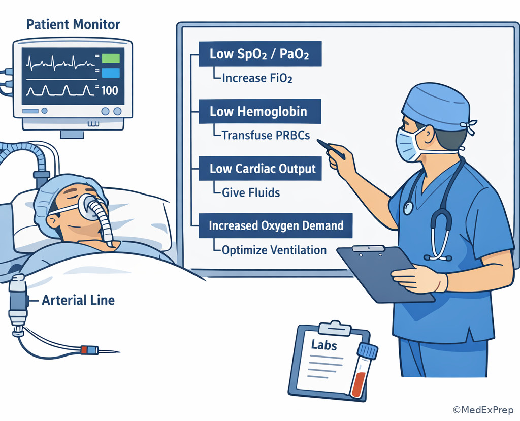
- Perioperative rescue depends on identifying the broken link in the chain: oxygen loading in the lung, carriage by hemoglobin, flow by the circulation, or consumption at the tissue level.
- This framework is the physiologic bridge to upcoming topics on V/Q mismatch and cardiac output regulation.
Key Terms & Must-Know Facts
1.1.1.1 Key terms
- PaO2: partial pressure of oxygen dissolved in arterial plasma.
- SaO2: arterial hemoglobin oxygen saturation.
- SpO2: pulse oximeter estimate of arterial saturation.
- CaO2: arterial oxygen content.
- CvO2: mixed venous oxygen content.
- DO2: oxygen delivery to tissues per minute.
- VO2: oxygen consumption by tissues per minute.
- Oxygen extraction ratio: fraction of delivered oxygen removed by tissues.
- P50: PaO2 at which hemoglobin is 50% saturated.
- Oxyhemoglobin dissociation curve: relation between PaO2 and hemoglobin saturation.
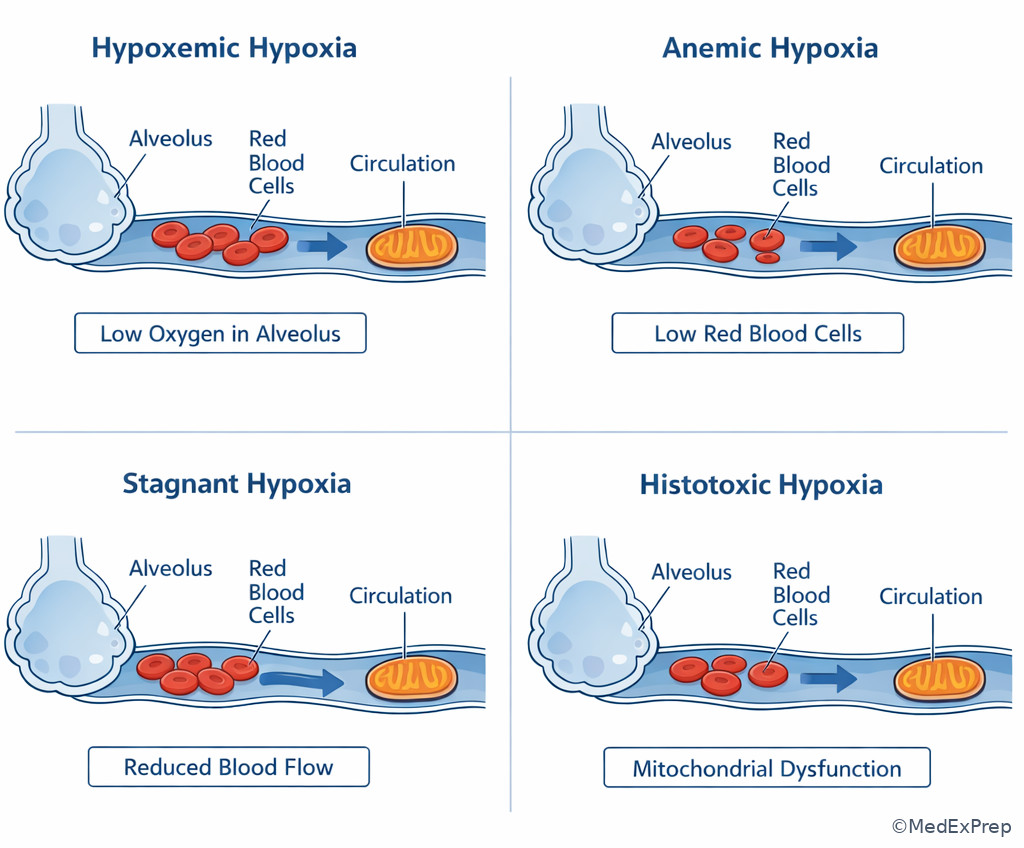
- Anemic hypoxia: low oxygen content due to reduced functional hemoglobin.
- Stagnant hypoxia: low tissue oxygenation from reduced blood flow.
- Histotoxic hypoxia: impaired tissue oxygen use despite delivery.
- Mixed venous oxygen saturation (SvO2): global balance of oxygen delivery and consumption.
1.1.1.2 Must-know facts
- The dominant term in arterial oxygen content is hemoglobin-bound oxygen, not dissolved oxygen.
- \( CaO_2 \approx 1.34 \times Hb \times SaO_2 + 0.003 \times PaO_2 \)
- \( DO_2 = CO \times CaO_2 \times 10 \)
- Raising PaO2 markedly increases dissolved oxygen only a little; it rarely compensates for major anemia.
- Pulse oximetry reflects saturation, not oxygen content and not cardiac output.
- On the flat portion of the dissociation curve, large PaO2 changes may cause little SpO2 change.
- On the steep portion, small PaO2 drops can cause abrupt desaturation.
- Fever, acidosis, and increased 2,3-DPG shift the curve right and promote unloading.
- Hypothermia, alkalosis, and low 2,3-DPG shift the curve left and impair unloading.
- Low SvO2 suggests either low delivery, increased consumption, or both.
Exam takeaway: Separate lung loading, hemoglobin carriage, and blood flow to identify the single best correction.
flowchart LR
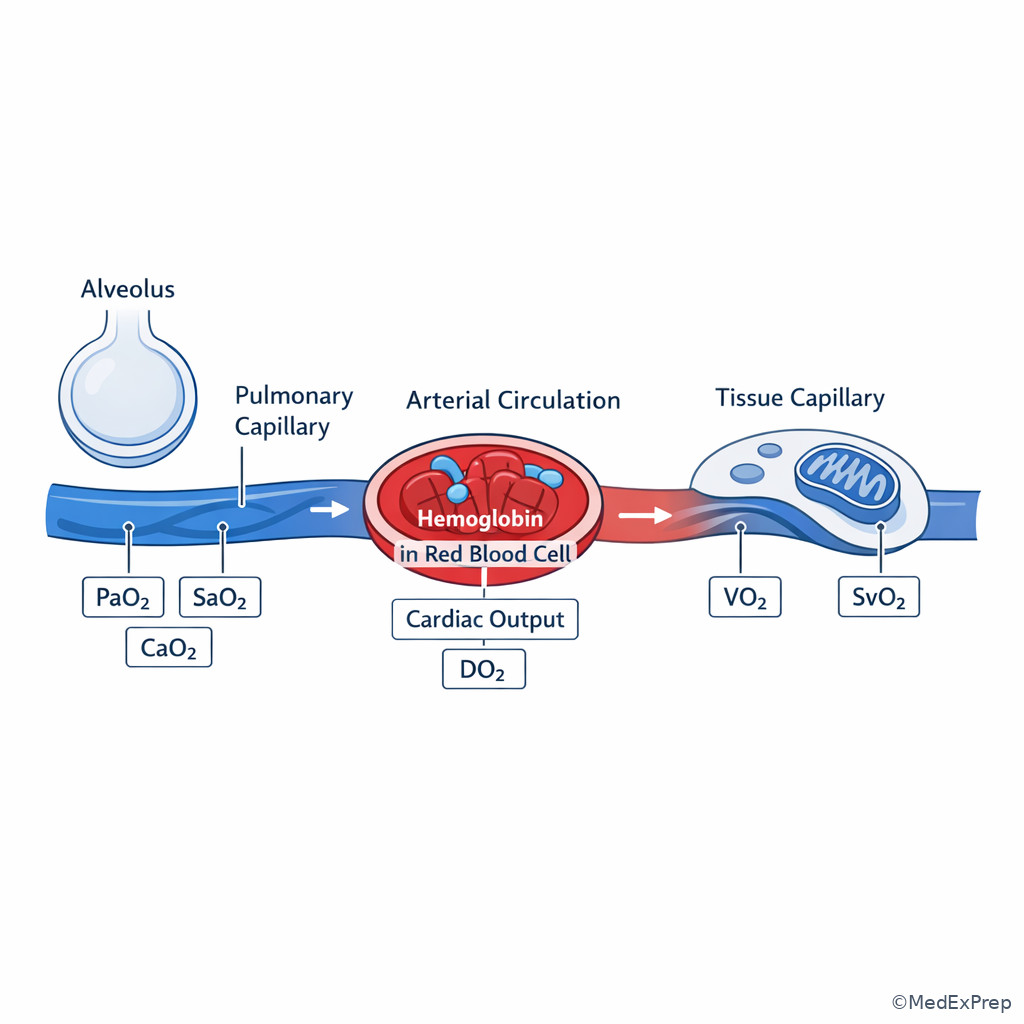
A[Inspired oxygen and alveoli] --> B[Arterial PaO2]
B --> C[Hemoglobin saturation]
C --> D[Arterial oxygen content CaO2]
E[Hemoglobin concentration] --> D
D --> F[Cardiac output]
F --> G[Oxygen delivery DO2]
G --> H[Tissue extraction]
H --> I[VO2 and SvO2]
Exam takeaway: The exam often hides oxygen transport failure in one step of the loading-carrying-flow pathway.
Core content
1.1.1.3 Foundational formula: oxygen content versus oxygen partial pressure
A Explanation
The central decision conflict is this: when oxygen transport is inadequate, should you respond to the number on the pulse oximeter/ABG or to the total oxygen carried in blood? The resolving rule is that tissues receive oxygen based primarily on oxygen content, and oxygen content is driven overwhelmingly by hemoglobin-bound oxygen. A patient with a PaO2 of 300 mm Hg and Hb of 5 g/dL may still have much lower oxygen content than a patient with PaO2 80 mm Hg and Hb 15 g/dL.
Use the board-level formula: \( CaO_2 \approx 1.34 \times Hb \times SaO_2 + 0.003 \times PaO_2 \). The first term dominates because each gram of hemoglobin carries about 1.34 mL O2 when fully saturated, whereas dissolved oxygen contributes only 0.003 mL O2/dL per mm Hg PaO2. That means even hyperoxia adds relatively little content unless hyperbaric conditions are used.
WHY THIS IS TESTED: exam stems often tempt you to equate “excellent oxygenation” with “adequate oxygen delivery.” Choose increased FiO2 when the problem is impaired arterial loading; reject FiO2 as the sole fix when severe anemia or low cardiac output is the limiting factor.
Key rule: PaO2 tells you how much oxygen is dissolved; CaO2 tells you how much oxygen is available to be delivered.
B Worked example
A 68-year-old man undergoing colectomy has coronary artery disease, chronic kidney disease, and well-controlled COPD. After blood loss, his SpO2 is 100% on FiO2 0.8, PaO2 is 240 mm Hg, Hb is 6.1 g/dL, lactate is 3.4 mmol/L, and blood pressure is stable on low-dose phenylephrine. A tempting clue is the excellent PaO2, which makes “increase FiO2 further” look appealing. The task is to identify the main determinant limiting tissue oxygen delivery.
Reasoning chain: identify task: choose the most appropriate correction of oxygen transport. Extract key facts: severe anemia, high PaO2 already, evidence of tissue stress, CAD increases ischemic risk. Apply rule: dissolved oxygen is a minor component of CaO2, so raising FiO2 further adds little. Eliminate distractors: bronchodilator therapy does not address the issue; permissive anemia is unsafe here; low-dose vasoconstrictor may support pressure but does not restore oxygen content. Verify: the safest next step is to address anemia and bleeding while maintaining oxygenation.
C Exam trap
Common wrong answer: “Increase FiO2 to 1.0 and observe.”
Why it is tempting: candidates overvalue PaO2 and assume hyperoxia can compensate for low oxygen-carrying capacity.
Single clue that eliminates it: Hb of 6.1 g/dL with high PaO2 means the limiting problem is oxygen content, not alveolar oxygen availability.
D Checkpoint
Question: A 54-year-old woman under general anesthesia has SpO2 99% on FiO2 0.6, PaO2 180 mm Hg, Hb 5.8 g/dL after acute surgical bleeding, and normal ETCO2. Which intervention most directly increases arterial oxygen content?
- Increase FiO2 to 1.0
- Transfuse packed red blood cells
- Increase minute ventilation
- Add PEEP
- A. Tempting because higher FiO2 raises PaO2; wrong because dissolved oxygen contributes minimally compared with hemoglobin-bound oxygen.
- B. Tempting because it seems slower than oxygen adjustment; right because increasing hemoglobin most directly raises CaO2 in severe anemia.
- C. Tempting because ventilation affects gas exchange; wrong because CO2 elimination does not meaningfully correct low oxygen content here.
- D. Tempting because PEEP can improve oxygenation; wrong because she is already well oxygenated and the limiting variable is hemoglobin.
1.1.1.4 Oxygen delivery: coupling oxygen content to cardiac output
A Explanation
The next decision conflict is whether oxygen transport failure is caused by too little oxygen per dL of blood or too little blood flow per minute. The resolving rule is the delivery equation: \( DO_2 = CO \times CaO_2 \times 10 \). If either cardiac output or arterial oxygen content falls, total delivery falls. In practice, low cardiac output can be just as dangerous as anemia or hypoxemia, and multiple deficits often interact.
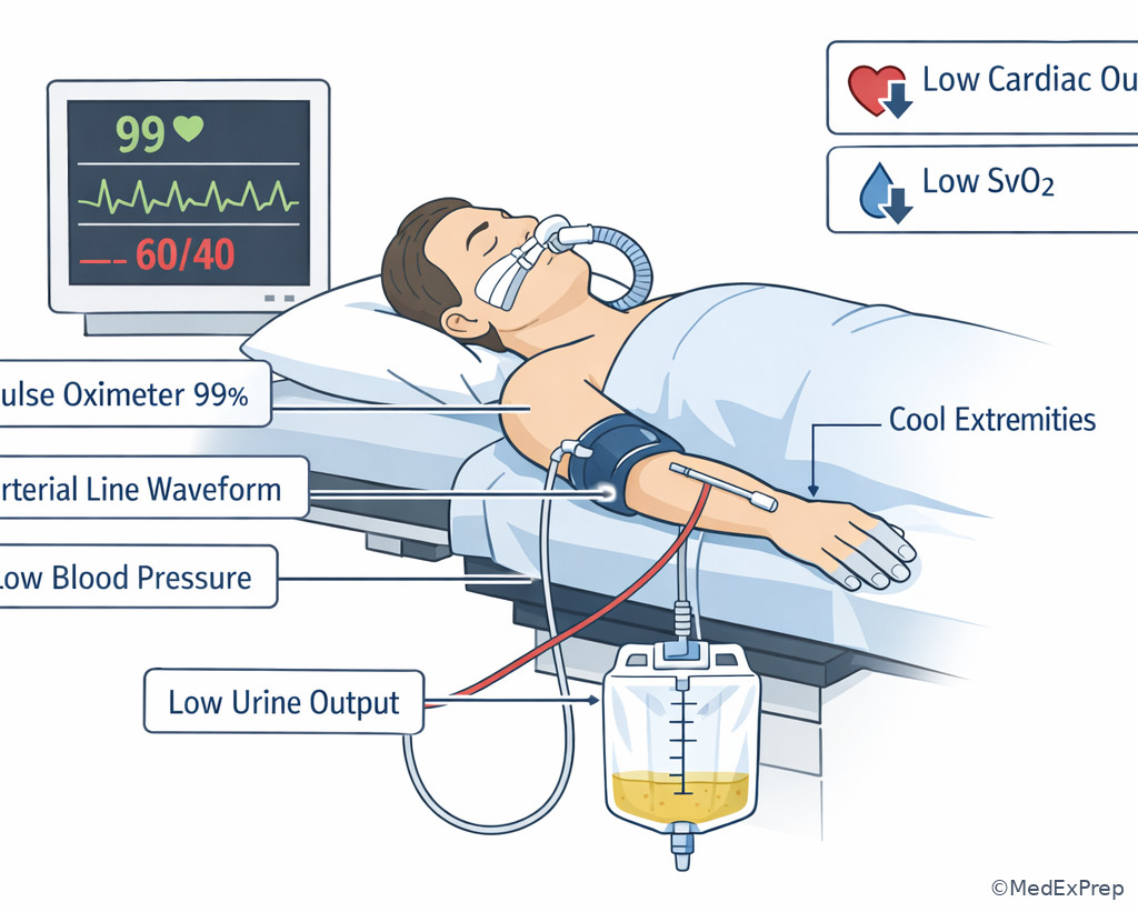
This is why pulse oximetry can look perfect while organs are underperfused. SpO2 says almost nothing about flow. A patient in cardiogenic shock may have normal oxygen saturation but profoundly low delivery. Conversely, a high cardiac output state can partially compensate for low content, at least until demand outstrips reserve.
WHY THIS IS TESTED: ABA-style stems ask you to choose between a “lung answer” and a “circulation answer.” When saturation is adequate but SvO2 is low, lactate is rising, urine output is falling, and perfusion is poor, think delivery failure from inadequate flow until proven otherwise.
B Worked example
A 72-year-old woman with severe aortic stenosis, diabetes, and chronic anemia undergoes urgent hip fracture repair. After induction, SpO2 remains 98% on FiO2 0.6, Hb is 9.2 g/dL, cardiac index falls, MAP is 48 mm Hg, urine output ceases, and SvO2 drops to 48%. A distracting finding is the acceptable hemoglobin, which may make transfusion look like the obvious answer. The task is to identify the dominant cause of low oxygen delivery.
Reasoning chain: identify task: choose the main intervention to restore oxygen transport. Extract key facts: normal saturation, hemoglobin not profoundly low, clear low-flow state after induction, low SvO2 and hypoperfusion. Apply rule: delivery equals content times output. Eliminate distractors: increasing FiO2 may marginally help but not enough; immediate transfusion is not the most direct fix; hyperventilation does nothing for flow. Verify: improve cardiac output and perfusion pressure while respecting aortic stenosis physiology.
C Exam trap
Common wrong answer: “Transfuse because SvO2 is low.”
Why it is tempting: low venous saturation triggers a reflex association with inadequate oxygen carrying capacity.
Single clue that eliminates it: the abrupt postinduction low-output state with preserved oxygenation points to reduced flow as the immediate dominant problem.
D Checkpoint
Question: A mechanically ventilated patient has SpO2 100%, PaO2 220 mm Hg, Hb 10 g/dL, cool extremities, low urine output, lactate 4.1 mmol/L, and low cardiac output after induction. Which change most directly increases oxygen delivery?
- Increase FiO2 from 0.6 to 1.0
- Improve cardiac output
- Reduce respiratory rate
- Administer methylene blue empirically
- A. Tempting because more oxygen feels universally helpful; wrong because PaO2 is already high and dissolved oxygen adds little.
- B. Tempting because it targets circulation rather than oxygenation; right because low flow is the limiting term in the delivery equation.
- C. Tempting if one overinterprets ventilator settings; wrong because the scenario is not ventilation failure.
- D. Tempting because tissue hypoxia can suggest abnormal hemoglobin states; wrong because the stem gives no evidence of methemoglobinemia or vasoplegia as the primary issue.
- The largest determinant of arterial oxygen content is ___.
- True or False: A normal SpO2 excludes inadequate oxygen delivery.
- Name the 2 major variables in the oxygen delivery equation.
Exam takeaway: Normal saturation does not exclude low oxygen delivery when hemoglobin or cardiac output is reduced.
flowchart TD
A[Normal SpO2] --> B{Is oxygen transport adequate?}
B -->|No| C[Check hemoglobin]
B -->|No| D[Check cardiac output and perfusion]
C --> E[Low Hb lowers CaO2]
D --> F[Low CO lowers DO2]
E --> G[Low SvO2 lactate ischemia]
F --> G Exam takeaway: A patient can appear well oxygenated on the monitor while organs remain underperfused.
1.1.1.5 Oxyhemoglobin dissociation curve: loading versus unloading
A Explanation
The decision conflict here is whether a shift in the dissociation curve helps or harms the patient. The resolving rule is to ask where the oxygen problem exists: pulmonary loading or tissue unloading. A right shift decreases hemoglobin affinity, making tissue unloading easier but requiring a higher PaO2 for the same saturation. A left shift increases affinity, preserving saturation at a given PaO2 but impeding release to tissues.
Right shift causes include acidosis, hypercapnia, hyperthermia, and increased 2,3-DPG. Left shift causes include alkalosis, hypocapnia, hypothermia, low 2,3-DPG, fetal hemoglobin, and carbon monoxide exposure. In exam questions, the trap is to think of the curve only in terms of saturation. In the OR, impaired unloading can matter clinically when perfusion is marginal and the patient is cold or alkalotic.
WHY THIS IS TESTED: the exam uses curve shifts to explain why a PaO2 or SpO2 value does not fully predict tissue oxygenation. Choose a right shift as beneficial for unloading in metabolically active tissues; reject it as a universal improvement because pulmonary loading may worsen if reserve is limited.
See Fig 3.
B Worked example
A 4-year-old child after long scoliosis surgery is cool to 34.5°C, ventilated with a respiratory alkalosis, SpO2 100%, PaO2 170 mm Hg, and has marginal urine output with rising lactate. The distracting clue is the excellent oxygenation data, which make inadequate oxygen transport seem unlikely. The task is to explain why tissue oxygen extraction may still be impaired.
Reasoning chain: identify task: determine why adequate PaO2 and SpO2 do not guarantee delivery to tissues. Extract key facts: hypothermia and alkalosis both shift the curve left; rising lactate suggests tissue-level mismatch. Apply rule: left shift increases affinity and impairs unloading. Eliminate distractors: occult shunt is less likely with high PaO2; severe anemia is not provided; ventilator malfunction does not fit the data. Verify: correcting hypothermia and excessive alkalosis improves release of oxygen to tissues.
C Exam trap
Common wrong answer: “A left shift is beneficial because saturation is higher.”
Why it is tempting: the monitor-centered view focuses only on hemoglobin loading in the lungs.
Single clue that eliminates it: evidence of tissue dysoxia despite high saturation means unloading, not loading, is the problem.
D Checkpoint
Question: Which perioperative condition most impairs tissue oxygen unloading despite preserved arterial saturation?
- Hyperthermia with acidosis
- Hypothermia with alkalosis
- Fever with elevated 2,3-DPG
- Hypercapnia during one-lung ventilation
- A. Tempting because it clearly changes the curve; wrong because it shifts right and favors unloading.
- B. Tempting because the pulse oximeter may look reassuring; right because it shifts left and makes tissue release harder.
- C. Tempting because the patient may still be unstable; wrong because elevated 2,3-DPG shifts right.
- D. Tempting because hypercapnia can be harmful; wrong because increased CO2 shifts right rather than left.
Exam takeaway: Right shift helps unloading, left shift impairs unloading, and the stem decides which matters more.
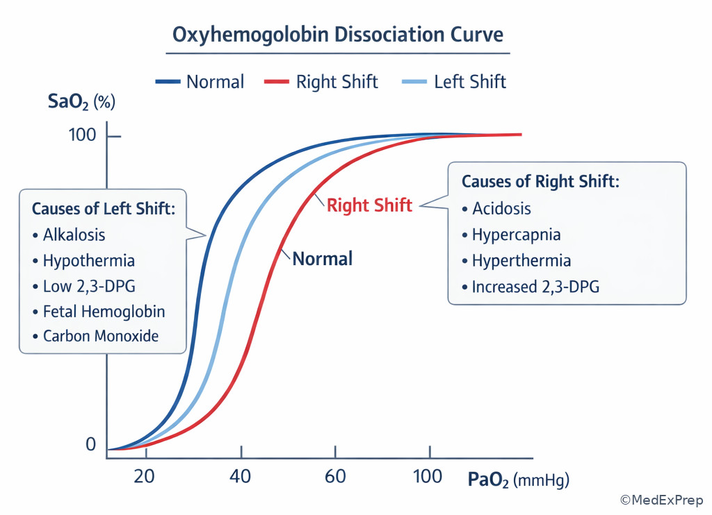
Exam takeaway: Match the acid-base or temperature disturbance to whether unloading improves or worsens.
flowchart LR
A[Right shift] --> A1[Acidosis]
A --> A2[Hypercapnia]
A --> A3[Hyperthermia]
A --> A4[High 2,3-DPG]
B[Left shift] --> B1[Alkalosis]
B --> B2[Hypothermia]
B --> B3[Low 2,3-DPG]
B --> B4[CO or fetal Hb]
1.1.1.6 Venous oxygen saturation, extraction, and oxygen debt
A Explanation
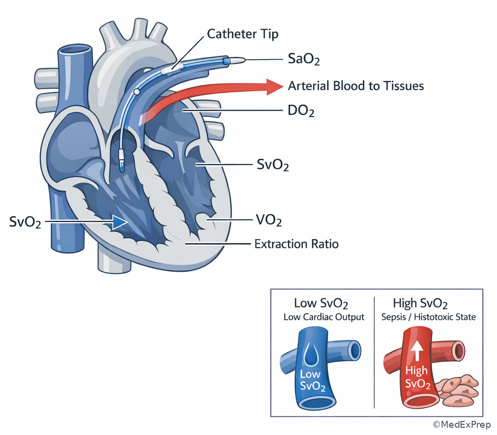
The decision conflict is whether low venous oxygen saturation means the patient is extracting appropriately or failing delivery. The rule is to interpret venous saturation as a balance variable: \( SvO_2 \) or central venous saturation falls when delivery decreases, consumption increases, or both. It rises when extraction is impaired or delivery exceeds demand.
Mixed venous oxygen saturation is not a stand-alone resuscitation target in every patient, but it is highly testable because it summarizes the delivery-consumption relationship. Low SvO2 in a bleeding patient under anesthesia suggests inadequate delivery from low content, low flow, or both. A very high SvO2 in septic shock or cyanide toxicity can indicate inability of tissues to extract or use oxygen. Rising lactate suggests oxygen debt or impaired oxidative metabolism, though lactate is not specific.
WHY THIS IS TESTED: exam stems often give a normal SpO2 but abnormal SvO2 or lactate. Choose a delivery-consumption interpretation, not a purely pulmonary one. See Fig 4.
B Worked example
A 59-year-old man with ischemic cardiomyopathy, obesity, and chronic beta-blocker use is in the ICU after major abdominal surgery. SpO2 is 99% on low ventilator support, Hb is 10.4 g/dL, SvO2 falls from 68% to 45%, lactate increases, and extremities are cool. A distracting clue is the acceptable hemoglobin, which may falsely reassure against oxygen transport failure. The task is to interpret the venous saturation change.
Reasoning chain: identify task: determine whether tissues are extracting more because delivery is inadequate. Extract key facts: preserved saturation, moderate Hb, signs of low flow, falling SvO2, rising lactate. Apply rule: low SvO2 means delivery is falling relative to demand. Eliminate distractors: primary V/Q mismatch is less likely with preserved oxygenation; dyshemoglobinemia is not suggested; increased FiO2 alone is not enough. Verify: treat the hemodynamic cause of reduced delivery.
C Exam trap
Common wrong answer: “High SvO2 always means adequate resuscitation.”
Why it is tempting: a higher venous saturation seems like more leftover oxygen and therefore better delivery.
Single clue that eliminates it: in sepsis or histotoxic states, tissues may fail to extract or use oxygen, so venous saturation can be deceptively high.
D Checkpoint
Question: A patient with stable SpO2 but falling SvO2 and rising lactate most likely has which physiologic change?
- Improved tissue extraction because delivery is inadequate
- Severe hyperoxia increasing dissolved oxygen
- Left-shifted curve improving tissue unloading
- Artifact from pulse oximetry failure alone
- A. Tempting because it sounds paradoxical; right because low SvO2 reflects greater extraction when delivery is insufficient.
- B. Tempting if one overweights PaO2; wrong because hyperoxia would not explain rising lactate and falling venous saturation in this pattern.
- C. Tempting because curve shifts affect transport; wrong because a left shift impairs unloading rather than improving it.
- D. Tempting because monitors can fail; wrong because the abnormality is in venous oxygen balance, not just SpO2 signal quality.
- Low SvO2 usually means delivery is ___ relative to demand.
- True or False: A high SvO2 always indicates adequate tissue oxygen use.
- Name 2 reasons lactate can rise in a patient with normal SpO2.
Exam takeaway: Low SvO2 points to low delivery or high demand, while high SvO2 can reflect failed extraction.
flowchart TD
A[SvO2 value] --> B{Low or High?}
B -->|Low| C[Low DO2]
B -->|Low| D[High VO2]
B -->|High| E[Excess delivery]
B -->|High| F[Impaired extraction or histotoxic state]
C --> G[Check CO Hb SaO2]
D --> H[Check fever shivering agitation pain]
F --> I[Think sepsis cyanide mitochondrial failure] Exam takeaway: Venous saturation reflects the global balance between oxygen delivery and tissue extraction.
1.1.1.7 Applied perioperative integration: when to think hypoxemia, anemia, low flow, or increased demand
A Explanation
The final decision conflict is practical: in the operating room or ICU, what should the anesthesiologist correct first when oxygen transport is threatened? The resolving rule is to identify the dominant broken determinant: arterial loading, hemoglobin content, cardiac output, or excessive consumption. The exam rewards answers that match the specific defect rather than choosing the most dramatic intervention.
If SpO2 and PaO2 are low, think impaired loading and anticipate the next topic on Ventilation Perfusion Relationships, where V/Q mismatch is the most common cause of hypoxemia during anesthesia. If SpO2 and PaO2 are adequate but Hb is severely reduced, anemia is the main content problem. If saturation and Hb are acceptable but perfusion markers are poor, low flow is the dominant issue. If delivery seems preserved but consumption rises due to shivering, fever, seizures, or agitation, reducing VO2 can be part of rescue.
WHY THIS IS TESTED: board stems are built around “most appropriate next step.” The best answer is the intervention that fixes the main impaired term first. See Fig 5.
Common traps & distractors: Do not anchor on a single monitor. SpO2 can hide anemia and low flow; hemoglobin can look acceptable in abrupt shock if dilution has not yet occurred; blood pressure can appear preserved on vasoconstrictors while cardiac output remains inadequate.
B Worked example
A 63-year-old man with coronary disease, obesity, and untreated OSA undergoes laparotomy. Forty minutes after induction, he develops tachycardia, rising peak inspiratory pressure, SpO2 86%, Hb 11.2 g/dL, and stable urine output. A tempting clue is his CAD, which may push you toward anemia or ischemia-focused answers. The task is to identify the determinant most likely failing first.
Reasoning chain: identify task: choose the immediate priority in oxygen transport rescue. Extract key facts: acute desaturation, high airway pressures, adequate hemoglobin, no primary evidence of low-flow shock. Apply rule: low arterial loading is now the main defect. Eliminate distractors: transfusion does not address abrupt hypoxemia; inotropy does not fix rising airway pressure; reducing VO2 alone is insufficient. Verify: investigate pulmonary oxygenation failure first, likely V/Q or ventilation-related, then circle back to the other determinants.
C Exam trap
Common wrong answer: “Treat the lactate number before examining which determinant is failing.”
Why it is tempting: lactate feels like the most urgent marker of oxygen debt.
Single clue that eliminates it: lactate is downstream; the exam wants you to correct the specific upstream defect in loading, content, flow, or demand.
D Checkpoint
Question: Which pattern most strongly suggests anemia rather than hypoxemia or low cardiac output as the main limiter of oxygen delivery?
- SpO2 84%, PaO2 52 mm Hg, rising ETCO2
- SpO2 99%, PaO2 160 mm Hg, Hb 5.5 g/dL
- SpO2 100%, PaO2 210 mm Hg, profound hypotension and cool extremities
- SpO2 98%, PaO2 110 mm Hg, fever and shivering after emergence
- A. Tempting because oxygen delivery is clearly impaired; wrong because the dominant defect is hypoxemia from poor loading.
- B. Tempting because the oxygenation numbers look fine; right because severe anemia most strongly lowers CaO2 here.
- C. Tempting because oxygenation is normal; wrong because the dominant problem is low-flow delivery failure.
- D. Tempting because oxygen demand matters; wrong because the pattern points more to increased VO2 than reduced oxygen content.
Exam takeaway: Match the rescue step to the failing determinant—loading, content, flow, or demand.
flowchart TD
A[Signs of poor oxygen transport] --> B{SpO2 or PaO2 low?}
B -->|Yes| C[Low arterial loading: think pulmonary cause]
B -->|No| D{Hb very low?}
D -->|Yes| E[Low CaO2 from anemia]
D -->|No| F{Perfusion low or SvO2 low?}
F -->|Yes| G[Low cardiac output or shock]
F -->|No| H[High demand or impaired extraction] Exam takeaway: The best next step depends on whether the failed determinant is loading, hemoglobin, flow, or demand.
Exam Traps & Differentiators
Most common wrong answer and why: choosing “increase FiO2” whenever oxygen transport is inadequate. This is tempting because oxygen is familiar, immediate, and usually part of first-line support. It is wrong when saturation and PaO2 are already adequate and the real failure is severe anemia or low cardiac output.
| Stem pattern | Think | Why | Do not anchor on |
|---|---|---|---|
| Low SpO2, low PaO2, rising airway pressure | Impaired arterial loading | Problem is before oxygen binds Hb | Transfusion as first fix |
| Normal SpO2, very low Hb, ischemic risk | Anemic hypoxia | CaO2 reduced despite normal saturation | Hyperoxia alone |
| Normal SpO2, poor perfusion, low SvO2 | Low-flow delivery failure | CO term in DO2 is failing | ABG-centered reasoning alone |
| Normal oxygenation, fever/shivering/seizure | Increased VO2 | Demand has risen relative to delivery | Assuming all lactate is from low PaO2 |
| High SvO2 with persistent lactic acidosis | Impaired extraction/use | Sepsis or histotoxic pattern | “High SvO2 means resuscitated” |
| Looks similar but isn’t | Shared features | Single discriminator | Most likely fix |
|---|---|---|---|
| Hypoxemia vs anemia | Tachycardia, dysoxia, lactate | PaO2/SpO2 low in hypoxemia; Hb low in anemia | Oxygen/airway-lung fix vs blood/content fix |
| Anemia vs low cardiac output | Normal SpO2, ischemia risk | Hb severely low vs shock/perfusion signs | Transfusion/source control vs hemodynamic rescue |
| Low cardiac output vs increased VO2 | Low SvO2, lactate may rise | Perfusion low in low CO; fever/shivering/agitation in high VO2 | Improve flow vs reduce demand |
| Right shift vs left shift issue | ABG may not look dramatic | Acid/heat/CO2 unload better; cold/alkalosis unload worse | Correct the physiologic driver |
- If the stem says “SpO2 is normal, but Hb is 5 g/dL,” think oxygen content failure.
- If the stem says “SpO2 and PaO2 are normal, but SvO2 is 45% with cool extremities,” think low-flow delivery failure.
- If the stem says “high PaO2 after FiO2 increase, but lactate still rises,” think hyperoxia did not fix the main determinant.
- If the stem says “hypothermic and alkalotic with high saturation but dysoxia,” think left shift with impaired unloading.
- If the stem says “abrupt intraoperative desaturation with airway changes,” think pulmonary oxygen loading problem; this overlaps with the upcoming V/Q topic.
- If the stem says “blood pressure is preserved on vasoconstrictor but organ perfusion is poor,” think output and delivery may still be inadequate; this overlaps with the upcoming cardiac output topic.
Tables
| Reference table: key equations and meanings | Expression | What it tells you | High-yield board use |
|---|---|---|---|
| Arterial oxygen content | \( CaO_2 \approx 1.34 \times Hb \times SaO_2 + 0.003 \times PaO_2 \) | Total oxygen carried in arterial blood | Separate anemia from hypoxemia |
| Oxygen delivery | \( DO_2 = CO \times CaO_2 \times 10 \) | Oxygen delivered to tissues per minute | Explain low-flow states with normal SpO2 |
| Venous oxygen content | \( CvO_2 \approx 1.34 \times Hb \times SvO_2 + 0.003 \times PvO_2 \) | Oxygen remaining after tissue extraction | Interpret extraction and balance |
| Oxygen consumption | \( VO_2 = CO \times (CaO_2 - CvO_2) \times 10 \) | Use of oxygen by tissues | Relate low SvO2 to high extraction |
| Extraction ratio | \( O_2ER = VO_2 / DO_2 \) | Fraction of delivered oxygen consumed | Understand compensation before oxygen debt |
| Comparison table | Hypoxemic hypoxia | Anemic hypoxia | Stagnant hypoxia | Histotoxic hypoxia |
|---|---|---|---|---|
| Primary defect | Low arterial oxygenation | Low functional Hb | Low blood flow | Impaired tissue oxygen use |
| SpO2/PaO2 | Usually low | Often normal | Often normal | May be normal |
| CaO2 | Low | Low | May be normal | May be normal |
| DO2 | Low | Low | Low | May be normal |
| SvO2 tendency | Low | Low | Low | High or normal-high |
| Typical fix | Improve oxygenation/ventilation | Restore hemoglobin or functional carrying capacity | Restore perfusion/cardiac output | Treat toxic/metabolic cause |
| Differential/classification table | Data pattern | Most likely interpretation | Best first anesthesiology move |
|---|---|---|---|
| Low SpO2, low PaO2, normal Hb | Loading problem | Pulmonary oxygenation failure | Increase oxygen, troubleshoot airway/lung mechanics |
| Normal SpO2, normal PaO2, very low Hb | Content problem | Anemic reduction in CaO2 | Control bleeding, transfusion strategy as indicated |
| Normal oxygenation, low SvO2, low output | Flow problem | Reduced DO2 from low CO | Hemodynamic optimization |
| Normal oxygenation, fever/shivering, low SvO2 | Demand problem | VO2 exceeds reserve | Treat agitation, fever, pain, shivering |
| High SvO2, lactic acidosis, vasodilatory shock | Extraction/use problem | Distributive or histotoxic pattern | Resuscitate and treat underlying cause |
Exam takeaway: Similar clinical dysoxia states are separated by which determinant in the transport pathway is broken.
Algorithm / Approach
Exam takeaway: Use a determinant-first algorithm to choose the safest next step in perioperative dysoxia.
flowchart TD
A[Concern for inadequate oxygen transport] --> B[Give supplemental oxygen and verify monitors]
B --> C{SpO2 or PaO2 low?}
C -->|Yes| D[Airway, ventilation, V/Q, shunt evaluation]
C -->|No| E{Hemoglobin severely low or bleeding?}
E -->|Yes| F[Control bleeding and restore oxygen-carrying capacity]
E -->|No| G{Perfusion low, SvO2 low, lactate rising?}
G -->|Yes| H[Optimize preload, rhythm, contractility, afterload]
G -->|No| I{Demand high or extraction impaired?}
I -->|High demand| J[Treat fever, shivering, pain, seizures, agitation]
I -->|Impaired extraction| K[Consider sepsis or toxic-metabolic cause] Use this algorithm exactly as the exam does: identify the transport failure, map it to loading, carrying, flow, or demand, then choose the intervention that most directly corrects the failing determinant. See Fig 6.
Rapid Review
- CaO2 → dominated by hemoglobin-bound oxygen, not dissolved oxygen.
- PaO2 → dissolved oxygen only; do not confuse with total oxygen content.
- SpO2 → saturation estimate, not a measure of hemoglobin concentration or cardiac output.
- DO2 → falls when either CaO2 or cardiac output falls.
- Severe anemia → can cause critical dysoxia with normal SpO2 and PaO2.
- Low cardiac output → suspect when SpO2 is normal but SvO2 falls and perfusion worsens.
- Right shift → favors unloading; think acid, heat, CO2, 2,3-DPG.
- Left shift → impairs unloading; think cold, alkalosis, low 2,3-DPG, fetal Hb, CO.
- Low SvO2 → delivery is inadequate relative to demand.
- High SvO2 in shock → think impaired extraction, not automatic adequacy.
- Hyperoxia → adds little to content compared with restoring hemoglobin.
- Lactate → downstream clue of dysoxia; identify the upstream determinant.
- Hypoxemia under anesthesia → preview V/Q mismatch as the common cause in the next topic.
- Pressure preserved on vasopressor → does not guarantee cardiac output or DO2 are restored.
Self-check quiz
1. A 58-year-old man undergoing hernia repair has COPD and hypertension. His SpO2 is 99% on FiO2 0.5, PaO2 is 160 mm Hg, and Hb is 5.9 g/dL after unexpected blood loss. Which variable is most directly reduced?
- Arterial oxygen content
- Alveolar oxygen tension
- End-tidal CO2 elimination
- Functional residual capacity
2. A 36-year-old woman in the PACU after laparoscopic surgery is shivering, alkalotic from hyperventilation, and mildly hypothermic. SpO2 is 100% on supplemental oxygen. Which change in hemoglobin affinity is most likely present?
- Right shift with improved unloading
- Left shift with impaired unloading
- Right shift with impaired loading
- No shift because saturation is normal
3. A 74-year-old man with ischemic cardiomyopathy and chronic kidney disease is anesthetized for bowel obstruction. SpO2 is 100% on FiO2 0.7, Hb 10.1 g/dL, MAP 50 mm Hg, extremities are cool, urine output has stopped, and SvO2 is 46%. What is the most appropriate next step to improve oxygen delivery?
- Increase FiO2 to 1.0
- Improve cardiac output and perfusion
- Transfuse 2 units of packed red blood cells immediately
- Lower the respiratory rate to raise PaCO2
4. A 65-year-old woman with coronary disease and chronic anemia has SpO2 100% on FiO2 0.8 during spine surgery. ABG shows PaO2 240 mm Hg, Hb 6.0 g/dL, and lactate 3.2 mmol/L after bleeding. Which intervention most directly raises arterial oxygen content?
- Add PEEP
- Increase minute ventilation
- Transfuse packed red blood cells
- Administer sodium bicarbonate
5. A 48-year-old trauma patient with obesity and asthma becomes acutely hypoxemic after induction. SpO2 falls to 84%, peak airway pressure rises, Hb is 12 g/dL, and blood pressure is preserved. Which determinant is most likely limiting oxygen delivery first?
- Arterial oxygen loading
- Hemoglobin concentration
- Cardiac output
- Tissue extraction failure
6. A 67-year-old man with sepsis, diabetes, and recent abdominal surgery remains intubated in the ICU. SpO2 is 99% on FiO2 0.5, Hb 9.8 g/dL, MAP is supported with norepinephrine, lactate is elevated, and SvO2 is 82%. Which explanation best fits this pattern?
- Severe anemia causing increased extraction
- Impaired tissue oxygen extraction despite delivery
- Occult hypoxemia from shunt as the only problem
- Left-shifted curve increasing tissue uptake
7. A 5-year-old child after a prolonged craniofacial reconstruction is hypothermic and alkalotic. SpO2 is 100%, PaO2 180 mm Hg, Hb 11 g/dL, lactate is rising, and capillary refill is acceptable. Which is the most likely explanation for persistent dysoxia?
- Right-shifted curve causing inadequate pulmonary loading
- Left-shifted curve impairing oxygen unloading to tissues
- Severe stagnant hypoxia from low cardiac output
- Anemic hypoxia from reduced carrying capacity
8. A 62-year-old woman with severe mitral regurgitation, chronic iron deficiency anemia, and pneumonia is in the OR for emergent laparotomy. SpO2 improves from 88% to 99% after recruitment and FiO2 increase, but SvO2 remains 44%, Hb is 7.1 g/dL, lactate is rising, and blood pressure is borderline with poor peripheral perfusion. Which is the best interpretation of the remaining oxygen transport problem?
- The problem is solved because oxygenation normalized
- Residual dysoxia is most likely due to combined low oxygen content and low flow
- The only remaining issue is left-shifted hemoglobin affinity
- High dissolved oxygen should compensate for the other abnormalities
Answer key
1. Correct answer: A. Arterial oxygen content
- A. Tempting because the stem gives Hb directly; correct because severe anemia lowers the hemoglobin-bound component of \( CaO_2 \), the dominant determinant of oxygen content. Single discriminator: very low Hb despite normal SpO2 and high PaO2. Sources: Miller’s Anesthesia; West’s Respiratory Physiology.
- B. Tempting because oxygen transport discussions often begin in the alveolus; incorrect because PaO2 is actually high, arguing against low alveolar oxygen tension as the main abnormality. Single discriminator: PaO2 160 mm Hg.
- C. Tempting because ventilation variables are common anesthesia distractors; incorrect because the question asks what is most directly reduced by anemia, and ETCO2 elimination is not the key oxygen transport defect here. Single discriminator: no ventilation failure is described.
- D. Tempting because COPD can alter lung volumes; incorrect because FRC is not the principal variable changed by acute blood loss in this stem. Single discriminator: abrupt hemorrhage, not positional or pulmonary mechanics issue.
2. Correct answer: B. Left shift with impaired unloading
- A. Tempting because shivering can raise metabolism; incorrect because the stem’s explicit alkalosis and hypothermia shift the curve left, not right. Single discriminator: hypothermia plus alkalosis.
- B. Tempting because normal saturation can conceal the problem; correct because left shift increases hemoglobin affinity and impairs release of oxygen to tissues. Single discriminator: alkalosis and mild hypothermia together. Sources: Costanzo, Physiology; Miller’s Anesthesia.
- C. Tempting because a right shift can affect loading; incorrect because right shift improves tissue unloading rather than representing the listed condition. Single discriminator: no acidosis, hyperthermia, or hypercapnia.
- D. Tempting because the pulse oximeter is normal; incorrect because saturation does not exclude a clinically important shift in affinity. Single discriminator: curve shifts alter unloading even when SpO2 is 100%.
3. Correct answer: B. Improve cardiac output and perfusion
- A. Tempting because oxygen is fast and safe to increase; incorrect because oxygenation is already maximal enough that dissolved oxygen gain will be trivial compared with the low-flow problem. Single discriminator: SpO2 100% on FiO2 0.7 with low SvO2 and shock signs. Sources: Miller’s Anesthesia; Marino’s The ICU Book.
- B. Tempting because it requires integrating hemodynamics rather than chasing oxygenation numbers; correct because reduced cardiac output is the failing term in \( DO_2 \). Single discriminator: MAP 50 mm Hg, cool extremities, anuria, SvO2 46%.
- C. Tempting because Hb matters for oxygen content; incorrect because Hb 10.1 g/dL is not the dominant problem compared with the abrupt low-flow state. Single discriminator: only moderate anemia, but severe perfusion failure.
- D. Tempting because CO2 shifts the dissociation curve; incorrect because changing PaCO2 is not the primary rescue for low-flow dysoxia. Single discriminator: shock physiology dominates the case.
4. Correct answer: C. Transfuse packed red blood cells
- A. Tempting because PEEP can improve oxygenation; incorrect because oxygen loading is already excellent and PEEP does not correct profoundly reduced hemoglobin content. Single discriminator: PaO2 240 mm Hg on high FiO2. Sources: West’s Respiratory Physiology; Miller’s Anesthesia.
- B. Tempting because minute ventilation is frequently adjustable in the OR; incorrect because CO2 clearance does not materially solve severe anemia. Single discriminator: the transport defect is content, not ventilation.
- C. Tempting because blood management can feel less immediate than oxygen changes; correct because increasing hemoglobin directly raises the largest term in \( CaO_2 \). Single discriminator: Hb 6.0 g/dL with preserved oxygenation.
- D. Tempting because lactate is elevated; incorrect because bicarbonate treats neither oxygen carrying capacity nor the bleeding source. Single discriminator: lactate is downstream from the true deficit.
5. Correct answer: A. Arterial oxygen loading
- A. Tempting because the answer seems too straightforward; correct because acute desaturation with rising airway pressure points to a pulmonary/ventilatory loading problem before hemoglobin carriage or flow becomes the first limiter. Single discriminator: sudden low SpO2 plus increased airway pressure. Sources: Miller’s Anesthesia; Barash, Clinical Anesthesia.
- B. Tempting because trauma raises concern for bleeding; incorrect because Hb is normal and the timeline suggests abrupt hypoxemia rather than content loss. Single discriminator: Hb 12 g/dL.
- C. Tempting because shock can complicate induction; incorrect because blood pressure is preserved and the first abnormality is oxygen loading. Single discriminator: no low-flow signs given.
- D. Tempting because extraction problems are advanced concepts; incorrect because the stem provides a proximal respiratory clue, not a histotoxic or septic pattern. Single discriminator: airway pressure rise.
6. Correct answer: B. Impaired tissue oxygen extraction despite delivery
- A. Tempting because anemia lowers content and can raise extraction; incorrect because Hb 9.8 g/dL does not explain an SvO2 of 82% in septic shock with high lactate. Single discriminator: abnormally high rather than low SvO2. Sources: Marino’s The ICU Book; Miller’s Anesthesia.
- B. Tempting because sepsis commonly appears on board exams as a delivery-consumption mismatch; correct because distributive or cellular-utilization failure can produce high venous saturation with persistent dysoxia. Single discriminator: high SvO2 despite elevated lactate.
- C. Tempting because shunt can persist in pneumonia; incorrect because the oxygenation numbers are acceptable and do not explain the high SvO2 pattern by themselves. Single discriminator: SpO2 99% on support.
- D. Tempting because affinity shifts affect tissue release; incorrect because left shift would not be the best explanation for septic high-SvO2 physiology. Single discriminator: sepsis with elevated SvO2 fits extraction failure more strongly.
7. Correct answer: B. Left-shifted curve impairing oxygen unloading to tissues
- A. Tempting because curve shifts can affect loading; incorrect because right shift would favor unloading, and the stem specifically gives hypothermia and alkalosis. Single discriminator: hypothermia plus alkalosis. Sources: Costanzo, Physiology; Miller’s Anesthesia.
- B. Tempting because oxygenation data look reassuring; correct because left shift can preserve saturation while limiting tissue release. Single discriminator: rising lactate despite normal PaO2, SpO2, and Hb.
- C. Tempting because stagnant hypoxia is an important differential; incorrect because capillary refill is acceptable and low-output evidence is not the key clue. Single discriminator: no strong low-flow markers.
- D. Tempting because children after long surgery may bleed; incorrect because Hb is 11 g/dL, making severe anemic hypoxia unlikely. Single discriminator: preserved carrying capacity.
8. Correct answer: B. Residual dysoxia is most likely due to combined low oxygen content and low flow
- A. Tempting because the SpO2 improved dramatically; incorrect because normalization of oxygenation does not guarantee restoration of oxygen content or flow. Single discriminator: SvO2 remains 44% with rising lactate. Sources: Miller’s Anesthesia; West’s Respiratory Physiology.
- B. Tempting because it requires integrating multiple determinants; correct because Hb 7.1 g/dL lowers content and poor perfusion/borderline pressure suggest persistent low-flow delivery failure. Single discriminator: low SvO2 despite corrected SpO2.
- C. Tempting because affinity shifts are subtle exam distractors; incorrect because the stem’s more powerful clues are anemia and poor perfusion, not temperature or alkalosis. Single discriminator: no left-shift trigger supplied.
- D. Tempting because PaO2 often rises sharply after recruitment and FiO2 increase; incorrect because dissolved oxygen cannot compensate for significant anemia plus low-flow physiology in ordinary clinical conditions. Single discriminator: persistent low SvO2 and lactate despite corrected oxygenation.
Keep going with the full ABA Anesthesiology Prep Course (Anesthesia)
You just studied the way members do, in the same lesson interface. Full access unlocks every lesson, the question bank and flashcards for a full year.
One payment · 1 year of access · No auto-renewal